Review Article
Austin J Cancer Clin Res 2015;2(7): 1061.
Costal Fibrous Dysplasia: A Proposal of a Managing Diagram based on a Review of the Literature
Mlika M1,4,5*, Boudaya S2,4,5, Chermiti F3,5, Marghli A2,4,5 and Mezni F EI1,4,5
1Department of Pathology, Abderrahman Mami Hospital, Tunisia
2Department of Thoracic Surgery, Abderrahman Mami Hospital, Tunisia
3Department of Pulmonology, Abderrahman Mami Hospital, Tunisia
4Research Unit: UR12SP16, Tunisia
5Tunis Medical School, Université Tunis El Manar, Tunisia
*Corresponding author: Mona Mlika, Department of Pathology. Abderrahman Mami Hospital, Postal code: 2037, Tunisia
Received: June 15, 2015; Accepted: August 09, 2015; Published: August 12, 2015
Abstract
Background: Fibrous dysplasia is a benign bone tumor with 2 major forms. The monostotic form is characterized by a unique bone localization and the polyostotic form which is characterized by multiple localizations. Clinical and radiological findings are non specific in most cases and the positive diagnosis is based on the microscopic examination of the surgical specimen or the biopsied samples. The management of these tumors remains non consensual.
Aim: We targeted to highlight the major recommendations to manage costal fibrous dysplasia based on a review of the literature.
2013. We focused only on costal tumors. We performed our research on Pub Med, Google and Google scholar. We used the key-words: « fibrous dysplasia, monostotic fibrous dysplasia, polyostotic fibrous dysplasia, prognosis, outcome, survival ».
Results: 20 articles were retained about 40 costal fibrous dysplasias. Most of them (70%) dealt with monostotic forms. Clinical signs consisted mainly in chest pain. Based on the radiological means, the diagnosis of fibrous dysplasia was suspected in only 2 cases. Surgical resection was performed in all cases even in asymptomatic lesions. The diagnosis was retained based on microscopic findings in all cases. A malignant transformation was reported in only one case.
Conclusion: The management of costal fibrous dysplasia is dependent on its size and symptomatic character. Lesions superior to 5 cm have to be resected even if non symptomatic. Abstention seems to be possible only in small asymptomatic lesion with a diagnosis of fibrous dysplasia performed on biopsied samples.
Keywords: Fibrous dysplasia; Rib; Surgery; Pathology; Prognosis
Introduction
Fibrous dysplasia of bone or Jaffé-Lichtenstein disease is a rare benign affection accounting for 0.8 to 7% of all bone benign tumors. Two forms have been identified: the monostotic one and the polyostotic one. The latter is defined by the multiplicity of bone localizations at diagnosis. It may be associated to cutaneous signs and endocrine symptoms when observed in the Mac Cune-Albright syndrome [1]. The former is defined by unique bone localization at diagnosis and is more frequent representing 70% of the cases. Polyostotic forms are observed in head, face, humerus, leg, upper limbs, iliac bone, ribs and vertebrae. On the other hand, monostotic forms are mainly observed in ribs. Which are concerned in about 40% of the cases. Radiological means allow to suspect the diagnosis; nevertheless, the positive diagnosis is based on microscopic examination [2]. In spite of the benign nature of this tumor, its management remains debated and no recommendations have been published concerning its treatment and follow-up. Some authors advocate the surgical resection; on the other hand, other authors advocate the abstention in asymptomatic tumors [2]. Our aim is to perform a review of the literature about costal fibrous dysplasias in order to highlight its management recommendations.
Review of the literature
We performed a review of the literature from 1996 to 2013. We focused only on costal tumors. We performed our research on Pub Med, Google and Google scholar. We used the key-words: « fibrous dysplasia, monostotic fibrous dysplasia, polyostotic fibrous dysplasia, prognosis, outcome, survival ».
– Inclusion criteria: We focused only on costal fibrous dysplasia in monostotic or polyostotic forms. Abstracts and articles with thorough clinical characteristics, management and follow-up information were included.
– Exclusion criteria: Articles and abstracts that don’t answer to the inclusion criteria were ruled out. Besides, unavailable articles or paying ones and articles published in other language than English or French were ruled out.
Results
Surprisingly and according to these criteria, 20 articles were retained about 40 costal fibrous dysplasia’s (Table 1).
Author
Year
Nbre of cases
Title
Reference
Van Rossem C
2013
1
Sarcomatous degeneration in fibrous dysplasia of the rib cage
[1]
Monge J
2013
1
Fibrous dysplasia in a 120,000 year old Neandertal from Krapina, Croatia
[2]
Turcu AF
2013
1
Fibrous dysplasia of bone associated with primary hyperparathyroidism
[3]
Mahadeva PP
2012
1
Monostotic fibrous dysplasia of the rib: a case report
[4]
Ramblade E
2013
1
SPECT/CT with mTc-MDP in a patient with monostotic fibrous dysplasia of the rib
[5]
Jakanani GC
2013
6
Percutaneous image-guided needle biopsy of rib lesions: a retrospective study of diagnostic outcome in 51 cases
[6]
Furukawa M
2012
1
Resection of the entire first rib for fibrous dysplasia usung a combined posterior-transmanubrial approach
[7]
Kemp CD
2012
1
Thoracic outlet syndrome caused by fibrous dysplasia of the rib
[8]
Traibi A
2012
7
Monostotic fibrous dysplasia of the rib
[9]
Rocco G
2011
1
Video assisted thoracic surgery rib resection and reconstruction with titanium plate
[10]
Chen G
2011
1
Polyostotic fibrous dysplasia of the thoracic spine: case review and review of the literature
[11]
Shim JH
2010
1
[12]
Hughes EK
2006
1
Begnin primary tumors of the ribs
[13]
Nadir A
2005
1
Coexisting fibrous dysplasia and bone cyst of the rib after labour trauma
[14]
Sabatel Hernande ZG
2004
1
Fibrous bone dysplasia in the context of a McCune-Albright Syndrome
[15]
Fang Z
2002
1
Establishment and characterization of a cell line from a malignant fibrous histiocytoma of bone developing in a patient with multiple fibrous dysplasia
[16]
Logrono R
1998
1
Fine needle aspiration cytology of fibrous dysplasia: a case report
[17]
Hiremaga IUR
1997
1
Fibrous dysplasia of the rib: an unusual cause of chest pain.
[18]
Jimenez CE
1996
1
SPECT imaging in a patient with monostotic rib fibrous dysplasia
[19]
Chang BS
1994
1
Fibrous dysplasia of the rib presenting as a huge chest wall tumor: report of a case
[20]
Table 1: The articles about the costal fibrous dysplasia.
70 % of the cases were monostotic. The age of the patients was mentioned in only 35 cases. The mean age was 40 years with extremes varying from18 to 70 years.
The sex of the patients was mentioned in only 29 cases and we noticed a male predominance with 18 men and 11 women with a sex-ratio of 1.63. Four patients had particular past medical history consistent with familial lung cancer [3], Down syndrome [4], rib fracture [5], deformation of the head bones and nose bones [6].
Signs and symptoms were mentioned in 27 cases. One case was asymptomatic discovered when exploring the extension of a breast cancer. The major symptoms consisted in chest pain (23 cases), cough (1 case), and the association of both signs (2 cases) and swelling of the chest pain (1 case).
Physical examination findings were mentioned in only 2 cases. It consisted in a sub-clavicular mass in 1 case and a right axillary mass in the second case [3,7].
Biologic investigations were mentioned in 8 cases [5,8]. They consisted in the calcaemia, phosphoremia, alcalin phosphatases levels and were within normal value. The chest-x-ray was performed in 19 cases and showed aspects favoring a fibrous dysplasia in 10 cases, a ground glass lesion in 7 cases, a cortical bone thinning in 7 cases, costal lesion with endo and extra-thoracic involvement in 1 case and an upper left lung lesion in 1 case.
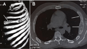
Chest CT-scan was performed in 15 patients. The mean size of the tumors was 42 mm. It showed a ground glass lesion with bone cortical thinning in 2 cases [9,10], a large mass of the hemothorax [7], a cystic lesions with a costal solid lesion [11], a benign costal lesion (Figure 1) [2,3,12,13] and multiple costal lesions [1].

    Figure 1: A/ 3-dimensional CT-scan showing a well-limited lesion of the
fourth rib, B/ Transversal CT-scan showing a costal lesion well limited with
cortical integrity.
Bone scintigraphy was performed in 3 cases [7,9,13] and showed an intense, heterogeneous mass from the third to the 9th rib [13], a heterogeneous and intense uptake of the fifth right rib [9], a heterogeneous fixation of the head, ribs and left trochanterian region [7].
Many diagnoses were supposed regarding the radiological and clinical findings including low grade sarcoma in 1 case [14], a benign bone lesion [10], a malignant bone lesion [4,10,15], bone metastases in 2 cases [10,16], fibrous dysplasia in 2 cases [5,8], vicious callus secondary to a thoracic trauma in 1 case [10].
Microscopic diagnosis was performed on biopsied samples in 9 cases [10,13,15,17] and on surgical specimen in 40 cases [1,4,5,8,9,10,12,14,16,17-20].
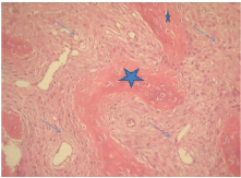
The mean size of the tumors was 9.5 cm. The cortical bone was thin in all cases and surgical limits were benign in all cases. Microscopic examination showed a double component lesion associating a fibrous component to an immature osseous component (Figure 2). An aneurismal cyst was observed in association to the tumor in 1 case. Surgical resection was performed in all cases. Surgical resection was performed through a posterior approach in 39 cases and through a video-assisted thoracoscopy in 1 case [10]. The follow up information’s were available in 22 cases. The mean follow-up period was 39 months. One patient died because of lung metastases secondary to an unknown primary cancer [6]. No complications were mentioned in the other patients. A low grade osteosarcoma was detected in 2 sites of polyostotic fibrous dysplasia in 1 patient [1].

    Figure 2:  Microscopic findings of fibrous dysplasia showing two componants:
a fibrous componant (arrow) and an immature osseous componant (star) (HE
x 400).
Discussion
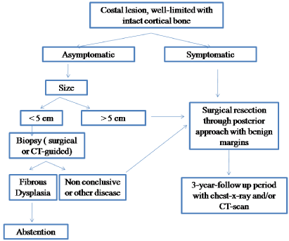
Through this review of the literature, we highlighted 2 facts that are the rarity of the costal fibrous dysplasia and the absence of consensual recommendations regarding its management. Symptoms and signs are non specific and dominated by chest pain. Although the necessity of the radiologic means in order to rule out a malignant a malignant disease, they seem to be insufficient to make the diagnosis. In fact, the diagnosis of fibrous dysplasia was suspected in only 2 cases. Surgical resection was performed in all cases even in asymptomatic ones. This fact may be due to large size of the lesions. In fact, the gross mean size of the lesions was 9cm. Surgical approach wasn’t clearly highlighted in the literature but it seems that posterior approach is widely approved. Surgical limits were benign in all cases and a malignant transformation was reported in only 1 case (2.5%) and all the patients were followed up with a mean period of 39 months. According to these findings, surgical resection seems to be necessary in large tumors (superior to 5 cm) even in asymptomatic ones. Posterior approach seems to be the best one because it was the most frequently performed without reported complications. Due to the rarity of the malignant transformation, a mean follow-up period of 3 years using chest-x-ray or CT-scan seems to be sufficient. Besides, microscopic examination is the only mean of positive diagnosis and abstention is possible only in biopsied cases.
Conclusion
Costal fibrous dysplasia is a rare benign bone tumor with non specific clinical and radiological findings. Positive diagnosis is based on microscopic findings. Surgical resection using a posterior approach is necessary especially in large lesions superior to 5cm. Abstention is possible only in asymptomatic small lesions depending on the establishment of the diagnosis which is based on the microscopic diagnosis performed on biopsies. The diagram represented in Figure 3 illustrates the main recommendations to manage these tumors according to this review of the literature.

Figure 3: Decisional diagram for the management of costal fibrous dysplasia.
References
- Van Rossem C, Pauwels P, Somville J, Camerlinck M, Bogaerts P, Van Schil PE. Sarcomatous degeneration in fibrous dysplasia of the rib cage. Ann Thorac Surg. 2013; 96: e89-90.
- Monge J, Kricun M, Radovčić J, Radovčić D, Mann A, Frayer DW. Fibrous dysplasia in a 120,000+ year old Neandertal from Krapina, Croatia. PLoS One. 2013; 8: e64539.
- Rambalde E, Parra A, Santapau A, Tardin L, Freile E, Banzo J. SPECT/CT with 99mTc-MDP in a patient with monostotic fibrous dysplasia of the rib. Rev Esp Med Nucl Imagen Mol. 2013; 32: 126-127.
- Shim JH, Chon SH, Lee CB, Heo JN. Polyostotic rib fibrous dysplasia resected by video-assisted thoracoscopic surgery with preservation of the overlying periosteum. J Thorac Cardiovasc Surg. 2010; 140: 938-940.
- Nadir A, Kaptanoglu M, Goze F. Coexisting fibrous dysplasia and bone cyst of a rib after labour trauma. Eur J Cardiothorac Surg. 2005; 27: 513.
- Fang Z, Mukai H, Nomura K, Shinomiya K, Matsumoto S, Kawaguchi N, et al. Establishment and characterization of a cell line from a malignant fibrous histiocytoma of bone developing in a patient with multiple fibrous dysplasia. J Cancer Res Clin Oncol. 2002; 128: 45-49.
- Kemp CD, Rushing GD, Rodic N, McCarthy E, Yang SC. Thoracic outlet syndrome caused by fibrous dysplasia of the first rib. Ann Thorac Surg. 2012; 93: 994-996.
- Sabatel Hernández G, Gómez Río M, Ortega Lozano S, Ramos Font C, Rodríguez Fernández A, López Ruiz JM, et al. [Fibrous bone dysplasia in the context of a McCune-Albright Syndrome]. Rev Esp Med Nucl. 2004; 23: 202-204.
- Furukawa M, Soh J, Toyooka S, Ozaki T, Miyoshi S. Resection of the entire first rib for fibrous dysplasia using a combined posterior-transmanubrial approach. Gen Thorac Cardiovasc Surg. 2012; 60: 584-586.
- Rocco G, Fazioli F, Martucci N, Cicalese M, La Rocca A, La Manna C, et al. Video-assisted thoracic surgery rib resection and reconstruction with titanium plate. Ann Thorac Surg. 2011; 92: 744-745.
- Mahadevappa A, Patel S, Ravishankar S, Manjunath GV. Monostotic fibrous dysplasia of the rib: a case report. Case Rep Orthop. 2012; 2012: 690914.
- Turcu AF, Clarke BL. Fibrous dysplasia of bone associated with primary hyperparathyroidism. Endocr Pract. 2013; 19: 226-230.
- Jakanani GC, Saifuddin A. Percutaneous image-guided needle biopsy of rib lesions: a retrospective study of diagnostic outcome in 51 cases. Skeletal Radiol. 2013; 42: 85-90.
- Traibi A, El Oueriachi F, El Hammoumi M, Al Bouzidi A, Kabiri el H. Monostotic fibrous dysplasia of the ribs. Interact Cardiovasc Thorac Surg. 2012; 14: 41- 43.
- Chen G, Yang H, Gan M, Li X, Chen K, Nalajala B, et al. Polyostotic fibrous dysplasia of the thoracic spine: case report and review of the literature. Spine (Phila Pa 1976). 2011; 36: E1485-1488.
- Hughes EK, James SL, Butt S, Davies AM, Saifuddin A. Benign primary tumours of the ribs. Clin Radiol. 2006; 61: 314-322.
- Logrono R, Kurtycz DF, Wojtowycz M, Inhorn SL. Fine needle aspiration cytology of fibrous dysplasia: a case report. Acta Cytol. 1998; 42: 1172-1176.
- Hiremagalur SR, Whitaker JH, Kumari NA, Roy TM. Fibrous dysplasia of the rib: an unusual cause of chest pain. Tenn Med. 1997; 90: 406-407.
- Jiménez CE, Moreno AJ, Pacheco EJ, Carpenter AL. SPECT imaging in a patient with monostotic rib fibrous dysplasia. Clin Nucl Med. 1996; 21: 491- 493.
- Chang BS, Lee SC, Harn HJ. Fibrous dysplasia of the rib presenting as a huge chest wall tumor: report of a case. J Formos Med Assoc. 1994; 93: 633-635.