
Review Article
Austin J Cancer Clin Res. 2016; 3(1): 1066.
Prognosis, Prevalence Trend and Different Treatment Options of Breast Cancer in Pakistan
Ayesha T1*, Muhammad AT2 and Azhar K3
1Department of Pharmaceutical Services, Ghurki Trust Teaching Hospital, Pakistan
2Department of Pharmaceutical Services, Shaukat Khanum Memorial Cancer Hospital and Research Centre, Pakistan
3Department of Pharmaceutical Services, Shaukat Khanum Memorial Cancer Hospital and Research Centre, Pakistan
*Corresponding author: JTariq Ayesha, Department of Pharmaceutical Services, Ghurki Trust Teaching Hospital, House #27, Muslim Street, Itahad Colony Samanabad, Lahore, Pakistan
Received: March 13, 2016; Accepted: July 11, 2016; Published: July 13, 2016
Abstract
Breast cancer is one of the malignant tumors that commences in the breast tissues. In this malignant tumor, cells of the breast tissue have potential of abnormal proliferation which is suspected by clinically findings such as thickening of breast, formation of breast lump, Nipple discharge and skin changes. Breast cancer is considered as heterogeneous disorder whose aetiology is multifactorial. Generally, triple assessment tool is used in clinical evaluation of breast abnormalities. This triple assessment tool comprises of: Clinical examination of presenting complaints such as breast mass, nipple discharge, breast pain, skin retraction and arm swelling; Imaging tests(mammography and ultrasound) and tissue sampling (biopsy, fine needle aspiration cytology and needle core biopsy). In Pakistan, breast cancer is the most common cancer and it ranks first among other malignancies. In 2014, only in the Lahore district, total malignancies reported were 5,521 in which 1,425 cases were of breast cancer with distribution of 1,393 and 32 among females and males respectively. In this study, we review the types, basic prognostic tools for the assessment of breast cancer and its prevalence trend from the last 10 years in Pakistan.
Keywords: Breast cancer; Prevalence; Risk factors
Introduction
The primary malignant neoplasm that starts in the cells of breast tissue is usually termed as breast cancer [1]. Cancer of breast usually starts in the ducts and lobule [2]. Cancer cells of breast can secede from its primary site and may proliferate. The proliferation of malignant cells is so fast to invade and metastasize in to different remote areas of body [3]. The prognosis and correct treatment of breast cancer is highly dependent on good perception about normal anatomy of breast [4]. The female breast is generally made up of milk producing glands (lobules), milk carrying ducts (from lobules to nipple) and stroma (comprises of fatty tissues and connective tissues enclosing lobules, tiny milk ducts, blood vessels and lymphatic vessels as well [5]. Mostly breast cancer starts in the cells lining of milk carrying ducts, called as ductal Ca breast [6]. Some breast cancers begin in the lobules (lobular Ca breast) while diminutive number commencing in other tissues of breast (Figure 1) [7].
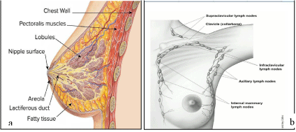
Figure 1: Anatomy of breast.
The interrelation of lobules and ducts form a closed system like twigs on a tree trunk. The cancer that is enclosed or restricted inside this closed system is termed as in-situ/non invasive breast cancer while the invasive breast cancer is that in which cancer proliferate from closed system of ducts-lobules and metastasizes in to surrounding tissues [8,9]. The lymphatic system of breast plays a vital role in the metastasis of cancerous cells [10]. The lymphatic system is usually comprises of lymph nodes (cells of immune system), lymphatic vessels (carrying clear fluid of lymph) and lymph (contain tissue fluid, waste products and immune system cells) [11]. In the breast, the majority of lymphatic vessels link to the lymph nodes underneath the arm (axillary nodes) while some of the lymphatic vessels attach to the lymph node contained by chest (internal mammary nodes) and either above the collarbone (supraclavicular nodes) or below the collarbone(infraclavicular nodes) [12].
Types of breast cancer
The breast cancer is of different types depending upon the way the cancerous cells appear under the microscope (carcinoma and sarcomas) and the proteins on cancer cells (hormone receptor positive/triple negative) [13]. Some important types of breast cancer are as follows:
Ductal carcinoma in situ (DCIS): Other name of this carcinoma is intraductal carcinoma. DCIS is usually belongs to non invasive type of cancer because it is limited to only ductal system and does not harm the neighboring tissues [14,15]. In this cancer, cells lining the milk ducts are altered to cancer cells that can’t grow outside the breast (Figure 2). DCIS is also contemplated as pre-cancer because in some cases it can progress to turn into invasive cancers [16]. Among non-invasive, DCIS is the most frequently diagnosed cancer with 1 in every 5 breast cancer patients [17].
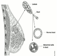
Figure 2: Abnormal duct cells in DCIS.
Invasive ductal carcinoma (IDC): Among invasive carcinomas, IDC is the most frequent type of breast cancer. The alternative term used for IDC is infiltrating ductal carcinoma [18]. This carcinoma also begins in the milk ducts but have capability to spread in the adjacent tissues via lymphatic system in to other body parts. According to American cancer society, it is roughly estimated that out of every 10 invasive Ca breast 8 patients are suffering from invasive ductal carcinoma [19,20].
Lobular carcinoma in situ (LCIS): Among non invasive breast carcinomas, LCIS is especially infrequent type of carcinoma which primarily considered as an indicator that cancer may develop in the breast. Some recent studies has been renamed the LCIS into lobular neoplasia because it is observed as peculiar growth in the number of cells milk glands (Figure 3) [21,22].
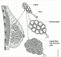
Figure 3: Abnormal cells in LCIS.
Invasive lobular carcinoma (ILC): Invasive lobular carcinoma is also known as infiltrating lobular carcinoma. It ranks 2nd after IDC. ILC begin in the lobules of breast (as a delicate thickening) and has greater ability to metastasize in to other body organs [23,24]. Rather than IDC, it might be difficult to observe ILC by means of mammogram. Its prevalence among other invasive breast cancer is 1 out of ten breast cancer patients diagnosed as ILC [25].
Inflammatory breast cancer (IBC): This cancer is an unusual and extremely destructive type of invasive breast cancer that is found to account for 1-3% of all breast cancers [26]. It is responsible to block the lymphatic vessels of breast so the breast looks distended, enlarged, red and inflamed [27]. Rather than a restricted dense mass/ lump, IBC is typically developed in layers. Additionally, it could confer the breast skin a wide, potholed appearance [28].
Paget disease of nipple: This breast cancer also starts in the ducts because it is continually linked with either DCIS or IDC but expand to the areola of the nipple and the nipple skin become irritating and scaly with area of bleeding and discharge [29]. It is very uncommon type of cancer because out of all breast cancers it is account for only 1% [30,31].
Other types of breast cancers are Phyllodes tumor (cancer of connective tissues of breast) [32], Angiosarcoma (cancer of blood and lymph vessels of breast) [33], Adenoid cystic (or adenocystic) carcinoma [34], Low-grade adenosquamous carcinoma (this is a type of metaplastic carcinoma) [35], Medullary carcinoma, Mucinous (or colloid) carcinoma, Papillary carcinoma, Tubular carcinoma, Metaplastic carcinoma (most types, including spindle cell and squamous), Micropapillary carcinoma and Mixed carcinoma (has features of both invasive ductal and lobular) [5,36].
Intrinsic subtypes of breast cancer: Classification of breast cancer on the basis of presence or absence of receptor [37,38].
A. ER positive
- Luminal A: ER and PgR positive, HER2 negative, Ki-67 ‘low’.
- Luminal B (HER-2 NEGATIVE): ER positive, HER2 negative, and at least one of Ki-67 ‘high’, PgR ‘negative or low’.
- Luminal B( HER-2 POSITIVE): ER positive, HER2 overexpressed or amplified, Any Ki-67, Any PgR.
- Erb-B2 over expression (HER2 positive ‘non-luminal’ ):
- HER2 over-expressed or amplified, ER and PgR absent.
- Basal-like(Triple negative ‘ductal’): ER and PgR absent, HER2 negative.
- Weigelt B, Peterse JL, Van't Veer LJ. Breast cancer metastasis: markers and models. Nat Rev Cancer. 2005; 5: 591-602.
- Rosen PP. Rosen's breast pathology. 2nd ed. Philadelphia: Lippincott Williams & Wilkins; 2001.
- Neal AJ, Hoskin PJ. Clinical Oncology Fourth Edition: Basic Principles and Practice: CRC Press; 2009.
- Rakha EA, Reis-Filho JS, Baehner F, Dabbs DJ, Decker T, Eusebi V, et al. Breast cancer prognostic classification in the molecular era: the role of histological grade. Breast cancer research. Breast Cancer Res. 2010; 12: 207.
- Tavassoli FA, Devilee P. Pathology and genetics of tumours of the breast and female genital organs. Iarc; 2003.
- Guray M, Sahin AA. Benign breast diseases: classification, diagnosis, and management. Oncologist. 2006; 11: 435-449.
- Peter A, Dervan. Understanding breast cancer. McFarland; 2001.
- Notingher I, Jell G, Lohbauer U, Salih V, Hench LL. In situ non‐invasive spectral discrimination between bone cell phenotypes used in tissue engineering. J Cel Biochem. 2004; 92: 1180-1192.
- Carlson RW, Allred DC, Anderson BO, Burstein HJ, Carter WB, Edge SB, et al. Invasive breast cancer. J Natl Compr Canc Netw. 2011;9: 136-222.
- Skobe M, Hawighorst T, Jackson DG, Prevo R, Janes L, Velasco P, et al. Induction of tumor lymphangiogenesis by VEGF-C promotes breast cancer metastasis. Nat Med. 2001; 7: 192-198.
- Olszewski WL. The lymphatic system in body homeostasis: physiological conditions. Lymphat Res Biol. 2003; 1: 11-21.
- Berger K, Bostwick J. A Woman's Decision: Breast Care, Treatment & Reconstruction. St. Martin's Press: 1998.
- Hernandez L, Magalhaes M, Coniglio SJ, Condeelis JS, Segall JE. Opposing roles of CXCR4 and CXCR7 in breast cancer metastasis. Breast Cancer Res. 2011; 13: R128.
- Mbeunkui F, Johann Jr DJ. Cancer and the tumor microenvironment: a review of an essential relationship. Cancer Chemother Pharmacol. 2009; 63: 571-582.
- Fisher ER, Costantino J, Fisher B, Palekar AS, Mamounas E. Redmond C. Pathologic findings from the national surgical adjuvant breast project (NSABP) protocol B‐17. Intraductal carcinoma (ductal carcinoma in situ). Cancer. 1995; 75: 1310-1319.
- Kinch M. Diagnosis of pre-cancerous conditions using pcdgf agents. Google Patents; 2004.
- Orel SG, Schnall MD. MR Imaging of the Breast for the Detection, Diagnosis, and Staging of Breast Cancer. Radiology. 2001; 220: 13-30.
- Arpino G, Bardou VJ, Clark GM, Elledge RM. Infiltrating lobular carcinoma of the breast: tumor characteristics and clinical outcome. Breast cancer research. Breast Cancer Res. 2004; 6: R149-R56.
- Borst MJ, Ingold JA. Metastatic patterns of invasive lobular versus invasive ductal carcinoma of the breast. Surgery. 1993; 114: 637-641.
- Saslow D, Boetes C, Burke W, Harms S, Leach MO, Lehman CD, et al. American Cancer Society guidelines for breast screening with MRI as an adjunct to mammography. CA Cancer J Clin. 2007; 57: 75-89.
- Afonso N, Bouwman D. Lobular carcinoma in situ. Eur J Cancer Prev. 2008; 17: 312-316.
- Cummings MC, Chambers R, Simpson PT, Lakhani SR. Molecular classification of breast cancer: is it time to pack up our microscopes?. Pathology. 2011; 43: 1-8.
- Cristofanilli M, Gonzalez-Angulo A, Sneige N, Kau S-W, Broglio K, Theriault RL, et al. Invasive lobular carcinoma classic type: response to primary chemotherapy and survival outcomes. J Clin Oncol. 2005; 23: 41-48.
- Solin LJ, Orel SG, Hwang W-T, Harris EE, Schnall MD. Relationship of breast magnetic resonance imaging to outcome after breast-conservation treatment with radiation for women with early-stage invasive breast carcinoma or ductal carcinoma in situ. J Clin Oncol. 2008; 26: 386-391.
- Schelfout K, Van Goethem M, Kersschot E, Verslegers I, Biltjes I, Leyman P, et al. Preoperative breast MRI in patients with invasive lobular breast cancer. Eur Radiol. 2004; 14: 1209-1216.
- Mukhtar RA, Nseyo O, Campbell MJ, Esserman LJ. Tumor-associated macrophages in breast cancer as potential biomarkers for new treatments and diagnostics. Expert Rev Mol Diagn. 2011; 11: 91-100.
- Jungi W. The prevention and management of lymphoedema after treatment for breast cancer. Int Rehabil Med. 1981; 3: 129-134.
- Dushkin H, Cristofanilli M. Inflammatory breast cancer. J Natl Compr Canc Netw. 2011; 9: 233-241.
- Kopans DB. Breast imaging. Lippincott Williams & Wilkins: 2007.
- Bijker N, Rutgers EJ, Duchateau L, Peterse JL, Julien JP, Cataliotti L. Breast‐conserving therapy for paget disease of the nipple: a prospective European Organization for Research and Treatment of Cancer study of 61 patients. Cancer. 2001; 91: 472-477.
- Julien J-P, Bijker N, Fentiman IS, Peterse JL, Delledonne V, Rouanet P, et al. Radiotherapy in breast-conserving treatment for ductal carcinoma in situ: first results of the EORTC randomised phase III trial 10853. Lancet. 2000; 355: 528-533.
- Tan PH, Jayabaskar T, Chuah KL, Lee HY, Tan Y, Hilmy M, et al. Phyllodes tumors of the breast: the role of pathologic parameters. Am J Clin Pathol. 2005; 123: 529-540.
- Schoppmann SF, Bayer G, Aumayr K, Taucher S, Geleff S, Rudas M, et al. Prognostic value of lymphangiogenesis and lymphovascular invasion in invasive breast cancer. Ann Surg. 2004; 240: 306-312.
- Arpino G, Clark GM, Mohsin S, Bardou VJ, Elledge RM. Adenoid cystic carcinoma of the breast: molecular markers, treatment, and clinical outcome. Cancer. 2002; 94: 2119-2127.
- Van Hoeven K, Drudis T, Cranor ML, Erlandson RA, Rosen PP. Low-Grade Adenosquamous Carcinoma of the Breast: A Clinocopathologic Study of 32 Cases with Ultrastructural Analysis. Am J Pathol. 1993; 17: 248-258.
- Elston CW, Ellis IO. Pathological prognostic factors in breast cancer. I. The value of histological grade in breast cancer: experience from a large study with long-term follow-up. Histopathology. 1991; 19: 403-10.
- Parker JS, Mullins M, Cheang MC, Leung S, Voduc D, Vickery T, et al. Supervised risk predictor of breast cancer based on intrinsic subtypes. J Clin Oncol. 2009; 27: 1160-1167.
- Goldhirsch A, Wood W, Coates A, Gelber R, Thürlimann B, Senn HJ, et al. Strategies for subtypes—dealing with the diversity of breast cancer: highlights of the St Gallen International Expert Consensus on the Primary Therapy of Early Breast Cancer 2011. Ann Oncol. 2011: 1736-1747.
- Key TJ, Verkasalo PK, Banks E. Epidemiology of breast cancer. Lancet Oncol. 2001; 2: 133-140.
- Yancik R, Wesley MN, Ries LA, Havlik RJ, Edwards BK, Yates JW. Effect of age and comorbidity in postmenopausal breast cancer patients aged 55 years and older. JAMA. 2001; 285: 885-892.
- Antoniou A, Pharoah PD, Narod S, Risch HA, Eyfjord JE, Hopper JL, et al. Average risks of breast and ovarian cancer associated with BRCA1 or BRCA2 mutations detected in case series unselected for family history: a combined analysis of 22 studies. Am J Hum Genet. 2003; 72: 1117-1130.
- Boyd NF, Martin LJ, Bronskill M, Yaffe MJ, Duric N, Minkin S. Breast tissue composition and susceptibility to breast cancer. J Natl Cancer Inst. 2010; 102: 1224-1237.
- Black MM, Barclay TH, Cutler SJ, Hankey BF, Asire AJ. Association of atypical characteristics of benign breast lesions with subsequent risk of breast cancer. Cancer. 1972; 29: 338-343.
- Cancer CGoHFiB. Breast cancer and hormonal contraceptives: collaborative reanalysis of individual data on 53 297 women with breast cancer and 100 239 women without breast cancer from 54 epidemiological studies. Lancet. 1996; 347:1713-1727.
- Kelsey JL, Gammon MD, John EM. Reproductive factors and breast cancer. Epidemiol Rev. 1993; 15: 36-47.
- Doherty LF, Bromer JG, Zhou Y, Aldad TS, Taylor HS. In utero exposure to diethylstilbestrol (DES) or bisphenol-A (BPA) increases EZH2 expression in the mammary gland: an epigenetic mechanism linking endocrine disruptors to breast cancer. Horm Cancer. 2010; 1: 146-155.
- Skegg DC, Noonan EA, Paul C, Spears GF, Meirik O, Thomas DB. Depot medroxyprogesterone acetate and breast cancer: a pooled analysis of the World Health Organization and New Zealand studies. JAMA. 1995; 273: 799-804.
- McNeely ML, Campbell KL, Rowe BH, Klassen TP, Mackey JR, Courneya KS. Effects of exercise on breast cancer patients and survivors: a systematic review and meta-analysis. CMAJ. 2006; 175: 34-41.
- Rakha EA, Reis-Filho JS, Ellis IO. Combinatorial biomarker expression in breast cancer. Breast cancer research and treatment. 2010; 120: 293-308.
- McPherson K, Steel C, Dixon J. ABC of breast diseases: breast cancer—epidemiology, risk factors, and genetics. BMJ. 2000; 321: 624-628.
- Thompson D, Easton DF, Consortium BCL. Cancer incidence in BRCA1 mutation carriers. J Natl Cancer Inst. 2002; 94: 1358-1365.
- Aloraifi F, Alshehhi M, McDevitt T, Cody N, Meany M, O'Doherty A, et al. Phenotypic analysis of familial breast cancer: Comparison of BRCAx tumors with BRCA1-, BRCA2-carriers and non-familial breast cancer. Eur J Surg Oncol. 2015; 41: 641-646.
- Visvader JE, Lindeman GJ. Cancer stem cells: current status and evolving complexities. Cell Stem Cell. 2012; 10: 717-728.
- Robertson FM, Bondy M, Yang W, Yamauchi H, Wiggins S, Kamrudin S, et al. Inflammatory breast cancer: the disease, the biology, the treatment. CA Cancer J Clin. 2010; 60: 351-375.
- Nicholson BT, Harvey JA, Cohen MA. Nipple-Areolar Complex: Normal Anatomy and Benign and Malignant Processes 1. Radiographics. 2009; 29: 509-523.
- Jung BF, Ahrendt GM, Oaklander AL, Dworkin RH. Neuropathic pain following breast cancer surgery: proposed classification and research update. Pain. 2003; 104: 1-13.
- Brachtel EF, Rusby JE, Michaelson JS, Chen LL, Muzikansky A, Smith BL, et al. Occult nipple involvement in breast cancer: clinicopathologic findings in 316 consecutive mastectomy specimens. J Clin Oncol. 2009; 27: 4948-4954.
- Armer JM, Radina ME, Porock D, Culbertson SD. Predicting breast cancer-related lymphedema using self-reported symptoms. Nurs Res. 2003; 52: 370-379.
- Mannello F. Analysis of the intraductal microenvironment for the early diagnosis of breast cancer: identification of biomarkers in nipple-aspirate fluids. Expert Opin Med Diagn. 2008; 2: 1221-1231.
- Fentiman IS, Fourquet A, Hortobagyi GN. Male breast cancer. The Lancet. 2006; 367: 595-604.
- Jemal A, Bray F, Center MM, Ferlay J, Ward E, Forman D. Global cancer statistics. CA Cancer J Clin. 2011; 61: 69-90.
- Sujindra E, Elamurugan T. Knowledge, attitude, and practice of breast self-examination in female nursing students IJEPR. 2015; 1: 71-74.
- Siegel RL, Miller KD, Jemal A. Cancer statistics, 2015. CA Cancer J Clin. 2015; 65: 5-29.
- Medina-Franco H, Vasconez LO, Fix RJ, Heslin MJ, Beenken SW, Bland KI, et al. Factors associated with local recurrence after skin-sparing mastectomy and immediate breast reconstruction for invasive breast cancer. Ann Surg. 2002; 235: 814-819.
- Banning M, Hafeez H, Faisal S, Hassan M, Zafar A. The impact of culture and sociological and psychological issues on Muslim patients with breast cancer in Pakistan. Cancer Nurs. 2009; 32: 317-324.
- www.punjabcancerregistry.org.pk/reports/PCR_2014.pdf.
- Shaukat Khanum Memorial Cancer Hospital & Research Centre.
- Warner E, Plewes DB, Hill KA, Causer PA, Zubovits JT, Jong RA, et al. Surveillance of BRCA1 and BRCA2 mutation carriers with magnetic resonance imaging, ultrasound, mammography, and clinical breast examination. JAMA. 2004; 292: 1317-1325.
- Organization WH. Guidelines for management of breast cancer. 2006.
- Hammond ME, Hayes DF, Dowsett M, Allred DC, Hagerty KL, Badve S, et al. American Society of Clinical Oncology/College of American Pathologists guideline recommendations for immunohistochemical testing of estrogen and progesterone receptors in breast cancer. J Clin Oncol. 2010; 28: 2784-2795.
- Paruthiyil S, Parmar H, Kerekatte V, Cunha GR, Firestone GL, Leitman DC. Estrogen receptor β inhibits human breast cancer cell proliferation and tumor formation by causing a G2 cell cycle arrest. Cancer Res. 2004; 64: 423-428.
- Konecny G, Pauletti G, Pegram M, Untch M, Dandekar S, Aguilar Z, et al. Quantitative association between HER-2/neu and steroid hormone receptors in hormone receptor-positive primary breast cancer. J Natl Cancer Inst. 2003; 95:142-153.
- Tanner M, Gancberg D, Di Leo A, Larsimont D, Rouas G, Piccart MJ, et al. Chromogenic in situ hybridization: a practical alternative for fluorescence in situ hybridization to detect HER-2/neu oncogene amplification in archival breast cancer samples. Am J Pathol. 2000; 157: 1467-1472.
- Scholzen T, Gerdes J. The Ki-67 protein: from the known and the unknown. J Cell Physiol. 2000; 182: 311-322.
- DeRisi JL, Iyer VR, Brown PO. Exploring the metabolic and genetic control of gene expression on a genomic scale. Science. 1997; 278: 680-686.
- Rowland JH, Desmond KA, Meyerowitz BE, Belin TR, Wyatt GE, Ganz PA. Role of breast reconstructive surgery in physical and emotional outcomes among breast cancer survivors. J Natl Cancer Inst. 2000; 92: 1422-1429.
- Fisher B, Costantino J, Redmond C, Poisson R, Bowman D, Couture J, et al. A randomized clinical trial evaluating tamoxifen in the treatment of patients with node-negative breast cancer who have estrogen-receptor–positive tumors. N Engl J Med. 1989; 320: 479-484.
- Fisher B, Bauer M, Margolese R, Poisson R, Pilch Y, Redmond C, et al. Five-year results of a randomized clinical trial comparing total mastectomy and segmental mastectomy with or without radiation in the treatment of breast cancer. N Engl J Med. 1985; 312: 665-673.
- Pawitan Y, Bjöhle J, Amler L, Borg A-L, Egyhazi S, Hall P, et al. Gene expression profiling spares early breast cancer patients from adjuvant therapy: derived and validated in two population-based cohorts. Breast Cancer Res. 2005; 7: R953-R964.
- Burger RA, Sill MW, Monk BJ, Greer BE, Sorosky JI. Phase II trial of bevacizumab in persistent or recurrent epithelial ovarian cancer or primary peritoneal cancer: a Gynecologic Oncology Group Study. J Clin Oncol. 2007; 25: 5165-5171.
- Fisher B, Redmond C, Legault-Poisson S, Dimitrov NV, Brown AM, Wickerham DL, et al. Postoperative chemotherapy and tamoxifen compared with tamoxifen alone in the treatment of positive-node breast cancer patients aged 50 years and older with tumors responsive to tamoxifen: results from the National Surgical Adjuvant Breast and Bowel Project B-16. J Clin Oncol. 1990; 8: 1005-1018.
- Mauri D, Pavlidis N, Ioannidis JP. Neoadjuvant versus adjuvant systemic treatment in breast cancer: a meta-analysis. J Natl Cancer Inst. 2005; 97: 188-194.
- Thomas E, Holmes FA, Smith TL, Buzdar AU, Frye DK, Fraschini G, et al. The Use of Alternate, Non–Cross-Resistant Adjuvant Chemotherapy on the Basis of Pathologic Response to a Neoadjuvant Doxorubicin-Based Regimen in Women With Operable Breast Cancer: Long-Term Results From a Prospective Randomized Trial. J Clin Oncol. 2004; 22: 2294-2302.
- Pusic AL, Klassen AF, Scott AM, Klok JA, Cordeiro PG, Cano SJ. Development of a new patient-reported outcome measure for breast surgery: the BREAST-Q. Plast Reconstr Surg. 2009; 124: 345-353.
- Bhatti ABH, Jamshed A, Shah MA, Khan A. Breast conservative therapy in Pakistani women: Prognostic factors for locoregional recurrence and overall survival. J Cancer Res Ther. 2015; 11: 300-304.
- Baskar R, Lee KA, Yeo R, Yeoh KW. Cancer and radiation therapy: current advances and future directions. Int J Med Sci. 2012; 9: 193-199.
- D'Amico AV, Whittington R, Malkowicz SB, Schultz D, Blank K, Broderick GA, et al. Biochemical outcome after radical prostatectomy, external beam radiation therapy, or interstitial radiation therapy for clinically localized prostate cancer. JAMA. 1998; 280: 969-974.
- Shaukat Khanum Memorial Cancer Hospital & Research Centre.
- Munro A, Potter S. A quantitative approach to the distress caused by symptoms in patients treated with radical radiotherapy. Br J Cancer. 1996; 74: 640-647.
- Davis BJ, Horwitz EM, Lee WR, Crook JM, Stock RG, Merrick GS, et al. American Brachytherapy Society consensus guidelines for transrectal ultrasound-guided permanent prostate brachytherapy. Brachytherapy. 2012; 11: 6-19.
- Hiddemann W, Martin W, Sauerland C, Heinecke A, Büchner T. Definition of refractoriness against conventional chemotherapy in acute myeloid leukemia: a proposal based on the results of retreatment by thioguanine, cytosine arabinoside, and daunorubicin (TAD 9) in 150 patients with relapse after standardized first line therapy. Leukemia. 1990; 4: 184-188.
- Cheung K. Endocrine therapy for breast cancer: an overview. The Breast. 2007; 16: 327-343.
- Veronesi U, Cascinelli N, Mariani L, Greco M, Saccozzi R, Luini A, et al. Twenty-year follow-up of a randomized study comparing breast-conserving surgery with radical mastectomy for early breast cancer. N Engl J Med. 2002; 347: 1227-1232.
- Fisher B, Anderson S, Bryant J, Margolese RG, Deutsch M, Fisher ER, et al. Twenty-year follow-up of a randomized trial comparing total mastectomy, lumpectomy, and lumpectomy plus irradiation for the treatment of invasive breast cancer. N Engl J Med. 2002; 347: 1233-1241.
- Veronesi U, Paganelli G, Galimberti V, Viale G, Zurrida S, Bedoni M, et al. Sentinel-node biopsy to avoid axillary dissection in breast cancer with clinically negative lymph-nodes. Lancet. 1997; 349: 1864-1867.
B. ER negative
Risk Factors for the Breast Cancer
Something that increases the possibility to acquire a cancer is called risk factor. Risk factors that have influence on breast cancer are:
Non-genetic factors
Females have greater chance (100 times) to develop breast cancer as compared to man because females have more estrogen and progesterone hormones which encourage the cancer cells growth in breast [39]. The probability of having breast cancer is higher with older age [40]. In addition, having family history of breast cancer [41], personal history of breast cancer, chest region including breast expose to radiation, dense breast tissues [42], certain benign breast lesions (nonproliferative lesions, proliferative lesions without atypia and proliferative lesions with atypia) [43], female who had their 1st menstrual period before the age 12 yrs, use of oral contraceptives [44], obesity after the period of menopause [45], lack of physical exercise, exposure to diethylstilbestrol [46], use of Depotmedroxyprogesterone acetate (DMPA) [47], use of combinational hormonal therapy after menopause, alcohol drinking, inadequate diet (less intake of fibrous food, fruits and vegetables) and exposure to environmental chemicals (Pesticides such as DDE, polychlorinated biphenyls) are major contributing risk factors for breast cancer [48,49].
Genetic risk factors
It has been reported in some recent studies that approximately 5-10% cases of breast cancer are the results of genetic mutation (that inherited from their parents) [50,51]. The mutation in the genes of BRCA1, BRCA2, ATM, TP53, CHEK2, PTEN, CDHI, STK11 and PALB2 are the recognized contributors toward breast cancer [52].
Signs and Symptoms of Breast Cancer
Generally in the primitive stage, breast carcinomas don’t exhibit any signs but as the tumor cells proliferate and metastasize, it can cause some changes in the breast tissues [53]. The most common symptoms relating to breast cancer are: development of lump/mass [10], distension and inflammation of breast [54], skin of nipple and areola becomes itchy and crusty [55], painful feeling in the breast [56], nipple retraction [57], discharge of fluid other than milk from the nipple and breast skin show crumpling and indentation [58,59].
Prevalence of Breast Cancer in Pakistan
Both males and females are affected by this group of neoplasm but in males it is rarely reported and does not happen usually [60]. Among women, breast cancer is most commonly diagnosed cancer both in developed and less developed countries. According to the global health estimates of WHO 2013, it is reported that worldwide breast cancer was the death reason of over 508,000 women in 2011 [61]. Incidence rates for the breast cancer vary significantly from less developed to developed countries (such as 19.3/100,000 in eastern Africa and 89.7/100,000 in Western Europe).
From some recent studies, it has been reported worldwide that in year 2011, incidence rates for the breast cancer among men and women were 1.3/100,000, and 124.3/100,000 respectively [61,62]. In contrast to incidence rate, 0.3/100,000 and 21.5/100,000 were mortality rates that were recorded in that year for men and women respectively [63]. WHO said in one of report that “Breast cancer rates are getting worse and it is not sparing even younger age group.” In less developed countries like Pakistan, women tend to die at greater rates than in developed countries because in general breast cancer is detected when it is in advanced stages [64].
In Pakistan, breast cancer is the most commonly diagnosed cancer. According to Punjab cancer registry 2014, total malignancies reported in the year 2014 is 5521, out of which 1425 cases were contributed by breast cancer with prevalence rate of 25.8% that is quite higher from the previous year’s [65,66]. According to Cancer registry report of Shoakat Khanum memorial cancer hospital and research center, total number of the cases of breast cancer reported from the last 10 years (2004-2014) are 13992, out which 13882 and 110 cases were contributed by females and males breast cancers respectively [67]. Graphical representation (Graph 1 and 2) given at the end, show the prevalence trend of breast cancer in Pakistan from the last 10 years.
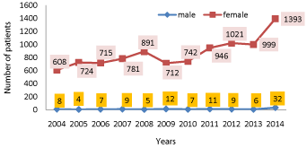
graph 1: Prevalence trend of breast cancer in both genders.
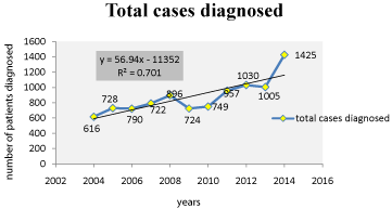
graph 2: Overall survival, autologous stem cell transplant (ASCT) versus no ASCT (p=0.12).
Detection and Prognosis of Breast Cancer
Generally, triple assessment tool is used in clinical evaluation of breast abnormalities. This triple assessment tool comprises of: Clinical examination of presenting complaints such as breast mass, nipple discharge, breast pain, skin retraction and arm swelling; Imaging tests (mammography and ultrasound); And tissue sampling (biopsy, fine needle aspiration cytology and needle core biopsy).
In order to diagnose the breast cancer, following diagnostic tests are used such as Clinical examination of breast (breast self examination and physical examination), imaging tests to differentiate benign and malignant carcinomas and biopsies [68]. Other laboratory investigations are also used such as complete blood count with differentials (CBCD), renal and liver profile, chest X-ray with CT, abdominal ultrasound with CT of abdomen, bone scan, ECG and multiple gated acquisition (MUGA) scan if age >60, positron emission tomography (Figure 4) [69]. Receptors test is also employed to evaluate the breast cancer type [70]. In this test removed part of tumor (during biopsy/surgery) is assessed for the presence or absence of receptors (estrogen, progesterone, and HER-2). The presence of estrogen receptors on breast cancer is termed as estrogen positive cancers on the other hand cancers will be PR +ve if the receptor test is positive for progesterone and the tumors with over expression of HER-2 is characterized as HER-2 positive [71,72].
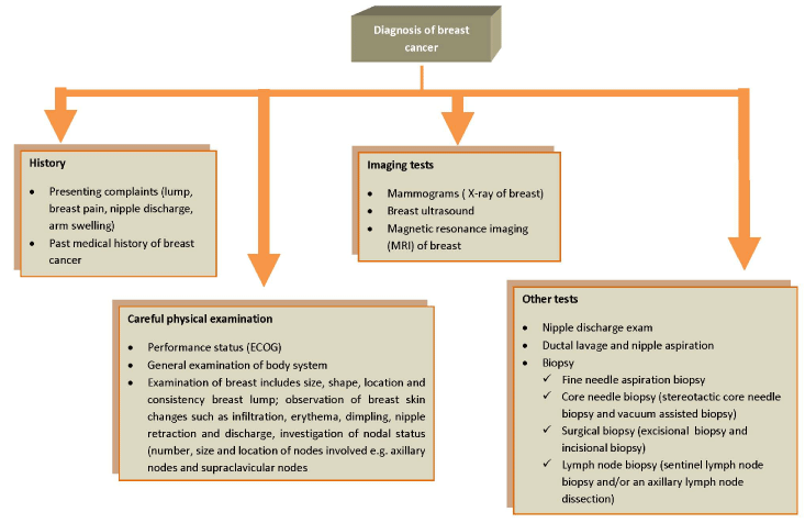
Figure 4: Flow chart for the breast cancer diagnosis.
Tests usually employed to analyze the biopsy/surgery samples are immunohistochemistry test (IHC), Fluorescent in situ hybridization (FISH) and Chromogenic in situ hybridization (CISH) [73]. In addition to above tests, test of ploidy and cell proliferation rate are utilized to assess the abnormal DNA in the cells and rate of cell division by estimating the ki-67 labeling index that help in better prognosis [74]. Gene expression profiling is used to find the pattern of different genes [75].
Treatment of Breast Cancer
Different therapies are used to treat the breast cancer and treatment is highly depending upon the better prognosis and staging of cancer [76]. Different options for breast cancer are surgery, radiation therapy, chemo therapy, hormonal therapy, targeted therapy and bone directed therapy [77]. These treatment modalities are categorized in to two main groups such as:
Local and systemic therapies
Surgery and radiation therapies are the example of local therapy because their purpose is to treat the localized tumor without affecting the rest of the body parts [78]. While chemotherapy, hormone therapy and targeted therapy belong to systemic therapy because they are directly introduced in to blood stream to attain access toward cancerous cells.
Adjuvant and neoadjuvant therapy
Administration of additional treatment to exterminate and restrain the clinically unnoticeable cancerous cells after primary surgery is termed as adjuvant therapy [79]. The primary objective of this therapy is to prevent the recurrence of cancer [80]. This prudent approach has been recognized to improve the survival in both node -ve and node +ve breast cancer in combination with systemic and hormonal therapy (optional). Through adjuvant therapy, survival of patients could be enhanced up to 10 years by 7%–11% in premenopausal women with early stage breast cancer and by 2-3% in women having age above 50 years [81].
Neoadjuvant therapy is defined as administration of treatment (chemotherapy or hormonal therapy) prior to surgery to reduce the tumor size [82]. The possibility of cancer recurrence become also reduces by using this approach. In the treatment of locally advanced breast cancer, this pre-operative induction chemotherapy is considered as reasonable approach because of having higher response rate [83].
Surgery for breast cancer
Surgery has been considered as most primitive and frequently used treatment option for breast cancer [84]. Types of surgeries to remove breast tumors are mentioned in Table 1.
    Breast Surgery
    Description
    Possible side effects of surgeries
    References


    Breast conserving surgery
    Other common names for this surgery are partial    mastectomy and Lumpectomy. This surgery is performed to amputate a part of    breast that is confined to cancer.
    Pain, transient inflammation, soreness, bleeding,    tough scar tissue and infection at the site of surgery are the possible side    effects associated with theses surgeries.
    [93]


    Mastectomy
    Mastectomy is of following types
      i)total mastectomy
      ii) skin sparing mastectomy
      iii)modified radical mastectomy iv) radical    mastectomy
    In addition to pain and apparent change in the    breast shape, the potential side effects of mastectomy include wound    infection, hematoma and seroma.
    [94]


    Lymph node surgery
    Lymph node surgery is the removal of lymph nodes    (under arm) if breast cancer has spread and invade to lymph nodes. This    surgery are of two types:
      i) Axillary lymph node dissection
      ii) Sentinel lymph node biopsy
    Burning pain, arm swelling, bleeding, infection    Lymphedema, frozen shoulder, lack of sensation of the skin on the upper and    inner arm and lymphatic cording
    [95]
Table 1: Types of surgeries to remove breast tumor.
One of the established methods for treating the early stage invasive breast cancer is Breast conservative therapy (BCT).one of the recent study carried out at Shoakat Khanum memorial hospital and research center reported that BCT has satisfactory long-term results in Pakistani women [85].
Radiation therapy for breast cancer
In radiotherapy, high energy rays are used to exterminate the cancerous cells. Radiotherapy is frequently used subsequent to breast surgery [86]. The main purpose of this therapy is to lessen the possibility of cancer re-emergence in the breast tissues and neighboring lymph nodes. This therapy can be given by two ways: external beam radiation and internal radiotherapy (Figure 5) [87].
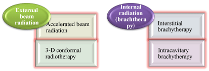
Figure 5: Types of radiotherapy.
Radiotherapy available in Pakistan:
A. External Beam Radiation Therapy
Intensity Modulated Radiation Therapy (IMRT), Conformal Radiation Therapy and Image-Guided Radiation Therapy (IGRT) are the External Beam Radiation Therapies used in Shoakat Khanum memorial cancer hospital and research center, Pakistan by Radiation Oncologists in order to treat various types of cancers according to subspecialties such as head and neck, brain, prostate, lung breast, and gynecological cancers. IMRT delivers doses of radiation with different intensity levels within 2mm square segments, which optimizes the radiation dose to the irregularly shaped tumors while minimizing the dose to surrounding structures, further reducing side effects. The IGRT permits the radiation oncologist to target the tumor by adjusting the radiation beam so that radiation with higher dose can be delivered safely [88].
B. Brachytherapy (Internal Radiotherapy)
In brachytherapy, radioactive seeds are carefully placed inside cancerous tissue and positioned to attack the cancer most effectively. A brachytherapy is an outpatient procedure that is effective in treating esophagus, lung, gynecologic, and breast cancers, among some others. SKMCH&RC is the only 3D HIGH DOSE RATE brachytherapy provider in Pakistan [88].
There are several side effects of radiotherapy such as Inflammation and heaviness in the breast; fatigue; skin changes in the treated area (redness, wound and peeling); brachial plexopathy; numbness, pain and weakness in the arm, shoulder and hand; cracking of the ribs; lymphedema and angiosarcoma are associated with external beam radiation [89]. In addition Breast pain, redness of breast skin, bruising, infection, weakness and fracture of the ribs are related with brachytherapy [90].
Chemotherapy for breast cancer
Chemotherapy (anticancer treatment) is intended to kill the cancerous cells/abnormal growth in the body that could be administered either orally or by intravenously. This treatment is frequently delivered in cycles and continued for several months depending upon the recovery [91]. Chemotherapeutic agents (Figure 6) are usually recommended for following three situations: after surgery (as adjuvant therapy), before surgery (as neoadjuvant therapy) and for advanced stage breast cancer (as palliative therapy). Chemotherapy is commonly used in combination of one or more drug [92].
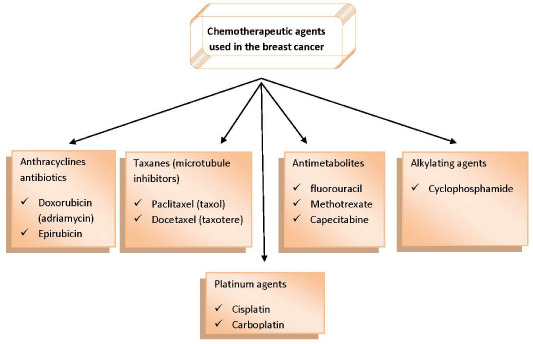
Figure 6: Chemotherapeutic agents to treat the breast cancer.
Conclusion
Globally, carcinoma of breast is one of the most common malignancies among females. In Pakistan, breast cancer is most frequently diagnosed at young age with highest incidence rate of 50/100,000 as compared to west where it is more common after the age of 60. Approximately one in every nine Pakistani women is likely to suffer from breast cancer. This review summarized the types of breast cancer, potential causative agents, diagnostic tools, different treatments for this chronic disease and its prevalence trend in Pakistan from the last 10 years. Annual Punjab cancer registry report 2014 described that only in the Lahore district; total malignancies reported from Jan 2014-Dec 2014 were 5,521 in which 1,425 cases were of breast cancer with distribution of 1,393 and 32 among females and males respectively. Rigorous research is required in this field to aware the females because breast cancer management at advanced stages is a big challenge for developing countries.
References