
Case Report
Austin Cardio & Cardiovasc Case Rep. 2018; 3(1): 1018.
A Case of Primary Cardiac Angiosarcoma Presenting with Hemorrhagic Pleural and Pericardial Effusions in a Young Woman
Dayanand P¹*, Kabach M², Alrifai A², Lovitz L² and Nores M³
¹Department of Internal Medicine, University of Miami/ JFK Medical Center, Florida, USA
²Department of Cardiology, University of Miami/JFK Medical Center, Florida, USA
*³Department of Cardiothoracic Surgery, JFK Medical Center, Florida, USA
*Corresponding author: Pradeep Dayanand, Department of Internal Medicine, University of Miami/ JFK Medical Center, 5301 South Congress Ave Atlantis, FL 33462, USA
* DePersis M, Division of Cardiology, Robert Packer Hospital, Guthrie Health Systems, Sayre PA, USA
Received: February 06, 2018; Accepted: February 23, 2018; Published: March 02, 2018
Abstract
In today’s medical practice, young and relatively healthy patients presenting with recurrent hemorrhagic pleuro-pericardial effusions in a short time span are seldom encountered. The authors of this article present a case of a 39-year-old woman who presented with recurrent pleuro-pericardial hemorrhagic effusions, an extensive work-up was undertaken subsequently which revealed a possible mass arising from the right atrial free wall and compressing the right ventricle. At this point, a primary cardiac malignancy was high on our differential and we debated cardiac biopsy versus conservative management with her family and proceeded with cardiac biopsy to establish a definitive diagnosis. The biopsy resulted in hemorrhagic shock and her unfortunate death despite multiple blood transfusions. Histopathology of the biopsy confirmed the diagnosis of primary cardiac angiosarcoma after her death. Primary cardiac malignancies are extremely rare and patients have a wide array of non-specific symptoms at presentation, either due to associated effusions or mass effect from the tumor. The prognosis is grave and treatment options are limited, the median survival is only about 6-9 months. Cardiac biopsy is required to make the diagnosis of primary cardiac angiosarcoma but it can be associated with unfavorable outcomes because of the highly vascular nature of the tumor.
Keywords: Biopsy; Angiosarcoma; Tumor
Introduction
Primary cardiac tumors are extremely rare, with malignant tumors comprising one fourth of all cardiac tumors, majority of which are angiosarcomas [1]. They tend to occur in patients between 30-40 years old and are thrice as frequent in men as compared to women [2]. They mostly emerge from the right atrium although they might also involve other cardiac chambers less frequently [3]. Recurrent pericardial effusions in the setting of elevated troponins poses a dilemma both in diagnosis and management as the presentation can be vague and the lack of effective treatment modalities further compounds the problem especially in the context of a rapidly enlarging primary cardiac vascular tumor.
Case Presentation
Here in, we present a case of a young woman who presented with shortness of breath, chest pain & mild troponin elevation.
A 39-year-old female with no known medical co-morbidities presented with one week of pleuritic chest pain, dyspnea on exertion and orthopnea. She had a similar presentation to an outside facility two weeks prior to this, where she was found to have a pericardial effusion which was drained.
On exam, she was tachypneic, in significant distress, hypotensive with blood pressure of 80/60, tachycardic with pulse of 106, she also was found to have distended jugular veins, and muffled heart sounds. The rest of her exam was within normal limits. Subsequent work up included an electrocardiogram which was unremarkable except for sinus tachycardia. Chest X ray (Figure 1) revealed an enlarged cardiac silhouette and a left sided pleural effusion. A Transthoracic Echocardiogram (TTE) was undertaken next which revealed a large pericardial effusion with tamponade physiology (Figure 2). She then underwent emergent pericardiocentesis along with thoracentesis which yielded hemorrhagic fluid with an exudative cytology and a chest tube was left in place. During her hospital course, she kept draining continuously from her chest tube and eventually underwent a pericardial window. Coronary angiography was performed next to determine a possible culprit lesion for elevated troponins which demonstrated non obstructive disease but there was noticeable contrast filling a mass on the right atrial free wall and abutting the right ventricle (Figures 3, 4). Transesophageal Echocardiogram (TEE) was performed to evaluate the mass which revealed a large pericardial mass attached to the right atrium and compressing the right ventricle (Figures 5, 6). Cardiac Magnetic Resonance (CMR) test was attempted to further identify the mass, however it could not be completed due to patient’s instability and tachycardia. A primary cardiac vascular tumor was thought to be the underlying cause, incisional biopsy was planned to establish definitive diagnosis. Incisional biopsy (Figure 7, 8) was performed as a definitive option but massive uncontrollable hemorrhagic shock ensued which finally resulted in the unfortunate death of the patient despite multiple blood transfusions. The histopathology results of the biopsy specimen were interpreted as primary cardiac angiosarcoma (Figures 9,10) after the patient’s death.
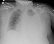
Figure 1: Chest X-ray showing large pleuro- pericardial effusion.
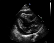
Figure 2: TTE depicting pericardial effusion with tamponade.
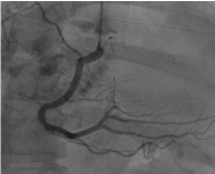
Figure 3: Cardiac catheterization illustrating vascular tumor.
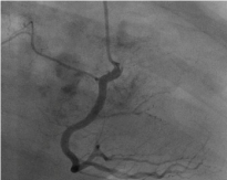
    Figure 4:  Vascular tumor blush upon injection of contrast into right coronary
artery.
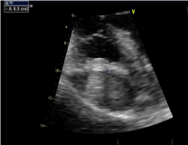
Figure 5: TEE revealing cardiac mass arising from right atrium.
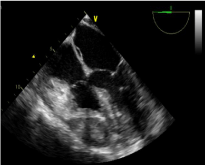
    Figure 6:  TEE depecting compression of the right ventricle by the cardiac
mass.

Figure 7: Mediastinal exploration revealing tumor extent.
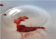
Figure 8: Gross specimen obtained upon incisional biopsy.
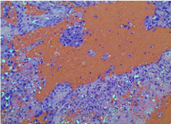
    Figure 9:  Histopathology of the biopsy specimen showing spindle-shaped
cells with pleomorphic nuclei lining anastomosing vascular spaces. Mitotic
figures and areas of hemorrhage and necrosis can also be visualized.
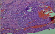
    Figure 10:  Histopathology of the biopsy specimen showing spindle-shaped
cells with pleomorphic nuclei lining anastomosing vascular spaces. Mitotic
figures and areas of hemorrhage and necrosis can also be visualized.
Discussion
Cardiac angiosarcomas are highly vascular and aggressive primary cardiac tumors [4]. They can cause recurrent pericardial effusions [5] in affected patients and it is conceivable that they might also cause a troponin leak by invading inwards into the myocardium.
The clinical presentation of angiosarcomas is determined by the location of the tumor rather than its histopathology, and the initial presentation might vary from superior vena cava syndrome or peripheral edema to heart failure or arrhythmias correspondingly [6,7].
Echocardiogram, cardiac CT and cardiac MRI are the diagnostic modalities used to identify the presence of a primary cardiac tumor [4] and its relationship to surrounding structures, echo being the most widely used and economically feasible as it not only shows the location and extent of the tumor but also reveals hemodynamic changes if any [8]. Trans-esophageal echo offers a slight advantage over transthoracic echo in delineating tumor anatomy and valvular anomalies especially in tumors involving the posterior wall [9].
Microscopically this is a high-grade necrotizing and hemorrhagic tumor comprised by irregular vascular spaces lined by cells with high grade nuclear atypia [4] Immunohistochemically they usually stain for CD 31, CD 34 and factor VIII related protein [10].
They frequently pose a dilemma in diagnosis and management because of the absence of specific treatment and poor prognosis [11,12]. Though surgery is the mainstay of treatment [13,14], it fails to improve survival because complete resection is often not possible give the location of the tumor, complications post-surgery, and its strong predisposition for metastasis as more than three fourth of the patients have widespread metastasis upon presentation [15].
References
- McAllister HA. Benign tumors and cysts of the heart and pericardium, myxoma. Tumors of the cardiovascular system, in atlas of tumor pathology. 1978: 5-20.
- Sabatine M, Colucci W, Schoen F. Primary tumors of the heart. Braunwald’s heart disease. A Textbook of Cardiovascular Medicine. 2005: 1741-1756.
- Herrmann MA, Shankerman RA, Edwards WD, Shub C, Schaff H. Primary cardiac angiosarcoma: a clinicopathologic study of six cases. J Thorac Cardiovasc Surg. 1992; 103: 655-664.
- Kurian KC, Weisshaar D, Parekh H, Berry GJ, Reitz B. Primary cardiac angiosarcoma: case report and review of the literature. Cardiovascular Pathology. 2006; 15: 110-112.
- Lee CH, Chan GS, Chan WM. Unexplained recurrent pericardial effusion: a lethal warning? Heart. 2003; 89: e11.
- Shanmugam G. Primary cardiac sarcoma. European journal of Cardiothoracic surgery. 2006; 29: 925-932.
- Schwartzman P, White R. Imaging of cardiac and pericardial masses. J Thorac Imaging. 2000; 15: 265-273.
- Meng Q, Lai H, Lima J, Tong W, Qian Y, Lai S. Echocardiographic and pathologic characteristics of primary cardiac tumors: a study of 149 cases. Int J Cardiol. 2002; 84: 69-75.
- Grebenc ML, Rosado de Christenson, Melissa L, Burke AP, Green CE, Galvin JR. Primary cardiac and pericardial neoplasms: radiologic-pathologic correlation. Radiographics. 2000; 20: 1073-1103.
- Donsbeck AV, Ranchere D, Coindre JM, Le Gall F, Cordier JF, Loire R. Primary cardiac sarcomas: an immunohistochemical and grading study with long-term follow-up of 24 cases. Histopathology 1999; 34: 295-304.
- O’Byrne K, Steward W. The role of chemotherapy in the treatment of adult soft tissue sarcomas. Oncology. 1999; 56: 13-23.
- Bittira B, Tsang J, Huynh T, Morin J, Hüttner I. Primary right atrial synovial sarcoma manifesting as transient ischemic attacks. Ann Thorac Surg. 2000; 69: 1949-1951.
- Erpolat OP, Icli F, Dogan OV, Gokaslan G, Akmansu M, Erekul S, et al. Primary cardiac angiosarcoma: a case report. Tumori. 2008; 94: 892-897.
- Poole GV, Meredith JW, Breyer RH, Mills SA. Surgical implications in malignant cardiac disease. Ann Thorac Surg. 1983; 36: 484-491.
- Bear PA, Moodie DS. Malignant primary cardiac tumors: the Cleveland Clinic experience, 1956 to 1986. Chest. 1987; 92: 860-862.
- Llombart-Cussac A, Pivot X, Contesso G, Rhor-Alvarado A, Delord J, Spielmann M, et al. Adjuvant chemotherapy for primary cardiac sarcomas: the IGR experience. Br J Cancer. 1998; 78: 1624-1628.
- Putnam JB, Sweeney MS, Colon R, Lanza LA, Frazier O, Cooley DA. Primary cardiac sarcomas. Ann Thorac Surg. 1991; 51: 906-910.
- Conklin LD, Reardon MJ. Autotransplantation of the heart for primary cardiac malignancy: development and surgical technique. Tex Heart Inst J. 2002; 29: 105-108.
- Burke AP, Cowan D, Virmani R. Primary sarcomas of the heart. Cancer. 1992; 69: 387-395.