
Case Report
Austin Cardio & Cardiovasc Case Rep. 2019; 4(1): 1031.
Myocardial Bridging of Coronary Artery in Adolescent
Edem Ziadinov*
Department of Thoracic and Cardio-Vascular and Surgery, Hacettepe University Hospital, Turkey
*Corresponding author: Edem Ziadinov, Department of Thoracic and Cardio-Vascular and Surgery, Hacettepe University Hospital, Turkey
Received: March 25, 2019; Accepted: May 01, 2019; Published: May 08, 2019
Abstract
We discuss the surgical treatment of the coronary arterial myocardial bridging in adolescent and possible challenges one may encounter.
Keywords: Myocardial bridge; Left anterior descending artery; Surgical myotomy
Introduction
The Myocardial Bridging (MB) is a condition when coronary artery takes intramyocardial course, instead of subepicardial. It is common in general population with rate 5%-86% as per autopsy data [1]. The patients with MB usually present in adulthood with median age 56, 2. According to coronary angiographic records there is female predominance over males with ratio 3:1 [2]. Symptomatic patients with MB present with chest pain, sweating, weakness, syncope and other clinical signs related to myocardial ischemia. These symptoms usually observed during physical exertion [3] and might be confused with atherosclerotic coronary artery disease. It is still not well understood why some patients have symptoms and others do not and have MB found accidentally. Usual location of MB is middle segment of the left anterior descending coronary artery (LAD) [4]. The surgical myotomy is optional treatment for symptomatic patients, however careful customizing is required [5].
Case Presentation
17 years old male patient for last 7 years had chest discomfort on physical exertion and had regular follow-up with a cardiologist. Patient was receiving calcium channel blocker therapy with temporary effect. Last year patient’s chest discomfort complaints increased and he had several episodes of exercise induced syncope. Patient sent to our hospital for detailed examination and treatment. Physical examination and initial blood results did not reveal pathological changes. Electrocardiogram (ECG) showed sharpening of QRS waves.
Transthoracic echocardiography showed mild concentric hypertrophy of left ventricle, no gradient at left ventricle outflow tract and normal origin of coronary arteries. Ejection fraction was 75%. Treadmill stress test with modified Bruce protocol started with baseline blood pressure 135/90mmHg which after 15 minutes increased till 200/90mmHg. At the end of the test patient complained for numbness in hands, darkening in eyesight, dizziness and feeling losing of consciousness. Cardiac computed tomography angiography showed 5cm length MB over LAD and no atherosclerotic changes found. 99mTc-MIBI myocardial perfusion scintigraphy at rest did not reveal any pathological changes in myocardium. Coronary angiography showed normal sized lumen LAD in diastole (Figure 1), and severely diminished at proximal-mid part in systole (Figure 2). Patient’s condition discussed on the meeting between cardiologists and cardiac surgeons. Taking in account clinical presentation, data from stress test and coronary angiography result decision made for surgical intervention.
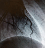
Figure 1: Coronary angiogram of left coronary artery during diastole.
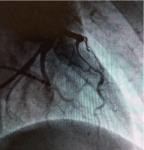
Figure 2: Coronary angiogram of left coronary artery during systole.
Under general anesthesia midline incision and sternotomy performed. Due to deep location of proximal-mid LAD we decided to proceed with cardiopulmonary bypass (CBP). Conventional cannulation done with aortic cannula and double staged single venous cannula. Cardiac arrest induced by cold bloody cardioplegic solution. Surgical gauze placed beneath the left ventricle. The intramyocardial part of LAD located, and using surgical knife, epicardium and myocardium transected over the artery (Figure 3).
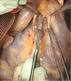
Figure 3: Intraoperative view of surgical myotomy.
The deepest location of LAD considered as one cm below epicardium (Figure 3). Side walls of myocardium sewn up to epicardium with prolene 6/0 with continuous suture, in places with single stitches (Figure 4). Patient rewarmed and weaned from CPB. CPB time was 15 minutes. Patient transferred to intensive care unit with stable hemodynamics and extubated 4 hours postoperatively. He transferred to in-patient service next morning and discharged from hospital five days later. Patient received prescription for antiplatelet treatment with aspirin for next three months.
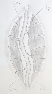
Figure 4: Myocardial reinforcement.
Discussion
The MB more often encountered in adults [2]. Nevertheless, children and adolescents with MB may also have clinical symptoms and despite good long term prognosis of MB [6], it ultimately may result in myocardial ischemia, myocardial infarction, arrhythmias and even sudden death [7]. Thus, Zhu et al (2012) reported a case of male adolescent athlete with a myocardial infarction caused by MB [7].
There is still controversy regarding treatment approach to patients with MB. Earlier preference was given to medical treatment with beta blockers or calcium channel blockers [6]. In 2009 Schwartz et al classified MB for three types, where they claimed that patients with type C MB (patients with symptomatic course of disease) may have surgical revascularization [8]. Tarantini et al (2016) published similar management strategy for MB with surgical treatment reserved for cases where medical treatment is failed [9]. Overall, in last decades, treatment approach moves towards preventive surgical revascularization [7]. There are two surgical procedures performed to patients with MB - myotomy and bypass grafting [10]. The choice made mainly depending of MB anatomy and surgeon’s preference. The indication for surgical myotomy in our center is patient’ complains resistant to medical therapy, and/or evidence of myocardial ischemia (e.g. ECG changes/increased levels of cardiac enzymes), and existence of MB on coronary angiography.
Below we would like to describe the reasoning of some surgical step of myotomy. The identification of artery is first and important step where detailed preoperative study of coronary angiography movie is essential. Generally the myotomy can be performed without CPB (off pump) and with CPB (on arrested or beating heart). In our case we decided to proceed with CPB as safer option because of deep and more proximal location of LAD. Usually LAD located to the right from the vein, however the distance between two vessels may vary. If whole coronary artery lies within the myocardial layer then it may be found by retrograde passage of a fine atraumatic vascular probe inserted through the most distal arteriotomy where LAD usually reappers. Another commonly used tool is epicardial doppler which shows blood flow in the coronary artery. There are several other techniques of coronary artery identification available and well described in the literature [11]. The epicardiotomy and myotomy is made by surgical blade N15 as it gives the finest feeling during dissection of underlying tissues. The myotomy made careful for not to damage the artery and its branches and not to penetrate into right ventricle. If penetration occured then horizontal mattress stitch with pledgets should be placed to close the hole. This stitch should be placed in a way not to disturb flow in overlying LAD, otherwise coronary bypass with internal mammary artery to distal LAD would be prudent. We also recommend to reinforce myotomy muscular walls to prevent disbanding of myocardial fibers during strenious cardiac contraction (Figure 4). Finally, we advise patient to receive antiplatelet therapy for 3-6 months in order to reduce the risk of coronary artery thrombosis due to possible microtrauma of arterial walls.
Conclusion
Surgical myotomy of myocardial bridge is optional and safe treatment for adolescents with MB and evidence of myocardial ischemia.
- Mohlenkamp S, Hort W, Ge J, Erbel R. Update on myocardial bridging. Circulation. 2002; 106: 2616-2622.
- Ciçek D, Kalay N, Müderrisoglu H. Incidence, clinical characteristics, and 4-year follow-up of patients with isolated myocardial bridge: a retrospective, single-center, epidemiologic, coronary arteriographic follow-up study in southern Turkey. Cardiovasc Revasc Med. 2011; 12: 25-28.
- Chiara R, Maria CM, Fabiola O, Roberto A. Myocardial bridging: A ‘forgotten’ cause of acute coronary syndrome - a case report. Int J Angiol. 2007; 16: 115-118.
- Mavi A, Sercelik A, Ayalp R, Karben Z, Batyraliev T, Gumusburun E. The angiographic aspects of myocardial bridges in Turkish patients who have undergone coronary angiography. Ann Acad Med Singapore. 2008; 37: 49- 53.
- Ekeke CN, Noble S, Mazzaferri E Jr, Crestanello JA. Myocardial bridging over the left anterior descending: Myotomy, bypass, or both? J Thorac Cardiovasc Surg. 2015; 149: 57-58.
- Ural E, Bildirici U, Celikyurt U, Kilic T, Sahin T, Acar E, et al. Long-term prognosis of non-interventionally followed patients with isolated myocardial bridgeand severe systolic compression of the left anterior descending coronary artery. Clin Cardiol. 2009; 32: 454-457.
- Zhu CG, Liu J, Liu WD, Xu YL, Wu NQ, Guo YL,et al. Myocardial infarcti caused by myocardial bridging in a male adolescent athlete. J Cardiovasc Med (Hagerstown). 2012; 13: 138-140.
- Schwarz ER1, Gupta R, Haager PK, vom Dahl J, Klues HG, Minartz J, et al. Myocardial bridging in absence of coronary artery disease: proposal of a new classification based on clinical-angiographic data and long-term follow-up. Cardiology. 2009; 112: 13-21.
- Tarantini G, Migliore F, Cademartiri F, Fraccaro C, Iliceto S. Left Anterior Descending Artery Myocardial Bridging: A Clinical Approach. J Am Coll Cardiol. 2016; 68: 2887-2899.
- De Winter R, Kok W, Piek J. Coronary atherosclerosis within a myocardial bridge, not a benign condition. Heart. 1998; 80: 91-93.
- Ziadinov E, Al-Sabti H. Localizing intramyocardially embedded left anterior descending artery during coronary bypass surgery: literature review. J Cardiothorac Surg. 2013; 8: 202.