Case Report
Austin Cardio & Cardiovasc Case Rep. 2019; 4(1): 1033.
Burp Angina
Yarows SA1*, Smith F2 and Szyniszewski A3
¹Clinical Professor Internal Medicine, Michigan Health 128 Van Buren, Chelsea, Michigan, USA
²Medical Director, Intensive Cardiac Rehabilitation, St. Joseph Mercy Hospital, Ann Arbor, Michigan, USA
³Director of Structural Heart Program, St Joseph Mercy Hospital, Ann Arbor, Michigan, USA
*Corresponding author: Steven A Yarows, Clinical Professor Internal Medicine, Michigan Health 128 Van Buren, Chelsea, Michigan, USA
Received: July 19, 2019; Accepted: August 27, 2019; Published: September 03, 2019
Introduction
Classic angina pectoris is described as a pressure, heaviness, tightness, or constriction in the center or left of the chest that is precipitated by exertion and relieved by rest, typically over 5-15 minutes [1]. It is generally not described as sharp pain (although this is often asked by providers), or needles and pins. Atypical angina symptoms are exertional shortness of breath, nausea, diaphoresis, fatigue, or indigestion. Generally, symptoms of angina do not occur over ‘moments’, nor relieved by position, burping, or pressing on the precordium. Although angina in females has been reported as presenting differently than males, the predominant difference is that angina may more likely in women to be induced by rest, sleep, and mental stress, in addition to or instead of physical exertion [2].
Case Presentation
A 68-year-old male physician gradually noticed burping during the initiation of exercise over 4-6 months. He exercised 5-6 days per week for 1½ hours per session with weight lifting and 30-35 minutes of aerobic exercise, usually at approximately 8 METS. The belching intensity and frequency progressed over the preceding month occurring at the beginning of exercise or even usual walking. The burp frequency occurred once every approximately 5 seconds and lasted 5-10 minutes. As the exercise or walking continued, the belching diminished, and his full exercise program was unimpeded. There was also noted left precordial discomfort completely resolved upon burping and slight increasing dyspnea with exercise or talking. No unusual diaphoresis, nausea, nor generalized chest discomfort was noted.
Current medical problems included well controlled hypertension (home readings 120-125/70-75mmHg) and irritable bowel syndrome. His daily medications included irbesartan 75mg daily and Aspirin 81mg daily.
His lipids over the past 11 years included a total cholesterol of 217-246mg/dl (latest 228mg/dl, 4 months prior), HDL 66-90mg/dl (latest 83mg/dl, 4 months prior), LDL 143-150mg/dl (latest 140mg/ dl, 4 months prior), triglycerides 17-32mg/dl (latest 25mg/dl, 4 months prior).
A Calcium Score CT was performed to exclude coronary artery disease on May 23, 2019 showing moderate coronary artery calcification with an Agatston’s score of 200. The calculated arterial age was 78 years with the age and gender matched percentile were 60%. The Left Main Artery calcium score was 0, Right Coronary Artery Calcium Score was 10, the Left Anterior Descending Calcium Score was 185, the Circumflex Calcium Score was 5, the Posterior Descending Artery Calcium Score was 0, and the Other Calcium scores were 0.
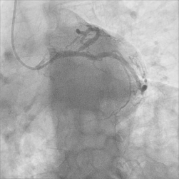
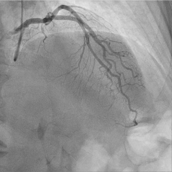
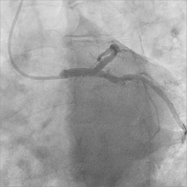
A left heart catheterization was performed May 24, 2019 due to the exertional symptoms and moderate Calcium Score CT findings. The catheterization showed the LV end-diastolic pressure was normal at 12mmHg. There was no aortic valve stenosis. The left ventriculogram in the RAO view demonstrated normal contractility, EF 65%.The left main coronary artery arose normally and had a distal 20% lesion that tapered into the ostial LAD. The LAD had an 80% ostial lesion (Figure 1,2). It was a very large vessel and wrapped around the apex of the heart feeding the distal and mid inferior wall. Flow wire of the left anterior descending was performed with adenosine, the Pd/ Pa was 0.85 indicating a significant lesion. This would account for the belching angina. The diagonal was free of significant disease. The circumflex artery and obtuse marginal were free of significant disease. The right coronary artery arose normally and fed the PDA, posterior ventricular and a small posterolateral branch. This system was free of any significant disease. There was successful stenting of the left anterior descending 80% stenosis to 10% residual TIMI grade 3 flow pre and post (Figure 3). The left main circumflex unjailed after stenting of the LAD with a stent back in the left main coronary artery.

Figure 1: Ostial 80% LAD occlusion.

    Figure 2: RAO view of left coronary artery showing diaphragmatic proximity
to distal LAD.

Figure 3: LAD post-angioplasty and stent.
The patient subsequently resumed his normal exercise routine with absence of exertional belching. He did still have normal, intermittent belching independent of exercise.
Discussion
Belching is a daily recurrent occurrence for most people, which naturally increases with the ingestion of carbonated beverages. Excessive belching is a commonly observed complaint in clinical practice. It occurs more commonly in patients with gastroesophageal reflux disease or functional dyspepsia [3]. Impedance monitoring has revealed that there are two mechanisms through which belching can occur: the gastric belch and the supragastric belch. The gastric belch is the result of a vagally mediated reflex leading to relaxation of the lower esophageal sphincter and venting of gastric air.
Belching associated with chest pain or indigestion is a recognized symptom of myocardial ischemia or infarction in the emergency room literature [4]. Belching has been correlated with inferior wall myocardial infarctions (OR=1.57, 95% CI=1.21-2.03) [5]. In 1978, 27 patients with inferior myocardial infarction were noted to have the urge to burp, which was described as “Eructonesius” [6]. The combination of sweating, nausea, belching and vomiting has been associated with an inferior wall myocardial infarction with a predictive value of 91% [7].
Belching without indigestion as a form of angina pectoris has been rarely reported. The only documented case reports are subsequently discussed.
An 83-year-old woman with several decades of ischemic heart disease was reported with a new angina symptom of loud, uncontrollable belching required cardiac intervention (anatomy not reported) [8]. Partial relief of belching angina chest pain was described in patients with at least 70% or greater obstruction in at least a single coronary artery [9]. A 63- year old male, who had been healthy all his life, presented with a two-month history of belching episodes as the chief and the only complaint [10]. The symptoms were confined mainly to effort and emotions, which were the clues for the diagnosis. A stress test electrocardiogram was strongly positive for angina pectoris. Coronary angiography showed 50% narrowing at the left anterior descending (LAD), both branches of the circumflex artery and 98% blockage of the Right Coronary Artery (RCA) and its branch, the posterior descending artery (PDA). The patient underwent coronary artery bypass grafting surgery.
So, with limited literature, what should a practicing physician do when a patient presents with excessive belching? This case demonstrated rapid, progressive belching at the initiation of exercise with a frequency of approximately every 5 seconds. There was non-specific dyspnea, but his regular exercise capacity did not diminish. This should be considered a potential angina equivalent. An appropriate cardiac workup could be considered.
Take home messages
• Rapid (i.e.-every 5-10 seconds), progressive belching with exercise may be an anginal equivalent.
• Stimulation of the vagal nerve by a dominant artery supplying the inferior myocardial wall is postulated to be the mechanism of the exertional belching in this angina equivalent.
- Mahler, Simon A. Angina Pectoris: Chest Pain Caused By Coronary Artery Obstruction, Uptodate, Literature Review Current Through. 2019.
- Douglas Pm, Pagidipati, N. Clinical Features and Diagnosis of Coronary Heart Disease in Women, Uptodate, Literature Review Current Through. 2019.
- Kessing Bf, Bredenoord Aj, Smout Ajpm. The Pathophysiology, Diagnosis and Treatment of Excessive Belching Symptoms. Am J Gastroenterol. 2014; 109: 1196-1203.
- Smith J, Carley S. Belching as a Symptom of Myocardial Infarction. Emerg Med J. 2001; 18: 467.
- Culic V, Miric D, Eterovic D. Correlation Between Symptomatology and Site of Acute Myocardial Infarction. Int J Cardiol. 2001; 77: 163-168.
- Darsee, Jr. Eructonesius with Inferior Myocardial Infarction. Nejm. 298: 221- 222.
- Logan Rl, Wong F, Barclay J. Symptoms Associated With Myocardial Infarction: Are They Of Diagnostic Value? Nz Medj. 1986; 99: 276-278.
- Turi-Kovats, Nora; Arabadzisz, Hrisula; Zsoldos, Andras; Tomcsanyi, Janos. [Atypical Complaint in Myocardial Ischaemia: Belching]. [Hungarian]. Orvosi Hetilap. 2017; 158: 183-186.
- Bauman, D. Partial Relief of Anginal Chest Pain by Belching. Jama. 1977; 328: 481.
- El-Shafie, Kawther, Belching as a Presenting Symptom of Angina Pectoris. Sultan Qaboos University Medical Journal. 2007; 7: 257-259.