
Case Report
Austin Cardio & Cardiovasc Case Rep. 2019; 4(1): 1034.
Traumatic Pulmonary Pseudocyst - An Unusual Complication of Closed Thoracic Trauma
Simoglou C*, Skarmoutsou A and Gymnopoulos D
Surgical Department, St. Loukas, Thessaloniokii, Greece
*Corresponding author: Christos Simoglou, Surgical Department, St. Loukas, Thessaloniki, Greece
Received: September 23, 2019; Accepted: October 22, 2019; Published: October 29, 2019
Abstract
The traumatic pseudocyst is a rare complication of closed thoracic trauma, in which increased pressure causes endopulmonary parenchymal laceration without rupture of the side. We describe a young man who presented with a history of hemoptysis and radiological pulmonary cavity 2 weeks after closed thoracic trauma from a fall. Computed tomography showed picture compatible with post-traumatic pseudocyst and bronchoscopy evidence of inflammation, a pathological flora in the culture of bronchial washings. The administration of antibiotic therapy on the basis of antibiograms resulted in rapid and complete clinical and radiographic healing.
Keywords: Lung pseudocyst; Lung contusion; Closed chest trauma
Introduction
Pulmonary contusion is usually a consequence of closed chest injury, which most often resolves with simple supportive measures and without special intervention. In some cases, contusion of the lung may be complicated by the formation of cavities in the literature referred to as post-traumatic pulmonary pseudocyst and such a situation is then.
Description Patient
Male 42 years old, dairy worker, non smoker, with a free souvenir, fell climbing down a ladder and sustained a closed injury to the dorsal surface of the right hemithorax. After 2 days had back pain, dry cough, and then had a small fresh haemoptysis, without fever or other symptoms. Because haemoptysis stopped short, did not seek medical care. After 2 weeks went to pathology department hospitals for allergic reaction (urticaria, angioedema) after taking an antibiotic for periodontal inflammation. Under control there was chest radiograph (Figure 1) which showed thickening in the region right upper lobe with a cavity ydraeriko level. The blood test showed leukocytosis (20,800 leuk./mm3) and thrombocytosis (680,000 platelets/mm3). The patient was recommended lung control. When examined the next day, the patient had normal vital signs and saturation by pulse oximeter was 98% in ambient air.
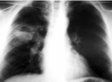
    Figure 1: Chest X-ray (2 weeks after injury) showing cavity water gas level
average level in the right lung field.
There had clubbing, lymph nodes or rashes. The findings from the lungs and neck were normal. Was computed tomography (Figure 2) which showed the following: “Presence Round soft tissue mass in the posterior part of the right upper lung lobe diameter about 3,5cm with a relatively fuzzy boundaries and infiltration of the proximal pulmonary parenchyma. After intravenous administration of soluble contrast media observed peripheral enhancement of the structure with non-enriched core content that has local low densities, indicative of degeneration hydric central mass. Mild peripheral filtration extend to the pleural thickening with localized pleural locally. Small bronchial lymph nodes swollen right, without other glands in the mediastinum. No pleural reaction or fluid collection. “The patient underwent fiber vrochoskopisi who showed erythema, edema of the mucosa and purulent secretions in the right upper bronchus lovaio without bleeding or other pathological evidence. Biopsies obtained showed evidence of inflammation without any evidence of malignancy or other specific disease process.
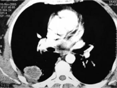
    Figure 2: Intersect by chest CT scan (after intravenous administration of
soluble contrast media), which shows image Round mass with peripheral
enhancement and thickening content.
Bronchial lavage were negative in staining for mycobacteria, while growing for common germs developed Enterobacter cloacae, and Streptococcus mitis. The dermoantidrasi Mantoux was negative. Based on antibiograms the patient received treatment with ciprofloxacin 500mg x2 for 2 weeks. Remained apyretic, without much cough or expectoration and had no back hemoptysis. At retesting after antibiotic treatment, the chest radiograph showed reduction in size of the cavity. The blood count showed 6100 white and 420,000 platelets with normal hemoglobin and hematocrit. Three weeks later the chest radiograph was normal (Figure 3), and the patient was completely well.
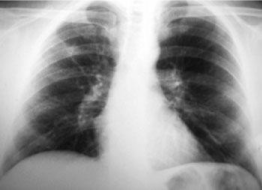
Figure 3: Normal chest radiograph 3 weeks after initiation of therapy.
Discussion
Reference is very limited or absent altogether The traumatic cavities of the lungs may result from an open or closed chest injury. Unlike open or perforating trauma always receiving urgent medical care, closed injuries and their consequences may go unnoticed or cause problems with differential diagnosis and treatment for pulmonologists. But their descriptions in the pulmonary literature. The described situation reported in the literature by various names such as traumatic (or traumatic) cyst cavity pnefmatokili or pseudocyst, although there seems to be the last word. In blunt chest trauma increased intrapleural pressure upon impact causing dissociation (shearing) of the parenchyma without the visceral pleura ruptured, resulting in the formation of cavities. Another possible mechanism is the sharp increase and then decrease the pressure intrapleural upon impact, which if it exceeds the elasticity of the lung, causing rupture again parenchyma. Believed that the greater compliance of the chest wall to allow younger people to transfer greater force of impact in the pulmonary parenchyma, which justifies the early injured (see below). In most published reports of wells were one-room, while in one case from France described multiple cystic lesions 5 in the right lower lobe, not existing before the injury and disappeared within a few months. Has been described and Greek case with bilateral traumatic pseudocysts. The wall cavity varies in thickness and consists of connective tissue without endothelium, 1 which justifies the name ‘pseudocyst’. The exact incidence of traumatic lung cavities is not known. The Sorsdahl states that until 1965 had only 28 published cases. Even in recently published Greek seira8 traumatic pseudocyst characterized as “rare entity”. In all series, patients were generally male and young people (in an American series, the average age was 29 years, while other works were many children). In all of the pseudocyst was the result of closed thoracic trauma. The main specific symptom in most patients was hemoptysis, usually minor and self-limiting. Depending on the size of the cavity and the existence of other parenchymal lesions may coexist dyspnea, cough, chest pain and fever. The wound cavity may become apparent in the X-ray immediately after the trauma. But if it is full of hemorrhagic material, may be seen after a few days, when the material is evacuated through the tracheobronchial tree. The Moore et al. indicate that typical X-ray shows pulmonary infiltrates consistent with contusion showing cavity formation in about 1 week after the trauma. Computed tomography is more sensitive than simple aktinografia 9 to detect such detailed parietal cavity formations with gas level. It is speculated that many mild cases of pseudocyst may go unnoticed if not checked by CT scan. The outcome of traumatic pseudocyst is generally good, with spontaneous remission in most cases.
The time course can be from weeks to months (in a Japanese series of 12 patients persistent air leak in conventional mechanical ventilation, which was treated successfully with high frequency ventilation endobronchial bleeding and the mean duration was 1.8 months). The literature emphasizes the need to recognize the complication is to avoid unnecessary diagnostic and therapeutic maneuvers. 11 However, there are cases presented additional complications (infection and abscess, 2.12 persistent air leak in conventional mechanical ventilation, which was treated successfully with high frequency ventilation endobronchial bleeding and swelling of the bladder under mechanical ventilation led to urgent lobectomy. Lobectomy were also abscesses and persistent bladder. In the series of Kato et al, 10 out of 12 patients had concomitant hemothorax or hemopnefmothorax CT without concomitant drainage of the pseudocyst. All patients in this series received antibiotics, without mentioning if there was proven contamination of pseudocyst. The approach of the patient with traumatic lung pseudocyst should be guided by history and clinical and radiological picture. If the general condition is good, usually only symptomatic treatment with analgesia, respiratory physiotherapy, possibly oxygen (hypoxia on the pulmonary contusion), and a broad-spectrum antibiotic. Further investigation of sputum cultures, bronchoscopy, etc. should be sought where doubt exists as to the diagnosis or the patient’s course dictates. Where it changes to an abscess, the treatment should be commensurate with intensive antibiotic therapy based antibiograms. If this continues for 4-6 weeks with no change in clinical status, should be the resection cavity in the form lobectomy. Our patient had typical clinical and radiologic findings of traumatic pseudocyst. The microbiological examination of bronchial washings showed contamination of the bladder (without systemic manifestations of infection). The whole situation was treated successfully with antibiotics, which resulted in rapid clinical cure with complete resolution of the radiological image. On the occasion of this incident, we note the need for familiarity pulmonologist with clinical and radiographic images of the traumatic situations of the chest during exercise, which will lead to better cooperation with Thoracic and better deal with these situations.
References
- Wallace MJ, Krishnamurthy S, Broemeling LD, Gupta S, Ahrar K, Morello FA, Hicks ME. CT-guided percutaneous fine-needle aspiration biopsy of small (=1cm) pulmonary lesions. Radiology. 2002; 225: 823-828.
- Anderson JM, Murchison J, Patel D. CT-guided lung biopsy: factors influencing diagnostic yield and complication rate. Clin Radiol. 2003; 58: 791- 797.
- Larscheid RC, Thorpe PE, Scott WJ. Percutaneous transthoracic needle aspiration biopsy: a comprehensive review of its current role in the diagnosis and treatment of lung tumors. Chest. 1998: 114: 704-709.
- Odel MJ, Reid KR. Does percutaneous fine needle aspiration biopsy aid in the diagnosis and surgical management of lung masses? Can J Surg. 1999: 42: 297-301.
- Wong RS, Ketai L, Temes RT, Follis FM, Ashby R. Air embolus during complicating transthoracic percutaneous needle biopsy. Ann Thorac Surg. 1995; 59: 1010-1011.
- Sinner WN, Zajicek J. Implantation metastasis after percutaneous transthoracic needle aspiration biopsy. Acta Radiol Diagn (Stockholm). 1976; 17: 473-480.
- Seyfer AE, Walsh DS, Graeber GM, Nuno IN, Eliasson AH. Chest wall implantration of lung cancer after thin-needle aspiration biopsy. Ann Thorac Surg. 1989; 48: 284-286.
- Sawabata N, Ohta M, Maeda H. Fine-needle aspiration cytologic technique for lung cancer has a high potential of malignant cell spread through the tract. Chest. 2000; 118: 936-939.
- Kim IH, Kim YT, Lim HK, Kim YH, Sung SW. Management for chest wall implantation of non-small cell lung cancer after fine-needle aspiration biopsy. Eur J Cardio-thorac Surg. 2003; 23: 828-832.
- Raftopoulos Y, Furey WW, Kacey DJ, Podbielski FJ. Tumor implantation after computed tomography-guided biopsy of lung cancer. J Thorac Cardiovasc Surg. 2000; 119: 1288-1289.