
Clinical Image
Austin Cardio & Cardiovasc Case Rep. 2022; 7(1): 1044.
A Case of Isolated Pulmonary Valve Endocarditis: The Importance of Repeat Echography and Microbiology in Diagnosis
McCaughey C*, Wheen P, Murphy G, Pearson I and Murphy R
St James Hospital, Dublin, Ireland
*Corresponding author: Conor McCaughey, St James Hospital, Dublin, Ireland
Received: March 28, 2022; Accepted: April 05, 2022; Published: April 12, 2022
Clinical Image
A 64-year-old man presented to our centre with a one month history of fatigue, exertional dyspnoea, and worsening lower limb oedema on a background of known HFrEF. Following successful diuresis persistently raised inflammatory markers and intermittent fevers prompted investigation for infective endocarditis (IE). Serial blood cultures grew staphylococcus epidermis which were all, unusually, CO2 dependant and likely from a common source. An initial transthoracic echocardiogram (TTE) showed no evidence of endocarditis and normal Tricuspid (TV) and Pulmonary valves (PV). A subsequent transoesophageal echocardiogram (TOE) showed no significant new valvular abnormality.
Despite having only three minor Dukes criteria, the microbiological findings suggesting an uncontrolled source fostered a high clinical suspicion for IE. A second transthoracic echo performed seven days later revealed a large sessile vegetation on the ventricular aspect of the pulmonary valve; visualized entering into the pulmonary artery during systole (A) along with an associated stalk (B). The vegetation measured approximately 1.5cmx0.75cm. Despite this knowledge in retrospect and adequate visualization of the pulmonary valve on the initial TOE no vegetation could be seen. Subsequent TOE positively identified the isolated pulmonary vegetation. Unfortunately, despite appropriate antibiotic therapy, this patient died after a prolonged admission.
Isolated pulmonary valve endocarditis is rare, and it can present a diagnostic challenge. While the sensitivity of combined TTE and TOE in the diagnosis of IE overall is estimated to be 85-90% [1], this case highlights the importance of repeat transthoracic echocardiography after an appropriate interval in those cases where both investigations are negative but a high clinical suspicion remains [2]. This case also highlights the value of Microbiology expertise in the diagnosis of IE (Figure 1).
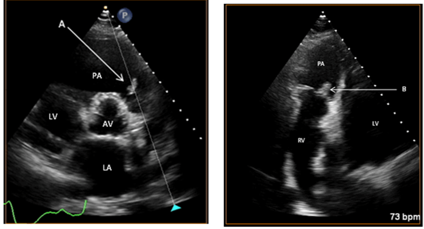
Figure 1:
References
- Evangelista A, Gonzalez-Alujas MT. Echocardiography in infective endocarditis. Heart (British Cardiac Society). 2004; 90: 614-617.
- Habib G, Badano L, Tribouilloy C, Vilacosta I, Zamorano JL, Galderisi M, et al. Recommendations for the practice of echocardiography in infective endocarditis. European Journal of Echocardiography: The Journal of the Working Group on Echocardiography of the European Society of Cardiology. 2010; 11: 202-219.