
Case Report
Austin Cardio & Cardiovasc Case Rep. 2023; 8(2): 1056.
Acute Cerebral Infarction following Ventricular Tachycardia Ablation
Le Dong*; Chen Long
Department of Cardiovascular Medicine, Jiangbei Campus, Zhongda Hospital Affiliated to Southeast University, China
*Corresponding author: Le Dong Department of Cardiovascular Medicine, Jiangbei Campus, Zhongda Hospital Affiliated to Southeast University, Nanjing 210000, Jiangsu Province, China. Email: donglejczh@zcxecl.com
Received: June 14, 2023 Accepted: July 13, 2023 Published: July 20, 2023
Abstract
Ablation is a more effective treatment than drug therapy for ventricular tachycardia, and its safety is increasingly emphasized. This case reports the treatment of acute cerebral infarction following ventricular tachycardia ablation, with prompt restoration of cerebral blood flow and a positive prognosis. These results provide valuable insights for cardiology colleagues.
Keywords: Ablation acute cerebral infarction; Ventricular tachycardia
Introduction
Patient Name: Li XX. Gender: Male. Age: 66. Ethnicity: Han. Admission Date: September 11, 2022.
Chief Complaint: Intermittent chest tightness, palpitations, and chest pain for 24 hours.
Medical History: The patient had no obvious cause of chest tightness and palpitations 24 hours ago, accompanied by pain behind the sternum, which was dull and lasted for about 10 minutes each time. The symptoms were relieved after rest, and amaurosis were accompanied during the attacks, but no syncope occurred.
Past Medical History: The patient had no history of hypertension, diabetes, or cerebral infarction.
Personal History: The patient had a history of occasional drinking and had smoked 3-4 cigarettes a day for 20 years.
Family History: Nothing noteworthy.
Physical Examination: T: 36.5°C, P: 80 bpm, R: 20 bpm, regular. BP: 155/92 mmHg.
The patient was conscious and alert. The breath sounds in both lungs were clear without dry or moist rales, and no pleural friction rub was heard. The heart size was normal, the heart rate was 80 bpm, and the rhythm was regular. The heart sounds were strong, and no pathological murmurs or extra heart sounds were heard in any auscultation areas of the valves. No pericardial friction rub was heard. No edema was found in both lower extremities.
Auxiliary Examination: Table 1
    Date

    Item

    Test Result


    12-Sep

    Cardiac biomarkers

    CK-MB <2.0ng/ml, TNI <0.01ng/ml


    MYO: 50ng/ml


    12-Sep

    Blood, urine, stool routine

    Hemoglobin: 126g/L, others are normal


    12-Sep

    D-Dimer

    333ug/L


    12-Sep

    Liver and kidney function, blood lipids,    electrolytes

    Potassium: 3.4mmol/lL, no other significant    abnormalities observed


    12-Sep

    Eight viral tests

    No significant abnormalities observed


    12-Sep

    Free thyroid function

    No significant abnormalities observed
Table 1:
ECG Examination: Figure 1
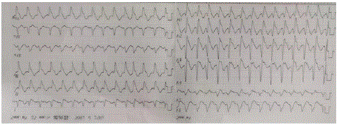
Figure 1: Ecg indicated ventricular tachycardia, heart rate 300 beats/min.
Echocardiography: Figure 2
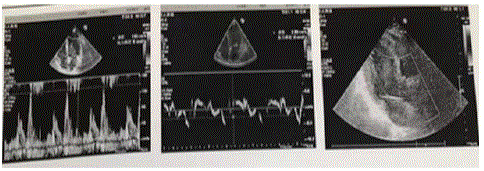
    Figure 2: Ultrasound Findings.

Cardiac Measurements:

A0: 23.8 mm LVD: 50.3 mm AV: 1.4 m/s

AA0: 33.4 mm LVS: 32.4 mm

LA: 41.1 mm LVPW:11.8 mm

RV: 26.2 mm RA: 40 × 53 mm MV: 1.4 m/s

IVS: 11.8 mm MPA: 23.1 mm

Cardiac Function:

EF: 65% FS: 35%

Description:

The connection and inner diameter of the main and pulmonary arteries are normal. The left atrium and right atrium have slightly enlarged diameters, while the diameters of the other chambers are within the normal range. The interventricular septum shows an interrupted echo. The ventricular walls do not show thickening, the motion is coordinated, the systolic amplitude is normal, and no obvious segmental movement abnormality is seen. The morphology, structure, and opening and closing movements of each valve are normal, and there is no obvious abnormality in the pericardial cavity.

Color Doppler and spectral Doppler: Mild regurgitation is visible in the mitral valve, tricuspid valve, and aortic valve. E/A: <1

Tissue Doppler suggests reduced mitral annular motion velocity. e’/a’ <1.

Ultrasound suggests:

Slight enlargement of the left and right atria.

Mild regurgitation is visible in the mitral valve, tricuspid valve, and aortic valve.

Reduced diastolic function of left ventricular.

(The above results are for reference only.)
Coronary Arteriography: Figure 3
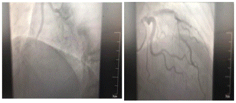
Figure 3: Coronary Arteriography.
Electrophysiological examination of the heart: The patient lies flat on the operating table, and the groin area on both sides is disinfected and covered with sterile towels. Under local anesthesia with 0.5% lidocaine, the Seldinger technique is used to puncture the left femoral vein. Two 7F sheaths are inserted and a quadripolar catheter is advanced through one of the sheaths to the right ventricle, and a coronary electrode is advanced through the other sheath to the coronary sinus. A complete intracavitary electrocardiogram is recorded. Ventricular stimulation shows ventriculo-atrial dissociation, and atrial stimulation shows decremental conduction. There is decremental conduction with no propagation of S1S2 at 600/530 ms. No jumping prolongation or echo is observed, and no inducible tachycardia is present. Ventricular stimulation with S1S1280 ms induces wide QRS complex tachycardia with a heart rate of 180 bpm. Intracavitary electrograms show ventriculo-atrial dissociation with a ventricular rate faster than the atrial rate. The electrocardiogram shows an RS pattern in lead I, a positive main wave in the inferior leads, an R wave in lead V1, and an RS pattern in leads V4-V6. The intracavitary electrophysiological diagnosis is ventricular tachycardia. A successful ablation of the ventricular tachycardia is performed at the base of the lateral aspect of the left anterior papillary muscle.
Postoperative ECG: Figure 4
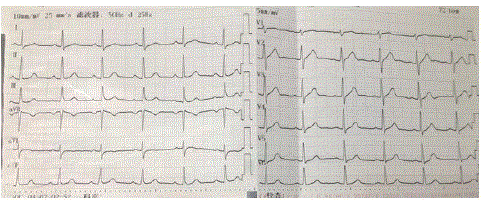
Figure 4: Electrocardiogram indicated sinus rhythm, ventricular rate 75 beats/mi.
Result
Postoperative Emergency: The patient developed hoarseness of voice, grade 1 muscle strength in the left upper limb, and grade 5 muscle strength in the left lower limb, with equal-sized and round pupils bilaterally, suggesting the possibility of cerebrovascular accident. Emergency neurovascular intervention performed mechanical thrombectomy and thrombolysis on the cerebral arteries (Figure 5 & 6).
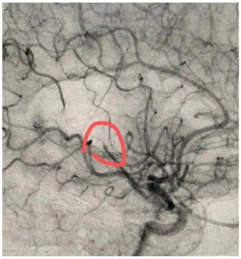
Figure 5: Middle cerebral artery M4 segment occlusion.
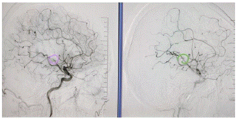
Figure 6: Overall survival, autologous stem cell transplant (ASCT) versus no ASCT (p=0.12).
Before discharge on September 21, 2022, the patient had muscle strength of level 5 in the left upper and lower extremities, with slightly poorer fine motor activity in the left upper extremity.
Discussion
1. Was the preoperative anticoagulation inadequate?
2. Was the surgery time too long?
3. Were there other reasons?
Anticoagulation Scheme: When the ablation catheter entered the left ventricle, 3000 U of heparin was injected, followed by an additional 1000 U per hour until the end of the surgery.
Operation Duration: 6 hours and 40 minutes, with a total of 9000 U of heparin used.
After atrial fibrillation ablation, 14% of patients developed asymptomatic cerebral infarction [1]. Left heart catheter ablation has a stroke risk, with a reported incidence of 0.8%-1.8%.
Possible mechanisms:
1. During ablation, thrombi and gas may be introduced into the left heart system, and prolonged stay of the catheter in the left heart system may lead to thrombus formation.
2. Thrombus formation may occur at the ablation site, especially at the tip of the catheter.
3. Recurrent ventricular tachycardia, low-flow state, poor left ventricular function, and local myocardial suppression can increase the risk of systemic embolism.
4. Ablation is inherently traumatic and can further activate coagulation during the ablation process.
Conclusion
How to prevent:
More precise operation, shortening the operation time, and sufficient heparinization of anticoagulants in the perioperative period can reduce the risk of stroke [1].
Once a stroke occurs, early thrombectomy and thrombolysis can result in generally good patient outcomes [2].
Author Statements
Acknowledgement
This article was authorized and agreed by Dr. Chen Long, the surgical doctor, and provided guidance for the article.
References
- Lu W, Pan L, Wang X, et al. Incidence and prognosis of asymptomatic cerebral infarction after cryoballoon or radiofrequency catheter ablation for atrial fibrillation. Chinese Circulation Journal. 2021; 36: 680-685.
- Zuo S, Bo X, Wu J, Fan C, Li S, et al. Chinese Journal of Cardiovascular Diseases. 2022; 50: 707-709.