
Case Report
Austin Cardio & Cardiovasc Case Rep. 2023; 8(4): 1064.
From Tachycardia to Lymphoma: A Case Report of CD30 Positive (ALK) T-Cell Lymphoma Initially Presenting with Cardiac Envolvement
Mohammed Al Thawabta, MD; Mutaz Karameh, MD; Mordechai Golomb, MD; OrenYagel, MD*
Heart Institute, Hadassah Medical Center, Faculty of Medicine, Hebrew University of Jerusalem, Jerusalem, Israel
*Corresponding author: Oren Yagel Heart Institute, Hadassah Medical Center, Faculty of Medicine, Hebrew University of Jerusalem, Ein Karem, POB 12000, Jerusalem 9112001, Israel. Tel: +972526105699 Email: oreny@hadassah.org.il
Received: October 24, 2023 Accepted: November 20, 2023 Published: November 27, 2023
Abstract
Anaplastic Large Cell Lymphoma (ALCL), Anaplastic Lymphoma Kinase-Positive (ALK+), is a rare subtype of large cell lymphoma. Cardiac involvement as the initial presentation leading to diagnosis is extremely rare. We present a young patient with atrial tachycardia as the presenting manifastation of ALCL.
Keywords: Anaplastic large cell lymphoma; ALK-positive; CD30; Interatrial septum hypertrophy; Atrial tachycardia
Abbreviations: ALCL: Anaplastic Large Cell Lymphoma; ALK+: Anaplastic Lymphoma Kinase-Positive; PET-FDG: Positron Emission Tomography Fluorodeoxyglucose; CT: Computed Tomography; CHP BV: Cyclophosphamide + Doxorubicin + Prednisone + Brentuximab + Vedotin
Background
ALCL-ALK+, a rare subtype of large cell lymphoma, are a CD30-positive neoplasm accounting for 2% to 8% of all lymphomas. It is characterized by the proliferation of predominantly large lymphoid cells and high expression of the cytokine receptor CD30 [1]. It is caused by chromosomal translocations involving the ALK gene. No potential predisposing factors have been reported [2]. Symptomatic involvement of the cardiovascular system is an uncommon entity [4]. Cardiac involvement as the initial presentation is extremely rare.
We report a unique case of CD30 positive T-cell lymphoma initially presenting with atrial tachycardia
Case Presentation
A 22-year-old male patient with no significant past medical history presented to our hospital due to palpitations.
A few weeks prior to his admission, the patient noted mild neck swelling and swallowing difficulty. He denied fever, night sweats, dizziness, or syncope. Of note, the patient was on a new dietary program with an intentional weight loss of 10 kg over two months.
On admission, his heart rate was 140 BPM, hemodynamically stable and afebrile. Physical examination revealed diffuse neck swelling and palpable supraclavicular mass and diffuse jugular lymph nodes enlargement, more prominent on the left. Cardiac auscultation was unremarkable. Electrocardiogram demonstrated narrow complex tachycardia of 140 BPM, suggestive of atrial tachycardia with no ST-T changes of acute ischemia (Figure 1). Laboratory workup was notable for elevated LDH. Other labs, including CBC, blood chemistry, Troponin, and CPK levels were within the normal limits.
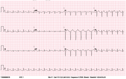
Figure 1: ECG - atrial tachycardia, sinus with STE in I with prolonged QT interval and no evedince of acute ischemia.
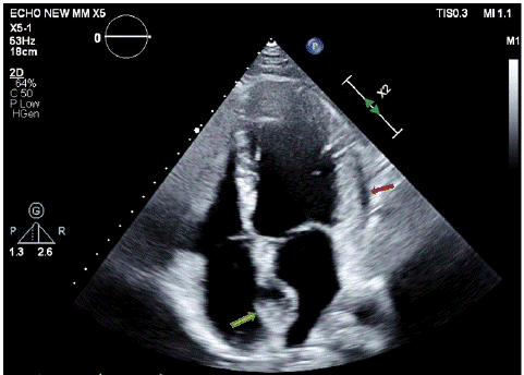
Figure 2: Echocardiogram- 4-chambers view - interatrial septum involvement (green arrow), pericardial effusion (red arrow).
Transthoracic echocardiogram demonstrated normal left ventricle and right ventricle size and function, no significant valvular dysfunction. Atria were of normal size with an interatrial septal mass of ~33mm, and mild pericardial effusion with no hemodynamic significance (Figure 3).
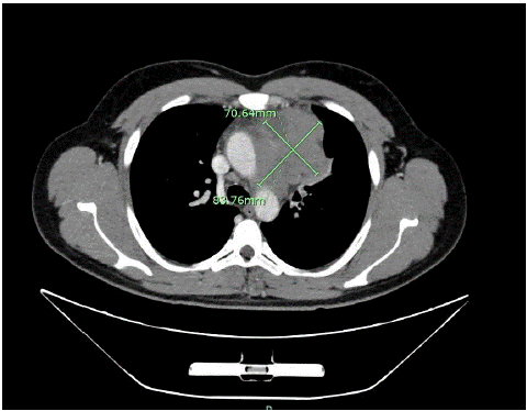
Figure 3: CT - Mediastinal lymph 70mm*83mm, with narrowing of the SVC and Pulmonary artery.
A Computerd Tomography (CT) exam of the chest demonstrated multiple lymph node enlargements in the left lower part of the neck and mediastinum in the interatrial septum, interventricular septum, with pressure on the atrium and pulmonary veins without evidence of liquefication or bone penetration. The mediastinal lymph nodes caused significant mechanical narrowing of the SVC and moderate narrowing of the pulmonary artery (Figure 4), the mediastinal mass was elongated to the interatrial septum with its involvement.
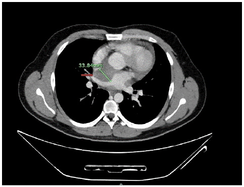
Figure 4: CT - interatrial septal thickening of 33mm (red arrow).
Due to the elevated LDH levels, lymphadenopathy, and an intra-atrial mass, a PET-FDG scan showed increased FDG uptake of cardiac structures, including the pericardium, intra-atrial septum, and a mass effect on atria and pulmonary vessels veins (Figure 5).
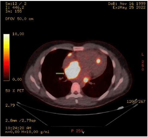
Figure 5: FDG-PET- intense uptake of the intra-atrial septum (green arrow).
For further evaluation, a tissue biopsy from the cervical lymph node revealed cores of lymphoid tissue infiltrated by sheets of large aypical lymphoid cells with round/oval nuclei, fairly prominent nucleolus and abundant cytoplasm with atypical cells highlighted strongly by immunohystochemical staining for CD30 and ALK-1, confirming the diagnosis of ALK-positive Anaplastic T-cell Lymphoma, CD30 positive.
The patient was admitted to the cardiac intensive care unit. Beta blockers were started for symptomatic relief, with consequent return to sinus rhythm. CHP-BV protocol was successfully initiated. The patient`s condition gradually improved with nofeeling of palpitation or shortness of breathing. A repeat echocardiogram after one week showed reduced effusion with no change in the mass size.
Discussion
ALCL-ALK+ is an aggressive lymphoma that generally occurs in young patients. Fever is the presenting symptom in 75% of cases [3]. Mediastinal and/or abdominal lymphadenopathy accures in 50-70% [2]. Extranodal involvement may occur in different tissues, including soft tissue, bone, bone marrow, gastrointestinal tract and lung [2]. Primary cardiac lymphoma is a rare entity, reported at less than 1% [4]. However, secondary cardiac involvement may be found in up to 30% of autopsy studies [4].
The clinical presentation of cardiac lymphoma depends on the site and type of involvement. The patient can present with chest pain, progressive heart failure, arrhythmia, or syncope [5].
Diagnosis of isolated cardiac lymphoma is difficult. Endomyocardial biopsy has a sensitivity of only 50% [6]. Cardic non-invasive imaging can help to diagnose cardiac lymphoma; however, due to the variable morphology of lymphomatous infiltrates, different cardiac imaging modalities are non-specific like cardiac MRI or CT. Therefore, a high clinical suspicion should be considered. In this case, a non-specific cardiac mass was found by echocardiogram and CT.
PET-FDG scan revealed diffuse increased uptake that can be infectious or malignant. Tissue diagnosis, preferably from a nodal or extracardiac site, is the gold standard of diagnosis [6] Combination chemotherapy, with or without adjuvant radiotherapy, is the backbone treatment of cardiac lymphoma; with side effect of treatment with cardiac involvement – for example necrosis and ASD/ free wall rupture heart failureatc, although there are no randomized trials, CHOP is currently the first-line treatment for ALK+ ALCL and has a favorable prognosis [2].
Conclusion
ALCL-ALK+ is an uncommon type of lymphoma, commonly presenting with fever and/or lymphadenopathy. Involvement of the cardiovascular system is unusual and an initial presentation with cardiac involvement is extremely rare. This unique case of a patient with ALCL-ALK+ initially presenting with atrial tachycardia secondary to cardiac involvolvent, demonstrates the wide differential diagnosis of cardiac tachycardia. It further highlights the importance of multimodality imaging facilitating proper diagnosis and guiding treatment.
Author Statements
Author Disclosures
The authors have no conflicts of interest to disclose. all authors had access to the data and a role in writing the manuscript.
References
- Tilly H, Gaulard P, Lepage E, Dumontet C, Diebold J, Plantier I, et al. Primary anaplastic large-cell lymphoma in adults: clinical presentation, immunophenotype, and outcome. Blood. 1997; 90: 3727-34.
- Tsuyama N, Sakamoto K, Sakata S, Dobashi A, Takeuchi K. Anaplastic large cell lymphoma: pathology, genetics, and clinical aspects. J Clin Exp Hematop. 2017; 57: 120-42.
- Ferreri AJM, Govi S, Pileri SA, Savage KJ. Anaplastic large cell lymphoma, ALK- positive. Crit Rev Oncol Hematol. 2012; 83: 293-302.
- Nascimento AF, Winters GL, Pinkus GS. Primary cardiac lymphoma: clinical, histologic, immunophenotypic, and genotypic features of 5 cases of a rare disorder. Am J Surg Pathol. 2007; 31: 1344-50.
- Nambiyar K, Gupta K, Debi U, Kant Sinha S, Kochhar R. ALK+ Anaplastic large cell lymphoma with extensive cardiac involvement: A rare case report and review of the literature Autopsy Case Report and Review. 2020.
- Punnoose LR, Roh JD, Hu S, Udell JA, Wagle N, Kirshenbaum JM, et al. Cardiac presentation of anaplastic large-cell lymphoma. J Clin Oncol. 2010; 28: e314-6.