Editorial
Austin J Cardiovasc Dis Atherosclerosis. 2014;1(1): 1.
An Aborted Left Ventriculogram
Daniel Ash1, Bryan Whitson2, Scott M Lilly2*
1Ohio State University College of Medicine, USA
2Department of Cardiovascular Medicine, Ohio State University Heart and Vascular Center, USA
*Corresponding author: Scott M Lilly, Division of Cardiovascular Medicine, Ohio State University Heart and Vascular Center, 473 W. 12th Avenue, Suite 200, Columbus, OH43210-1252, USA
Received: June 28, 2014; Accepted: July 25, 2014; Published: July 29, 2014
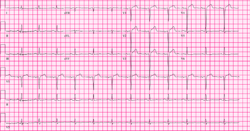
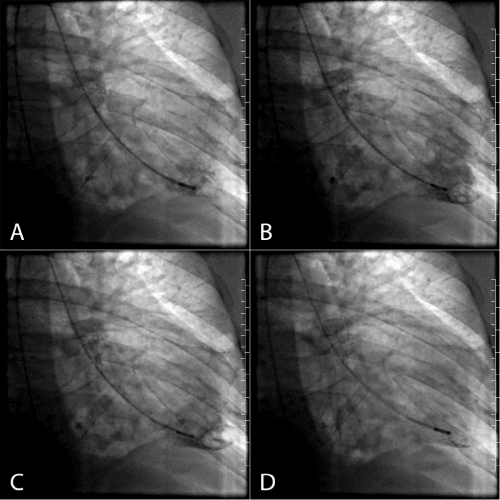
A 57-year-old male with coronary artery disease was admitted after 2 weeks of progressive dyspnea. His past medical history was notable for hypertension, type II diabetes mellitus, and hyperlipidemia. Initial electrocardiogram revealed Q waves in the anteroseptal leads (Figure 1). Coronary angiography revealed an obstructive lesion in the left anterior descending artery. A left heart catheterization was performed, and upon positioning the catheter for left ventriculography a large apical thrombus was identified (Figure 2A-D). The ventriculogram was aborted, and the patient was transferred to our hospital for further evaluation. Upon hospitalization, warfarin was initiated, with heparin bridging until a therapeutic level of anticoagulation could be achieved. Because cardiac MRI showed no anterior wall viability, he was determined to be a poor candidate for revascularization. He was discharged home for outpatient follow-up. As of this writing, his ejection fraction remains suppressed (35-40%) and he has NYHA class II heart failure symptoms without interval readmission. A recent echocardiogram demonstrated that the apical thrombus has dispersed.
Figure 1 :
Scheme 1 :
Assessment of left ventricular function is recommended in all patients presenting with acute ST-elevation myocardial infarction, and ventriculograms are often undertaken when non-invasive modalities are not immediately available or as a preamble to surgical referral. This case demonstrates the need for caution when undertaking left ventriculography in the setting of a recent myocardial infarction, particularly one presenting late. Although complications of left ventriculography are rare, balancing these with benefit of immediate incremental information is prudent.

