Case Report
Austin J Cardiovasc Dis Atherosclerosis. 2024; 11(1): 1062.
The Corporeality of Thromboaspiration: Introducing AVIS Protocol for Acute Coronary Syndromes with Large Thrombus Burden
Tanuj Bhatia1*; Sai Devrrat2; Aditya Kapoor3; Richa Sharma4; Roopali Khanna5; Abhishek Rastogi2
1Associate Professor & Cath Lab Director, Department of Cardiology, SGRR Medical College & SMI Hospital, Dehradun, India
2Senior Resident, Department of Cardiology, SGRR Medical College & SMI Hospital, Dehradun, India
3Professor & Head of the Department, Department of Cardiology, SGPGIMS, Lucknow, India
4Associate Professor, Department of Cardiology, SGRR Medical College & SMI Hospital, Dehradun, India
5Professor, Department of Cardiology, SGPGIMS, Lucknow, India
*Corresponding author: Tanuj Bhatia Associate Professor & Cath Lab Director, Department of Cardiology, SGRR Medical College & SMI Hospital, Dehradun, India. Email: tanujbhatia21@rediffmail.com
Received: January 25, 2024 Accepted: March 06, 2024 Published: March 13, 2024
Abstract
The Penumbra Cat Rx device has been shown to result in a safe and effective removal of thrombus, leading to improved outcomes even in high-risk patients. Here, we share our initial experience utilizing modern sustained mechanical thrombectomy in 21 patients with a significant thrombus burden. These patients were part of a diverse group with ST-Elevation Myocardial Infarction (STEMI) or STEMI equivalent conditions, and we also incorporated intracoronary imaging into the procedure. To the best of our knowledge, this represents the largest dataset from the Asia Pacific region involving the utilization of this innovative device and technique. Additionally, we introduce the “AVIS protocol” (routine mechanical thrombus Aspiration, intracoronary Vasodilators, Coronary Imaging followed by Stenting) as a potential method for managing acute STEMI cases with Large Thrombus Burden (LTB), which has the potential to greatly customize our approach for this specific group of patients undergoing percutaneous coronary intervention.
Introduction
Patients of Acute Coronary Syndromes (ACS), especially acute ST-Elevation Myocardial Infarction (STEMI) or STEMI equivalent often have Large Thrombus Burden (LTB). Though it is intuitive to consider that thrombosuction shall improve outcomes, earlier the practise of routine rheolytic thrombectomy has not shown to improve cardiovascular outcomes with thrombus aspiration [1-4]. The 2021 ACC/AHA/SCAI guidelines for Coronary Artery Revascularization [5] recommends thrombectomy as a “Class of Recommendation 3: No Benefit” on the basis of the trials (TOTAL, TAPAS, TASTE) [1-3]. However, the recent CHEETAH study [6] that utilised a sustained mechanical aspiration in contrast to the manual aspiration devices demonstrated an excellent angiographic outcome as well as no device related serious adverse events, in contrast to the TOTAL trial that demonstrated a small but statistically significant increased risk of stroke. On the other hand, the TOTAL trial did suggest additional dedicated studies focusing on high thrombus burden patients, with innovations in thromboaspiration might be specifically beneficial in LTB patients [1]. The Penumbra Cat Rx device [7] promises as safe and effective thrombus removal with better outcomes even in high-risk patients. Additionally, intravascular imaging especially Optical Coherence Tomography (OCT) promises to provide excellent resolution and documentation of residual thrombus burden as well as better post-procedure stent optimization [8]. Here we present our initial experience of the use with modern sustained mechanical thrombectomy in the 21 patients with large thrombus burden, in “all-comer” setting of STEMI or STEMI equivalent patients with Large Thrombus Burden (LTB), in conjunction with intracoronary imaging (IVUS or OCT), whenever feasible. All cases were performed between July 2023 and October 2023 at a high-volume centre running a 24 X 7 primary PCI programme. We also propose the “AVIS protocol” for the management of acute STEMI that may significantly tailor our approach for this subset of patients undergoing Percutaneous Coronary Intervention (PCI) that is almost always done on an “ad hoc basis” [9] and hence necessitates protocols in such situations.
Case Presentation
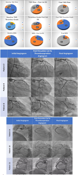
Out of total 21 cases, hereby we describe some of the complex cases in detail. Figures 1 and 2 show initial angiogram, post-Penumbra Cat Rx thromboaspiration angiogram and final angiogram of the six representative cases. Table 1 and central illustration represents grade of thrombus, TIMI flow and Myocardial Blush Grade for all 21 patients at initial angiogram, post-Penumbra Cat Rx thromboaspiration angiogram and at final angiogram. Table 2 outlines baseline characteristics of all the patients. Table 3 summarizes index myocardial infarction features of all patients. Table 4 provides angiographic details of all patients. Table 5 represents procedural details of all patients.
    Initial    Angiogram

    Post-Penumbra    Cat Rx thromboaspiration Angiogram

    Final    Angiogram


    Grade    of Thrombus


    5

    17

    -

    -


    4

    4

    -

    -


    3

    -

    5

    -


    2

    -

    13

    1


    1

    -

    3

    5


    0

    -

    -

    15


    TIMI    flow


    3

    -

    8

    18


    2

    3

    11

    3


    1

    2

    2

    -


    0

    16

    -

    -


    Myocardial    Blush Grade


    3

    -

    1

    17


    2

    1

    14

    3


    1

    4

    6

    1


    0

    16

    -

    -
Table 1: Grade of thrombus, TIMI flow and Myocardial Blush Grade of all patients.
Case No.
Age (in years)
Gender
Diabetic
HTN
F/h/o CAD
Obesity
Smoking
Dyslipi
demiaPast h/o CAD
1
53
M
ACS, 15 days back
2
45
M
Yes
Yes
3
46
F, post menopausal
Yes
Yes
Yes
Yes
4
36
M
Yes
Yes
Yes
Yes, PCI – 3 years back
5
76
M
Yes
Yes
Yes, PCI – 18 years back
6
56
M
Yes
Yes
Yes
Yes, PCI – 8 years back
7
37
M
Yes
Yes
Yes
Syncope
8
54
F, post menopausal
9
46
M
Yes
10
55
F, post menopausal
Yes
Yes
11
59
F, post menopausal
Preinfarct Unstable Angina
12
54
M
Yes
Yes
13
66
M
Yes
Yes
14
65
M
Yes, PCI – 2 years back
15
51
M
16
70
M
Yes
Preinfarct Unstable Angina
17
56
F, post menopausal
Yes
18
63
M
Yes
Yes
Yes
Preinfarct Unstable Angina
19
83
M
Chronic Stable Angina for 3 years
20
36
M
Yes
21
45
M
Yes
Table 2: Baseline characteristics.
Case No.
Type of MI
WP (hours)
Killip Class
TLT Status
Arrythmia
Post CPR, ROSC
Culprit Vessel
Baseline TIMI flow
AWMI
6
3
Yes, STK 15 days back
Ostial LAD
0
AWMI
3
4
Yes, VF
Yes
Ostial LAD
0
AWMI
12
4
Yes, VT
Yes
Proximal LAD
1
IWMI
8
2
Proximal LCx
0
STEMI equivalent
6
1
Proximal & distal LCx
0
IWMI
12
1
Proximal LCx
0
IWMI
12
2
Yes, TNK,
Rescue PCIYes, VT
Anomalous RCA (mid)
0
IWMI
6
2
CHB
Proximal RCA
0
IWMI
7
2
Distal RCA/ PLV
0
AWMI
16
2
Yes, STK, Pharmacoinvasive
Mid LAD
1
IWMI
14
3
Ostial LCx
0
IWMI
7
1
Distal RCA
2
IWMI
2
1
Mid RCA
2
IWMI
12
1
Proximal RCA
0
AWMI
24
2
Proximal LAD
0
AWMI
10
1
Proximal LAD
0
IWMI
18
2
Mid RCA
0
AWMI
12
3
Proximal LAD
0
IWMI
12
4
Junctional Rhythm
Proximal LCx
0
AWMI
6
4
VF
Yes
Proximal LCx & Proximal LAD
0
IWMI
6
1
Mid RCA
2
Table 3: Index myocardial infarction features.
    Case    No.

    Baseline    TIMI Flow

    TIMI    flow – Post Cat RX

    Final    TIMI flow

    Baseline    TIMI Thrombus Grade

    Thrombus    Grade Post Cat RX

    Final    TIMI Thrombus Grade

    Baseline    MBG

    MBG    – Post Cat RX

    Final    MBG


    0

    2

    3

    5

    2

    0

    0

    2

    3


    0

    2

    3

    5

    2

    0

    0

    2

    3


    1

    2

    3

    4

    2

    0

    1

    2

    3


    0

    2

    3

    5

    2

    0

    0

    2

    3


    0

    2

    3

    5

    2

    0

    0

    2

    3


    0

    2

    3

    5

    2

    0

    0

    2

    3


    0

    2

    3

    5

    3

    1

    0

    1

    3


    0

    3

    3

    5

    1

    0

    0

    2

    3


    0

    2

    2

    5

    3

    1

    0

    1

    2


    1

    3

    3

    5

    2

    0

    1

    2

    3


    0

    2

    3

    5

    2

    0

    0

    1

    2


    2

    3

    3

    4

    2

    1

    1

    2

    3


    2

    3

    3

    4

    1

    0

    2

    3

    3


    0

    3

    3

    5

    2

    0

    0

    2

    3


    0

    2

    3

    5

    2

    0

    0

    2

    3


    0

    3

    3

    5

    2

    0

    0

    2

    3


    0

    2

    3

    5

    3

    1

    0

    1

    3


    0

    1

    2

    5

    3

    1

    0

    1

    3


    0

    3

    3

    5

    1

    0

    0

    2

    2


    0

    1

    2

    5

    3

    2

    0

    1

    1


    2

    3

    3

    4

    2

    0

    1

    2

    3
Table 4: Angiographic details.
    Case    No.

    Access

    No.    of Passes of Cat RX

    Predilatation

    Imaging

    Imaging    technique

    Post    dilatation

    Additional    Comment


    7 F, Radial

    3

    -

    -

    -

    Yes

    -


    6 F, Femoral

    2

    -

    -

    -

    Yes

    -


    6 F, Radial

    2

    -

    -

    -

    Yes

    Efficacy in Thrombocytopenic    patient (Dengue)


    6 F, Radial

    2

    -

    Yes

    OCT    – Endothelial disruption minimal

    Yes

    Excellent Trackability


    6 F, Radial

    2

    Yes

    Yes

    OCT    – Plaque rupture at sites that were not identified on angiogram

    Yes

    Travels easier than Balloon


    6 F, Radial

    2

    Yes

    -

    -

    Yes

    Excellent Trackability


    6 F, Radial

    3

    Yes

    -

    -

    Yes

    Successful Rescue PCI


    6 F, Radial

    1

    -

    -

    -

    Yes


    6 F, Radial

    2

    Yes

    -

    -

    Yes

    Efficacy in distal small    vessels needs more assessment


    6 F, Radial

    3

    Yes

    -

    -

    Yes

    Role in Pharmacoinvasive    strategy with residual high thrombus burden


    6 F, Femoral

    3

    Yes

    -

    -

    Yes

    Trackability in Acute take off    (retroflexed) LCx


    6 F, Radial

    2

    Yes

    Yes

    OCT    – huge calibre RCA

    Yes

    OCT after thromboaspiration    reveals true calibre of vessel


    6 F, Radial

    1

    -

    -

    -

    Yes


    6 F, Radial

    2

    Yes

    Yes

    IVUS    – ISNA at proximal RCA stent

    Yes

    Thrombus aspiration improves    capability of IVUS images for defining reason of stent failure and dictates    further management


    6 F, Radial

    2

    -

    -


    Yes

    Efficacy in ectatic LAD


    6 F, Radial

    2

    Yes

    -

    -

    Yes

    0


    6 F, Radial

    2

    -

    -

    -

    No

    Efficacy in low calibre RCA


    6 F, Radial

    2

    Yes

    -

    -

    Yes

    CAT Rx did not cross the lesion    but still restored flow by “forward looking” mechanism of thromboaspiration


    6 F, Radial

    2

    -

    -

    -

    Yes

    Immediate restoration of rhythm    may point out lesser distal embolization with this strategy


    6 F, Radial

    2

    Yes

    -

    -

    Yes

    Multivessel thromboaspiration


    6 F, Radial

    2

    -

    Yes

    OCT    – Inflammatory Plaque

    Yes

    Efficacy in thrombocytopenic    patients and adequate thromboaspiration helps avoid aggressive antithrombotic    therapy
Table 5: Procedural details.

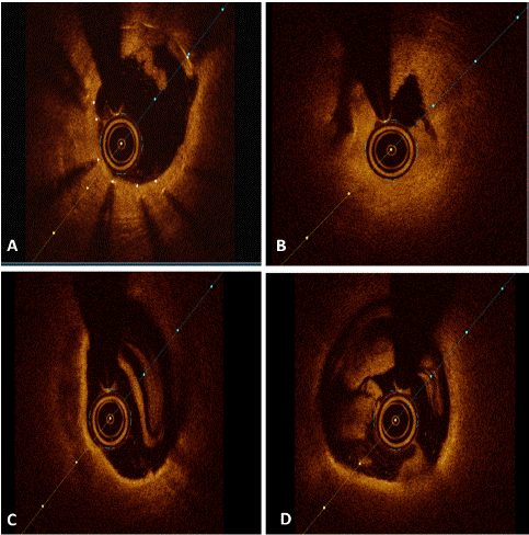
Figure 1: Optical coherence tomography findings in patient no. 4 (A) In-stent neo atherosclerosis with plaque rupture, (B) Catheter hugging thrombus, (C) Fibroatheroma with large necrotic core and thick cap except at 5 o’clock with thin cap, (D) Residual red & white thrombus.

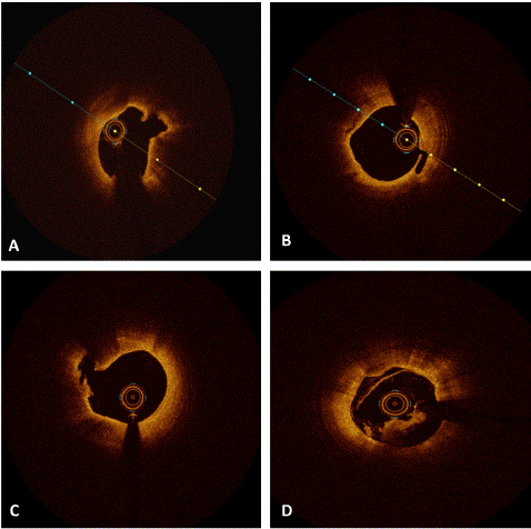
Figure 2: Optical coherence tomography findings in patient no. 6 (A) Thin cap fibroatheroma with plaque rupture at culprit site, (B) Plaque rupture at a site that unidentified as culprit on angiogram, (C) Plaque rupture with intraplaque cavity free from thrombus, (D) Endothelial disruption and residual red thrombus.
Case 2
A 45-year-old male with no conventional CAD risk factors presented to the ER in comatose situation, documented to have primary VF and cardioverted and shifted to the cardiac cath lab with the blood pressure of 80/50 mmHg. ECG showed hyperacute anterior wall MI and CAG revealed ostioproximal total occlusion of LAD with Grade 5 thrombus. The window period was two hours and primary PCI was done. A 6 Fr access was taken and just with 2 runs of Cat Rx aspiration, the thrombus reduced to Grade 2 and this helped us delineate the landing zones without additional need of predilation, especially at the time when we need precision and speed and with no thrombus to bother us and no concerns of slow flow. With this minimal thrombus, we could use a higher NC balloon of 3.5 mm diameter at higher pressures of 18 atmosphere and excellent final angiographic result was obtained in the next 10 minutes.
Case 4
A 36-year-old dyslipidemic male with a Low Density Lipoprotein (LDL) of 196 mg/dl and past history of coronary intervention to all three vessels LCx was totally occluded proximally whilst the LCx stent placed earlier was in the distal LCx. A single pass of the Cat Rx catheter restored TIMI 3 flow. There was a suspicion of small dissection and hence OCT pull back was taken. Of note, no pre-dilation had been performed till now. OCT revealed instent neo-atherosclerosis in the stent placed earlier in the distal LCx with a plaque rupture more proximally. There was a catheter hugging residual thrombus (both red and white) in proximal LCx. The DES was directly deployed with excellent angiographic and OCT results (Figure 3).

Figure 3: Image showing aspirated thrombus from the coronary artery of patient no. 7.
Case 6
A 56-year-old mail with acute inferior wall MI, presented during thewindow period of 12 hours. The CAG revealed total occlusion of LCx. Cat Rx thrombosuction restored TIMI 2 flow in one pass and residual 80% stenosis in the culprit vessel. OCT revealed plaque rupture at the culprit site as well as proximally where the vessel was looking angiographically free from significant disease. Proximal LCx also had eccentric calcium with an OCT calcium score of 2. The PCI was done thereafter in conventional fashion with OCT guidance for post-dilatation (Figure 4).

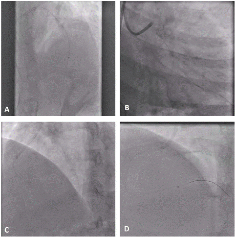
Figure 4: Images representing safe and effective navigation of Penumbra Cat Rx deep into coronary vasculature of (A) Mid left anterior descending artery, (B) Tortuous left circumflex artery, (C) Distal right coronary artery/posterior left ventricular artery, (D) Distal right coronary artery.
Case 14
A 65-year-old patient, underwent PTCA to RCA 2 years ago, presented with acute inferior wall MI at a window period of 12 hours. Coronary angiogram revealed total occlusion of RCA prior to the stent placed earlier. Thrombosuction with two runs of Penumbra catheter downgraded the thrombus from Grade 5 to Grade 2. Upon IVUS assessment, we could document the under expansion of the RCA stents in the distal part. There was neoatherosclerosis in proximal RCA with superficially attenuated plaque and features suggestive of vulnerable plaque with the MLA of 2.54 mm2. The under expanded portion of the stent was just dilated at high pressures with a 3.5 NC balloon and the region with neoatherosclerosis was also predilated and only a single stent was deployed in the proximal part of RCA that had associated neoatherosclerosis. Thrombosuction greatly helped us in appropriate assessment of the underlying aetiology for the stent failure by subsequent imaging and in this case, helped us avoid unnecessary stenting in the part that just needed post dilatation, that would have been treated by an additional stent if PCI would have been done in conventional fashion.
Case 18
A 63-year-old morbidly obese and diabetic male presented with acute AWMI with window period of 12 hours and in Killip III hemodynamic status. The CAG showed total occlusion of ostioproximal LAD with grade 5 thrombus and disease starting from distal left main. Ostial LAD also showed heavy calcium. Wire crossing was slightly difficult, and we had our apprehensions about crossing of the 5.3 Fr Cat Rx catheter owing to calcific disease at ostia. The thrombosuction reached upto the occlusion, did not cross it but was effective in sucking out the thrombus by its forward-facing suction effect. Post thrombosuction angiogram had already reduced the thrombus to grade 2. Predilatation was necessary with 2.5 mm NC balloon and a good result was achieved thereafter with stenting in conventional fashion.
Case 21
A 56-year-old diabetic male presented with acute IWMI of 6 hours window period. Patient was recovering from dengue fever and platelets were in rising trend, though patient was still thrombocytopenic with platelet count of 1,00,000 per microlitre. The CAG showed grade 4 thrombus in mid RCA and since we wished to manage with minimal antiplatelet and anticoagulation, we used Cat Rx to downgrade the thrombus from grade 4 to grade 1 with two passes.
The OCT evaluation thereafter documented an inflammatory plaque with cholesterol crystals, macrophage infiltration with large necrotic core and evidence of plaque rupture. This was followed by stenting in conventional fashion and OCT guided optimization with aggressive post dilatation, that was possible with minimal residual thrombus owing to effective thrombosuction in a thrombocytopenic individual.
Discussion
The 21 cases presented are of different presentations - very early, early and late, of different Killip class and involving different vessels, including tortuous anatomy. The use of Indigo Cat Rx aspiration system (Penumbra Inc, Alameda CA) for sustained mechanical aspiration thrombectomy before PCI showed excellent outcomes in all subsets irrespective of age, risk factors, time of presentation with excellent short-term outcomes and no major device related complications. Our series includes thrombosuction from all major vessels as well as small vessels like PLV (Case No 9). Moreover, we used radial access in 90% of our patients as compared to 52.8% in ROPUST study [7].
The Cat Rx aspiration catheter and Penumbra aspiration pump received FDA clearance in May 2017 for removal of soft thrombi in coronary and peripheral vasculature. This dedicated vacuum pump with Cat Rx Penumbra catheter that delivers a constant and consistent suction is an attractive innovation with excellent trackability with hypotube technology, allowing an inner diameter like large-bore catheters while maintaining a lower profile and a soft, atraumatic tip designed to help navigate the complex and delicate anatomy even in tortuous anatomy and anomalous vessels (Figure 5). Though the CHEETAH study [6] did exclude patients those who had received thrombolytic therapy for index coronary vessel occlusion, we did use it in two patients (Case No. 7 & 10), who were taken for rescue PCI and in a pharmacoinvasive strategy respectively, and both had residual LTB on the initial angiogram
Initially, coronary imaging was done to atleast evaluate the atraumatic nature of this forward-looking thrombosuction device with continuous vacuum suctioning. There were just minor dissections and intimal disruption documented in 2 cases (Case 4 and 5). There were no device related complications, no stroke and no significant drop of haemoglobin related to the device in any patient. Blood loss was just 10-25 ml. Though the role of thrombosuction in one of the metanalysis [4] has been conflicting, it is still a matter of active debate in LTB patients [10] especially worrying the users of a small increased risk of stroke. Stroke is a known complication of PCI with or without thrombosuction and strong predictors include carotid disease, old age, cardiogenic shock and atrial fibrillation [11]. The Cat Rx catheter was used in a technique and as described in the CHEETAH study [6] with the suction applied from the time the catheter is proximal to the thrombotic lesion or occlusion, all the time during navigation in and out of the coronary vessel and until it exists the Touhy-Borst valve. Since this technique doesn't employ clot fragmentation or saline jets, hence may not increase distal embolization and consequently microvascular obstruction and MBG, and may not increase the risk of stroke also [12].
We had two deaths in our series (patient no. 3 and 20). Both the patients had good angiographic result. Patient no. 3 was a survivor of cardiac arrest after prolonged CPR, had dengue fever with severe thrombocytopenia, sepsis and acute renal failure. Patient number 20 had a very prolonged cardiac arrest (>1 hour of CPR) in emergency.
The final thrombus grade 0/1 was seen in 20 out of 21 patients, TIMI grade 3 flow in 18 out of 21 patients and MBG grade 3 was seen in 17 out of 21 patients in the final angiogram (Table 1). Of note, none of the patients had slow flow at the completion of the procedure. All these figures demonstrate excellent thrombus debulking, in sync with the CHEETAH study [6] and this is an excellent advancement over the rates of 47.6% and 52.2% of MBG 3 documented with earlier thrombosuction devices [2-13].
We strongly recommend use of Penumbra device before any balloon angioplasty and usually there is no difficulty in crossing the lesion, because of the excellent neurotracking capability even in tortuous anatomy as well as its far field suctioning effect over a few millimetres ahead of the tip inside the vessel. This was our initial experience with Cat Rx thrombosuction, it prompts us to plan a careful observational study or randomised control trials with adequate power to document the utility of sustained mechanical thrombosuction in minimising infarct size and hastening recovery of LV function and reducing long term mortality. To the best of our knowledge, this is first largest data from the Asia Pacific region for the use of this novel device and technique. Future studies for documentation of improvement in ejection fraction, end diastolic volume, end systolic volume, global longitudinal strain and/or speed of recovery (speed of resolution of ST segment elevation) with the use of this device and AVIS protocol as compared to those undergoing conventional PCI may be planned. With the AVIS protocol being followed, intravascular imaging should also be more liberally used. Though this data practically demonstrates the potential of this therapy for appropriate thromboaspiration, echocardiographic and clinical studies shall put more light on this technique, embracing its entry into a real-world situation for all STEMI or STEMI equivalent patients with LTB.
Introducing the AVIS Protocol
We propose the “AVIS protocol” that employs sequential imaging after thrombo-aspiration in all patients with Large Thrombus Burden (LTB) presenting with STEMI or STEMI equivalent.
The AVIS protocol stands for – Aspiration, Vasodilators, Imaging (preferably OCT) and Stenting. We recommend welcoming the broader utilisation of this therapy and protocol. The sequence of the protocol is important as it tends to lessen the myocardial damage and assists in getting the precise result in an expedite fashion. Aspiration should be the foremost technique to reduce thrombus burden, after which endogenous vasodilatory mechanisms become more active and can be supplemented by exogenous vasodilators (nitroglycerin, nitroprusside, nicardipine and nicorandil). Adjunctive use of coronary imaging, especially OCT, is gaining ground due to improved peri- and post-procedure outcomes. This should be followed by predilatation (if required), stenting and optimization (if needed) at the final step. Of interest, EROSION III (14) had around 40% medically managed patients and it remains speculative whether having aspiration at top of the AVIS protocol followed by imaging may open door to a greater number of medically managed patients especially with the excellent pharamacotherapies (antiplatelet and anticoagulant regimes, non-statin drugs for hyperlipidemia) and directed therapies (like pelecarsen for elevated lipoprotein (a)) that may be available in future. We believe the AVIS protocol can dramatically improve the outcomes in STEMI patients. Large thrombus burden are highest risk individuals and conceptually should derive the maximum benefit with this therapy and protocol.
Conclusion
Thrombosuction with Penumbra promises to be more efficient, most reliable clot removal therapy with normalised microvascular function after subsequent PCI. It can help in de-escalation of the intensity of platelet inhibition by changing the type, dose, or number of antiplatelet drugs, in conjunction with better thrombosuction and reduced clot burden to handle and can be extremely helpful in being satisfied with low targets of ACT especially in HBR patients. It can also aid in improvement of visualisation of culprit vessels and avoiding PCI of uninvolved areas and implantation of shorter stents at more appropriate sites. Apt use of this innovative technology can lead to its incorporation into the protocol of management of acute STEMI.
References
- Jolly SS, Cairns JA, Yusuf S, Meeks B, Pogue J, Rokoss MJ, et al. Randomized trial of primary PCI with or without routine manual thrombectomy. New England Journal of Medicine. 2015; 372: 1389-98.
- Svilaas T, Vlaar PJ, van der Horst IC, Diercks GF, de Smet BJ, van den Heuvel AF, et al. Thrombus aspiration during primary percutaneous coronary intervention. New England Journal of Medicine. 2008; 358: 557-67.
- Fröbert O, Lagerqvist B, Olivecrona GK, Omerovic E, Gudnason T, Maeng M, et al. Thrombus aspiration during ST-segment elevation myocardial infarction. New England Journal of Medicine. 2013; 369: 1587-97.
- Jolly SS, James S, Džavík V, Cairns JA, Mahmoud KD, Zijlstra F, et al. Thrombus aspiration in ST-segment–elevation myocardial infarction: an individual patient meta-analysis: Thrombectomy Trialists Collaboration. Circulation. 2017; 135: 143-52.
- Lawton JS, Tamis-Holland JE, Bangalore S, Bates ER, Beckie TM, Bischoff JM, et al. 2021 ACC/AHA/SCAI guideline for coronary artery revascularization: executive summary: a report of the American College of Cardiology/American Heart Association Joint Committee on Clinical Practice Guidelines. Circulation. 2022; 145: e4-e17.
- Mathews SJ, Parikh SA, Wu W, Metzger DC, Chambers JW, Ghali MG, et al. Sustained Mechanical Aspiration Thrombectomy for High Thrombus Burden Coronary Vessel Occlusion: The Multicenter CHEETAH Study. Circulation: Cardiovascular Interventions. 2023; 16: e012433.
- Tashtish N, Chami T, Dong T, Chami B, Al-Kindi S, Mously H, et al. Routine Use of the “Penumbra” Thrombectomy Device in Myocardial Infarction: A Real-World Experience—ROPUST Study. Journal of Interventional Cardiology. 2022; 2022: 5692964.
- Higuma T, Soeda T, Yamada M, Yokota T, Yokoyama H, Izumiyama K, et al. Does residual thrombus after aspiration thrombectomy affect the outcome of primary PCI in patients with ST-segment elevation myocardial infarction? An optical coherence tomography study. Cardiovascular Interventions. 2016; 9: 2002-11.
- Rahman Z, Paul G, Choudhury A. Ad-hoc percutaneous coronary intervention and staged percutaneous coronary intervention. Mymensingh medical journal: MMJ. 2011; 20: 757-65.
- Levine GN, Bates ER, Blankenship JC, Bailey SR, Bittl JA, Cercek B, et al. 2015 ACC/AHA/SCAI focused update on primary percutaneous coronary intervention for patients with ST-elevation myocardial infarction: an update of the 2011 ACCF/AHA/SCAI guideline for percutaneous coronary intervention and the 2013 ACCF/AHA guideline for the management of ST-elevation myocardial infarction: a report of the American College of Cardiology/American Heart Association Task Force on Clinical Practice Guidelines and the Society for Cardiovascular Angiography and Interventions. Circulation. 2016; 133: 1135-47.
- Alkhouli M, Alqahtani F, Tarabishy A, Sandhu G, Rihal CS. Incidence, predictors, and outcomes of acute ischemic stroke following percutaneous coronary intervention. JACC: Cardiovascular Interventions. 2019; 12: 1497-506.
- Srinivasan M, Rihal C, Holmes DR, Prasad A. Adjunctive thrombectomy and distal protection in primary percutaneous coronary intervention: impact on microvascular perfusion and outcomes. Circulation. 2009; 119: 1311-9.
- Mongeon F-P, Bélisle P, Joseph L, Eisenberg MJ, Rinfret S. Adjunctive thrombectomy for acute myocardial infarction: A bayesian meta-analysis. Circulation: Cardiovascular Interventions. 2010; 3: 6-16.
- Jia H, Dai J, He L, Xu Y, Shi Y, Zhao L, et al. EROSION III: A Multicenter RCT of OCT-Guided Reperfusion in STEMI With Early Infarct Artery Patency. JACC Cardiovasc Interv. 2022; 15: 846-856.