Special Article – Clinical Case Reports
Austin J Cardiovasc Dis Atherosclerosis. 2016; 3(2):1025.
Long-term Favorable Course of Aspergillus Endo-, Myo-, and Pericarditis
Hayashi A¹, Kobayashi S², Hisauchi I¹, Komatsu T¹, Nakahara S¹, Sakai Y¹, Haruki K² and Taguchi I¹*
¹Department of Cardiology, Dokkyo Medical University Koshigaya Hospital, Japan
²Department of Infection Control, Dokkyo Medical University Koshigaya Hospital, Japan
*Corresponding author: Isao Taguchi, Department of Cardiology, Dokkyo Medical University Koshigaya Hospital, Minami-Koshigaya 2-1-50, Koshigaya, Saitama, Japan
Received: September 06, 2016; Accepted: September 14, 2016; Published: September 16, 2016
Abstract
Thirty year old healthy male with no medical history, who tested negative for human immunodeficiency virus antigens, was affected Aspergillus pancarditis. A case of this kind is extremely rare, and this is the first report of Aspergillus pancarditis with a long-term favorable clinical course that generally leads to very poor prognosis. The biopsy sample of hypertrophied atrial septum from the left atrium was essential for obtaining the definitive diagnosis and a longterm continuation of an effective antifungal oral agent might bring this successful result.
Keywords: Aspergillus; Pancarditis; Endocarditis; Biopsy
Case Presentation
We report a 30-year-old male patient with no significant past medical, family, or occupational history. The chief complaints of high fever and cough appeared in the latter third of March 2013; he was admitted to another hospital in April of the same year and received antibiotic therapy. However, his fever of 38°C and cough persisted, and he was admitted to our hospital for specialized workup and treatment.
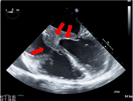
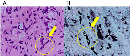
Malignant lymphoma or infective endocarditis was suspected at the time of his admission, based on test results indicating inflammation (C-reactive protein=4.2mg/mL, reference range <0.3mg/mL), chest computed tomography (CT) showing pericardial effusion, echocardiography showing vegetation like appearances, and gallium scintigraphy showing abnormal accumulation of gallium within the pericardial space. However, because of an abnormally high serum β-D-glucan level (612pg/mL, reference range < 20pg/ mL), which suggested fungal infection; we started an intravenous antifungal agent (voriconazole 400mg/day). Transesophageal echocardiography showed extensive thickening of both atrial walls, focused around the mitral annular ring, and including the atrial septum (Figure 1). In addition, malignant lymphoma was suspected because of the large pericardial effusion, and pericardiocentesis was performed. Approximately 800mL of yellow to faintly bloody pericardial fluid was obtained. The cytological diagnosis was negative for malignancy; therefore, we performed a myocardial biopsy. Under intracardiac ultrasound guidance, we obtained a sample of the hypertrophied atrial septum from the right atrium, near the oval window. The histopathological findings included scattered macrophages, lymphocytes, and eosinophils. Elongated figures scattered among the cellsappeared to be fungal filaments (Figure 2A). Grocott staining revealed the same filamentous appearance (Figure 2B), and blood tests were positive for the Aspergillus antigen (2.8ng/mL, reference range <0.5ng/mL). Aspergillus pancarditis (endocarditis, myocarditis and pericarditis) was diagnosed, and the antifungal agent was continued. However, because the abnormally elevated serum β-D-glucan levels persisted (1180 pg/mL), the patient’s treatment was changed from voriconazole 400 mg/day to amphotericin B 150mg/day on day 37. Beginning around day 50, the β-D-glucan level started to decrease. Echocardiography findings indicated improvement of the mural hypertrophy by day 50, and the mobile, vegetation-like shadow attached to the mitral annular ring had also disappeared. Three months after admission, hypertrophy affecting the base of the aorta; the walls of both atria, including the atrial septum; and the base of the posterior atrial wall had further improved to almost normal. In addition, the pericardial effusion did not return after pericardiocentesis.

    Figure 1:  Transesophageal echocardiogram at the time of admission Marked
hypertrophy of both atrial walls from the mitral annular ring to the atrial
septumis seen (red arrows).

    Figure 2:  Pathology findings AHE stain ×1,000 Scattered macrophages,
lymphocytes, eosinophils. Elongated shapes between the cells appear to
be fungal filaments (yellow arrows). B Grocott stain ×1,000The filaments are
short, friable, and swollen, suggestive of filamentous fungi (yellow arrows).
The electrocardiogram showed atrial flutter with 5:1 conduction and heart rate (HR) of 40 beats per minute (bpm)from the time of admission, and junctional rhythm (HR=48bpm) from day 50. Three months after admission, the patient’s sinus rhythm returned (HR=70bpm).
Renal dysfunction manifesting as a high serum creatinine level (1.5mg/dL, reference range <1.0mg/dL) was observed from around day 60, and because of nephrotoxicity associated with amphotericin B, the patient’s therapy was changed to oral voriconazole 400mg/ day. The patient’s renal function did not deteriorate further, and his serum creatinine level decreased to 1.2mg/dL. Although the patient was on oral voriconazole, his Aspergillus-related symptoms were not exacerbated, and his β-D-glucan values decreased to 144pg/ mL. Subsequent Aspergillus antigen testing was negative, and the patient was discharged on day 148 with continuous oral voriconazole administration.
Aspergillus infection usually affects the lungs, with rapid hematogenous dissemination to all organs and formation of micro abscesses [1]. When Aspergillus infects the heart, it causes an acute pathological state, including endocarditis, myocarditis and pericarditis [2]. Symptoms progress stealthily and rapidly during myocarditis, and patients commonly die within a short period of a few hours to a few days [3,4]. We believe that the favorable clinical outcome of our patient was accounted for by the patient’s normal left ventricular function, the absence of underlying disease that would cause impaired immunity, and the effectiveness of the antifungal agent against our patient’s infection.
Identifying the route of infection was a challenge in this case. Usually the pathogen reaches the lungs through inhalation; however, there was no lesion suggesting respiratory infection on the CT images of our patient. There was no history of penetrating trauma. There are various routes of infection, but usually Aspergillus does not spread hematogenously unless the patient has impaired immunity. Our patient was a healthy young man with no medical history, who tested negative for human immunodeficiency virus antigens.
The definitive diagnosis of Aspergillus infection by standard sputum cultures is difficult, and tissue biopsy is usually required. This invasive test provides the correct diagnosis in 10% to 30% of cases. The blood culture positivity rate is extremely low, at 6.4% to 11% [5]. Aspergillus was not isolated from our patient’s blood or pericardial fluid cultures, but was histopatholgically identified from myocardial tissue. The biopsy sample of hypertrophied atrial septum from the left atrium was essential for obtaining the definitive diagnosis in our patient.
Treatment for Aspergillus carditis is based on antifungal agents such as amphotericin B. However, systemic infection usually occurs during the time of diagnosis, and because the infection rapidly progresses, the prognosis is extremely poor. However, this case represents a favorable clinical course; more than 2 years have elapsed since the patient’s treatment was changed to oral voriconazole. Data on the duration of oral antifungal administration during maintenance therapy are scarce, and to date we have continued administration of voriconazole. This case is important, because to the best of our knowledge, this is the first report of Aspergillus pancarditis with a long-term favorable clinical course. Going forward, we will also need to consider the risk of reactivation of infection, and whether or not diligent follow-up observation, including echocardiography, ECG, and sample collection and testing is needed.
References
- Rinaldi MG. Invasive aspergillosis. Rev Infect Dis. 1983; 35: 1061-1077.
- Atkinson JB, Connor HA, Robinowitz M, McAllister HA, Virmani R. Cardiac fungal infections: review of autopsy findings in 60 patients. Human Pathology. 1984; 15: 935-942.
- Schønheyder H, Hoffmann S, Jensen HE, Hansen BF, Franzmann MB. Aspergillus fumigatus fungaemia and myocarditis in a patient with acquired immunodeficiency syndrome. APMIS. 1992; 100: 605-608.
- Rouby Y, Combourieu E, Perrier-Gros-Claude JD, Saccharin C, Huerre M. A case of Aspergillus myocarditis associated with septic shock. J Infec. 1998; 37: 295-297.
- El-Hamamsy I, Dürrleman N, Stevens LM, Perrault LP, Carrier M. Aspergillusendocarditis after cardiac surgery. Ann ThoracSurg. 2005; 80: 359-364.