Special Article – Clinical Case Reports
Austin J Cardiovasc Dis Atherosclerosis. 2016; 3(3): 1027.
Femoral Endarterectomy against Iliofemoral bypass-Case Series
Nagre SW*
Department of CVTS, Grant Medical College, Mumbai, India
*Corresponding author: Suraj Wasudeo Nagre, 31 Trimurti Building, JJ Hospital Compound, Byculla, Mumbai, Pin-400008, India
Received: September 06, 2016; Accepted: October 06, 2016; Published: October 10, 2016
Abstract
Background: The purpose of retrospective study is to evaluate the results of open endarterectomy in short atherosclerotic occlusion of the femoral artery against iliofemoral bypass grafting with vein graft in Asian population.
Methods: 10 male patients out of which 5 patients underwent endarterectomy of the femoral artery and other 5 underwent iliofemoral bypass grafting with vein graft between May 2013 and May 2015 are studied. The patients have a median age of 40+/−10 years. All patients underwent routine followup at 1, 3, 6 and 12 months and yearly thereafter. Routine clinical exami-nation, colour Doppler scan and if necessary, arteriogram were done to assess the outcome.
Results: There was no difference in results between both groups. The length of the lesion varied from 2 to10 cm, and the largest endarterectomy done was 10cm. The segments involved were external iliac artery extending to femoral artery. The patency rates were 100% at 1 year followup. Patency rate was equal in both groups without any complications. Time of harvesting vein graft was saved in endarterectomy patients. So the endarterectomy was better time and incision saving alternative whenever possible with equivocal results.
Keywords: Femoral artery; Endarterectomy; Iliofemoral bypass; Arteriogram
Introduction
The decision for an ideal vascular procedure for occlusive peripheral arterial disease depends on type of lesion involved, patient factors and the options available to deal with it. Despite a dramatic increase in the use of endovascular techniques to treat chronic limb ischemia secondary to femoral arterial disease (FAD), femoral endarterectomy (FE) and profundaplasty remain the procedures of choice because long-term patency results are still superior to any other intervention [1,2]. The American Heart Association [2,3] has recommended that angioplasty of the femoral artery be carried out only in single lesions of less than 10 cm, and better results are associated with short lesion length and good runoff, claudication, stenosis and the absence of diabetes. The immediate placement of a stent after angioplasty may address the issues of elastic recoil and dissections and improve the early success rate, but it has not been shown consistently to improve long-term patency [4]. Even though the endarterectomy was the one of the first described procedures, autogenous long saphenous vein bypass has surpassed it as the preferred procedure for occlusive disease in the infrainguinal region [1]. This is largely the result of several series reporting an inferior long term patency rate with endarterectomy [1,3]. Further studies were conducted to compare the efficacy of available surgical options (vein by-pass, open endarterectomy and semi-closed endarterectomy) to combat the femoro-popliteal occlusion and it showed that the immediate failures and late cumulative patency/survival were similar for all three procedures [3]. Studies supported the use of endarterectomy as available option for infrainguinal arterial diseases with very comparable results to vein grafts [1-5].
The long saphenous vein is a very demanding conduit if coronary or vein bypass is required in future [7,8]. Synthetic grafts and angioplasty studies has shown inferior results (high early and late restenosis rates) [9-11]. The present retrospective study is to evaluate the results of open endarterectomy in short atherosclerotic occlusion of the femoral artery against iliofemoral bypass grafting with long saphenous vein graft.
Patients and Methods
Endarterectomy of localized lesions of the iliofemoral artery was performed in 5 male patients between May 2013 and May 2015, with a proven diagnosis of chronic occlusion of isolated external iliac artery or iliofemoral artery. The diagnosis was confirmed by angiography. The median age of the patients was 40 +/− 10 years. Risk factors for peripheral atherosclerosis were diabetes, electrocardiogram changes of past myocardial is chaemia, smoking and hypertension.
All patients suffered from atherosclerosis, and had thrombosis or stenosis of the external iliac artery associated with/without adjoining femoral artery and profunda femoris artery. All endarterectomies were performed as a primary procedure and not as an adjunct to a bypass procedure. Selection of patients the indications for operation in this series were disabling intermittent claudication, rest pain and gangrene. Out of five in two cases, the claudication distance was 100m or less and in two cases had rest pain severe enough to interfere with sleep and one case had established gangrene of the toes [2,3].
Clinical Findings and Investigations
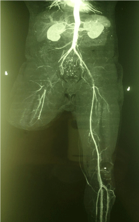
In those with occlusive disease localized to the iliofemoral artery, the patient’s usually had with no pulses in the limb distally on the affected side. Occasion-ally, weak pedal pulses were present. All patients were investigated by Colour Doppler scan by CE FDA 3D 4D Doppler ultrasound and femoral angiography (Figure 1). Where unequal femoral pulses were present, or bruits were audible over the iliac or common femoral vessels, suggesting more proximal lesions, lumbar orthography was performed and if present these patients were excluded from the study.

    Figure 1:  CT Angiogram showing occluded bilateral external iliac artery with
reformation of distal vessels.
Surgical Technique
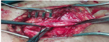
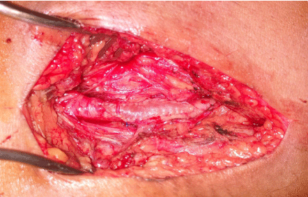
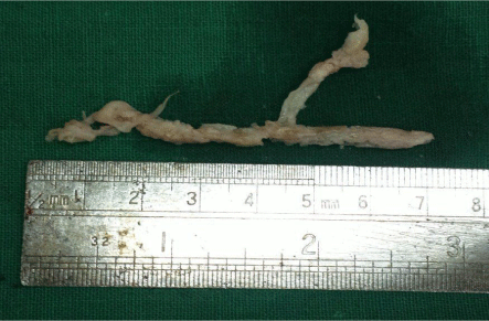
After adequate systemic heparinization, proximal and distal control of the involved artery was attained. A single longitudinal arteriotomy (Figure 2) was done extending both proximally and distally to the uninvolved segment to facilitate endarterectomy and also avoid the injury to the intima of the patent segment. After sharp division of the proximal and the distal ends of the plaque, the cut edge was secured with 7-0/8-0 polypropylene suture to avoid dissection of the artery. The base of the plaque was cleared of any debris and residual medial circular fibers with fine forceps and the vessel was irrigated with heparinised saline after removal of endarterectomy specimen (Figure 4). Fogarty embolectomy was done as a routine procedure for additional safety to evacuate any clot, ensure patency of their constructed artery and identify any significant residual stenosis requiring revision of the procedure. Arteriotomies were closed with a vein patch in all cases (Figure 3).

    Figure 2:  Two centimeter Femoral artiotomy across the profunda femoris
artery.

Figure 3: Femoral artiotomy closed with vein patch.

Figure 4: Femoral artery endarterectomy specimen.
The short saphenous vein from the operative site was generally used for the vein patch. A drain was always inserted during the closure without any negative pressure applied to the closed wound. Only those patients with absent distal pulse or reduced flow considered for postoperative angiogram. Post-operative anticoagulation was routine. Most patients were given 5,000 units of unfractionated heparin every 6 h for the first 24 h after surgery. They were also administered 75 mg each of table clopidogrel and aspirin in immediate postoperative period and there after once a day. Prior to discharge, usually on the fourteenth day, patients were shifted to a once daily dose of clopidogrel and aspirin. There were no hospital deaths, and no late deaths.
Results
There was clear evidence by restoration of pulses that the endarterectomy remained patent in all 5 patients (100 %). There was no difference in results between the limb side affected the length of the lesion varied from 2 to 12 cm, and the largest endarterectomy done was 10 cm. The segments involved were iliofemoral with profunda in all cases.
Effect of reconstruction on symptoms all patients in this group had complete relief from claudication after surgery. In patients having severe rest pain in addition to claudication, both symptoms were relieved. Patients presenting with to gangrene had relief from their symptoms after operation.
Late patency after Reconstruction
All cases in both groups have patency rate 100 % after one year of followup. Check angiogram done showing patent endarterectomy arteries. All patients got complete symptomatic relief with preservation of great saphaneous vein for further operative procedure if required like femoropopliteal bypass vein grafting and coronary artery bypass grafting. The extra time and incision for Great saphaneous vein harvesting was avoided with also avoiding the use of artificial PTFE graft.
Discussion
Bypass surgery has been the preferred initial treatment in patients with chronic critical is chemia and a femoropopliteal occlusion [1,7]. But the improvements in technique appear to allow endarterectomy to be performed below the adductor canal with equal success rate [7,8,13]. In our experience it is 100 % at 1year. There are a few reports dealing with the treatment of segmental occlusive disease of the femoral artery; but, In general, these reports have combined treatment of the external iliac artery with the femoral artery as a single entity [1,3]. Though the majority of surgeons prefer the saphenousve in bypass graft [1,2], a few advocate endarterectomy and reconstruction of the iliofemoral segment [7,8,14].
Endarterectomy has the advantage that it preserves the long saphenous vein. The vein patch graft was mainly taken from the easily accessible short saphenous vein. Preservation of the long saphenous vein has an advantage that it can be of use later for coronary, femoropopliteal and femoro-tibial grafts [4,5]. Angiography is often used to obtain an assessment of the length of artery requiring endarterectomy. However, this is frequently much longer than is apparent radio logically. The main information sought from the angiography is, however, the patency of the distal arterial bed [2].
When the superficial femoral artery a lone is the site of operation, the common femoral and popliteal arteries must be carefully scrutinized on the angiograms. If any doubt exists about the irate quacy they must be explored at operation, be-cause disease above or below the superficial femoral end artery-ectomy may cause primary failure. In short occluded segments we prefer to use direct end artery ectomy without stripping. In long occluded segments, if the theromatous material separates easily, we can use a stripper. It should be done under direct vision to ensure that no damage is done to the outer wall of the vessel. In all cases, whether stripping is done or not, the lowest limit of the endarterectomy is directly visualized through an arteriotomy, and the distal intima anchored where required. We believe that any arteriotomy distal to the common femoral artery should be closed with a vein or Dacron patch, and some common femoral arteriotomies also require this [13,14]. Some surgeons recommend angiography on the operating- table after stripping, to ensure that no athermanous fragments are left in the arterial lumen. It would certainly appear to be wise to do this especially when the stripping was difficult. However, we prefer open endarterectomy under direct vision in these conditions.
The primary results are encouraging, and all of those cases presenting with claudication were relieved of the irsymptoms. Possible complications given by Guys and St Thomas NHS foundation specific to a femoral endarterectomy are [15,16]:
- Hematoma and bleeding– some blood can collect under the skin after the procedure. As long as there is no ongoing bleeding this can often just be observed. Rarely, persistent and extensive bleeding occurs and requires urgent surgery.
- Leg swelling– leg swelling occurs in some patients after the operation. This usually resolves itself but take months to settle. Elevating the leg whilst sitting in a chair and walking will reduce the swelling.
- Skin numbness– some areas of skin numbness may occur due to the inevitable cutting of nerves when the incision is made to perform the surgery. At first this can be very noticeable but often fades with time. In the longer term it is not normally a problem for the vast majority of patients.
- Wound infection– should a wound infection occur, it usually only requires antibiotics to treat it. Occasionally the wound needs to be cleaned out under an aesthetic.
- Loss of blood supply to the legs– this may occur due to the blockage of the artery in the groin or pelvis or from dislodging loose material within the arteries that then passes down into the legs. This is rare but may require further surgery. Rarely amputation may be required.
- Infection of the synthetic patch– rare but usually requires removal of the patch. None of above complications occurred in our patients. But limitation of our study was number of cases are less. So further study with more number of cases was awaited.
Conclusion
The present study illustrates that endarterectomy of the femoral artery should be considered available option to bypass techniques in selected patients with localized disease. Endarterectomy provides revascularization without use of the long saphenous vein graft and sparing it for a future bypass, either coronary or femoro-popliteal or tibia, if required. It relieves symptoms of claudication effectively and amputation can be avoided in a vast majority of patients with threatened limbs. Femoro-popliteal/distal bypass with PTFE graft or long saphenous vein may be performed if an end artery atomized segment gets occluded. Thus time as well as incision required for harvesting Great saphaneous vein was avoided in addition to preserving it for further use. The use of artificial graft like PTFE was also avoided. The primary results are encouraging but further study with more number of cases was awaited.
References
- De Weese JA, Barner HB, Mahoney EB, Rob CG. Auto genousvenous bypass graft and thrombo endarterectomies for atherosclerotic lesions of the femoropopliteal arteries. AnnSurg. 1966; 163: 205–214.
- Adam DJ, Beard JD, Cleveland T, et al. BASIL trial participants bypass versus angioplasty in severe is chaemia of the leg (BASIL): Multicentre, randomised controlled trial. Lancet. 2005; 366: 1925–1934.
- Ahn SS, Rutherford RB, Becker GJ, et al. Reporting standards for lower extremity arterial endovascular procedures. Society for Vascular Surgery/ International Society for Cardiovascular Surgery. J Vasc Surg. 1993; 17: 1103–1107.
- Dieter RS, Pacanowski JR, Ahmed MH, Mannebach P, Nanjundappa A. FoxHollow atherectomy as a treatment modality for common femoral artery occlusion. WMJ. 2007; 106: 90–91.
- Edwards WS. Present status off emoro-popliteal arterial reconstruction. AnnSurg. 1968; 168: 1094–1096.
- Imparato AM, Bracco A, Kim GE. Comparisons of three techniques for femoralpopliteal arterial reconstruction, 1.Vein bypass, 2. Open endarterectomy, 3. Semi closed endarterectomy. Ann Surg. 1973; 177: 375–380.
- Inahara T, Toledo AC. Endarterectomy of the popliteal artery for segmental occlusive disease. Ann Surg. 1978; 188: 43–48.
- Abbas M, Claydon M, Ponosh S, et al. Open endarterectomy of the SPT segment: an experience. Ann Vasc Surg. 2007; 21: 39–44.
- Ahmadi R, Schillinger M, Maca T, Minar E. Femoropopliteal arteries: immediate and long-term results with a Dacron-covered stent-graft. Radiology. 2002; 223: 345–350.
- Hunink MGM, Wong JB, Donaldson MC, Meyerovitz MF, deVries JA, Harrington DP. Revascularization for femoropopliteal disease: a decision and cost-effectiveness analysis. JAMA. 1995; 274: 165–171.
- Wilson SE, Wolf GL, Cross AP. Percutaneous transluminal angioplasty versus operation for peripheral arteriosclerosis: report of a prospective randomized trial in a select group of patients. J Vasc Surg. 1989; 9: 1–9.
- Veith FJ, Gupta SK, Ascer E, et al. Six-year prospective multicenter randomized comparison of autologous saphenous vein and expanded polytetrafluoroethylene grafts in infrainguinal arterial reconstructions. J Vasc Surg. 1986; 3: 104–114.
- Irvine WT, Williams EJ, Plessas SN. Femoro-popliteal endarterectomy. Br Med J. 1965; 1: 1147–1149.
- Cambria RP, Ridge BA, Brewster DC, Moncure AC, Darling RC, Abbot WM. Delayed presentation and treatment of popliteal artery embolism. Ann Surg. 1991; 214: 50–55.
- Guys and St Thomas NHS foundation trust.
- Martin EC, Katzen BT, Benenati JF, Diethrich EB, Dorros G, Graor RA, et al. Multicenter trial of the Wall stent in the iliac and femoral arteries. J Vasc Interv Radiol. 1995; 6: 843–849.