
Case Report
Austin J Cardiovasc Dis Atherosclerosis. 2018; 5(1): 1035.
Reperfusion Injury with Bleeding Complication after Renal Stent Angioplasty
Hieronimus A1,2,3*, Syha R4, Amend B5, Zimmermann B1,2,3, Balletshofer B1, Häring HU1,2,3 and Randrianarisoa E1,2,3
1Department of Internal Medicine IV, Division of Endocrinology and Diabetology, Vascular Medicine, Nephrology and Clinical Chemistry, University Hospital of Tübingen, Tübingen, Germany
2Institute of Diabetes Research and Metabolic Diseases (IDM) of the Helmholtz Center Munich at the University of Tübingen, Tübingen, Germany
3German Center for Diabetes Research (DZD), Tübingen, Germany
4Department of Diagnostic and Interventional Radiology, University Hospital of Tübingen, Tübingen, Germany
5Department of Urology, University Hospital of Tübingen, Tübingen, Germany
*Corresponding author: Dr. Med. Anja Hieronimus, Department of Internal Medicine IV, Division of Endocrinology and Diabetology, Vascular Medicine, Nephrology and Clinical Chemistry, University Hospital of Tübingen, Tübingen, Germany
Received: April 24, 2018; Accepted: May 15, 2018; Published: May 22, 2018
Abstract
Renal percutaneous transluminal stent angioplasty (rPTA) of atherosclerotic renal artery stenosis remains controversial, but is a first-line treatment for stenosis related to fibromuscular dysplasia (FMD) in poor controlled arterial hypertension. We report on a reperfusion induced perirenal, retroperitoneal haematoma following rPTA of a renal artery stenosis in a woman with fibromuscular dysplasia. Bleeding complication of the kidney was successfully managed by endovascular coil embolization.
Keywords: Renal stent angioplasty, Fibro muscular dysplasia, Reperfusion syndrome, Bleeding complication
Abbreviations
rPTA: renal Percutaneous Transluminal Stent Angioplasty; FMD: Fibromuscular Dysplasia; CUS: Coloured Duplex Ultrasound; MRI: Magnetic Resonance Imaging; RR: Riva-Rocci; CT: Computer Tomography
Case Presentation
A 46 year old woman was admitted to our hospital for endovascular treatment of her poorly controlled blood pressure mediated by renal artery stenosis, which was diagnosed prior to her admission by coloured duplex ultrasound (CUS) following magnetic resonance imaging (MRI). Other causes for secondary hypertension have been excluded before.
CUS demonstrated an elevated flow velocity within the proximal (ostial) left renal artery with a maximal velocity of 380 cm/s as well as significant reduced intrarenal resistance indices comparing both kidneys (cumulative renal index left 0.4 vs. right 0.6). The renoaortal index was 4.9 and above > 3.5 suitable for a hemodynamical relevant renal artery stenosis. Comparing both kidneys in ultrasound as well as abdominal perfusion MRI, the left kidney presented a smaller size (left kidney 95 x 40 mm vs right kidney 112 x 52 mm) and was less perfused (Figure 1). Besides arterial hypertension, the only existing further cardiovascular risk factor was persistent nicotine consumption. No manifestation of atherosclerotic or plaque burden could be found in the CUS of the carotid arteries including intima media thickness (0.55 mm both sides) and the abdominal aorta. A vasculitis could also be ruled out by ultrasound and laboratory values. Thus, in particular taking into consideration the MRI, the stenosis was classified as a focal FMD.
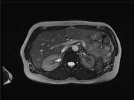
    Figure 1:  Abdominal perfusion magnetic resonance imaging in axial section
showing hypointense parenchyma indicative for delayed perfusion of the left
kidney.
The laboratory data (reference values shown in brackets) prior to intervention were: hemoglobin of 12.4g/dl (12.0 - 16.0g/dl), INR 1.0 and PTT 25sec (max. 40sec), potassium 2.9mmol/l (3.5 – 4.8 mmol/l), sodium 140mmol/l (136 - 148 mmol/l), creatinine 1.1mg/ dl (0.5 - 0.8mg/dl) and glomerular filtration rate 53.5ml/min/1.73 m2 (> 60ml/min/1.73m2), both related to Modification of Diet in Renal Disease formula. Total cholesterol was 165mg/dl (130 - 190 mg/dl), High density lipoprotein-cholesterol 47mg/dl (> 45mg/dl), Low density lipoprotein-cholesterol 112mg/dl (< 160mg/dl), glycated hemoglobin 5.4% (4.5 - 6.2%) and a microalbuminuria of 24 mg/g creatinine (< 20mg/g creatinine). The patient’s vital parameters were blood pressure according to Riva-Rocci (RR) 165/85mmHg, heart rate 60 beats per minute, temperature 36.6°C, amount of oxygen 98%.
The patient’s antihypertensive medication consisted of (daily dosage) Ramipril 5mg, Hydrochlorothiazide 25mg, Moxonidin 0.2μg and Bisoprolol 2.5mg. She did not tolerate calcium channel blockers because of a tendency to oedema. Her blood pressure was poorly controlled and she presented recurrent hypertensive crisis. In addition and as mentioned before, the left kidney already presented morphological changes with a smaller size and decreased blood flow. Particulary also in view of the impaired renal function, the indication for endovascular treatment by rPTA was given.
For access of the arterial system a retrograde puncture of the right arteria femoralis communis was performed and a 6F sheath (Terumo, Leuven, Belgium) was introduced. The origin of the left renal artery was entered with a 4F C2 (Cobra) configurated catheter and a 6F RDC (renal double curve) configurated guiding catheter. A successful passage of the high-grade renal artery stenosis of the left kidney with a 0.014-inch wire following a stent angioplasty was performed by a 6 mm balloon expandable stent (Dynamic Renal®, Biotronik, Berlin, Germany).
The intervention list decided for stenting because of the highgrade ostial stenosis of the renal artery. No residual stenosis or peripheral embolization of the left kidney could be detected in an angiography repeated instantly (Figure 2). Just before the completion of the intervention, the patient indicated a left sided back pain. A repeated angiography of the left kidney and related arteries showed no pathological finding. In particular, there was no early recoil, stent closure, perforation, dissection or signs of bleeding complication. The pain decreased spontaneously and therefore, the procedure was finalized. The patient received periinterventionally 5000 IE Heparin intra arterial as well as a dual antiplatelet therapy with Clopidogrel and Acetylsalicylic acid. The patient was transferred to normal ward.
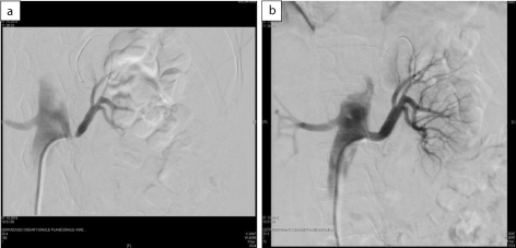
    Figure 2:  Angiography of both renal arteries:
a) Left renal artery preinterventional with high grade ostial localized renal
artery stenosis;
b) After angioplasty with stent implantation without any sign of early recoil,
stent closure, perforation, dissection or signs of bleeding complication.
Following, the patient complained about a rapidly increasing left flank pain once again. The flank pain became that severe that even opium-based analgesics couldn’t free her of symptoms. The vital parameters showed an arterial hypertension with RR 155/95 mmHg and heart rate 54 beats per minute. After analgetic and fluid administration, a blood sample was taken, showing stable hemoglobin (12.3g/dl) and creatinine (1.0 ml/min/1.73m2) and a normal lactate dehydrogenase with 135 U/l (< 250 U/l). Due to the consisting pain, a further examination was performed by ultrasound. It showed a noticeable finding with inhomogeneous echogenicity of the left kidney without any colour of the central parenchyma of the kidney in CUS. The capsula could not been detected and the kidney had an irregular shape and seemed distended. The upper and the lower pole showed a marginally colour signal with a resistance index of 1.0. The stent placed in the left renal artery was perfused regularly in CUS (Figure 3). A repeated blood sample showed already a dropping hemoglobin level (12.4g/dl to 11.1g/dl). In suspicion of a bleeding complication a contrast enhanced computer tomography (CT) was performed immediately. The CT showed an active parenchyma bleeding of the dorsal parenchyma lip of the left kidney as well as a retroperitoneal, perirenal haematoma (Figure 4). Following, the patient was transferred to the intensive care unit.
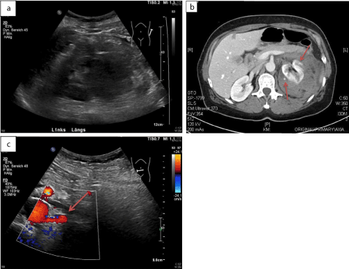
    Figure 3:  Sonographic examination of the left kidney after angioplasty and
stent implantation
a) Inhomogeneous irregular shape compatible with a haematoma.
b) The upper and the lower pole showed a reduced perfusion pattern in
coloured duplex ultrasound and a resistance index of 1.0
c) The stent of the left renal artery was perfused regularly(red arrow).
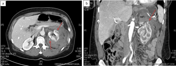
    Figure 4:  Contrast enhanced abdominal computer tomography
a) Axial and b) Coronar section showing a large perirenal haematoma of
the left kidney, which itself was distended, with active bleeding (red arrows),
capsular perforation and renal parenchymal compression.
To stop the active bleeding an endovascular strategy was planed because of an elevated blood pressure and the dual antiplatelet therapy including a prior loading dose and an application of heparin; the latter was interrupted before performing the ultrasound. The decision against a surgical procedure primarily was the hemodynamic stability and the increased perioperative risk including a potential nephrectomy for bleeding control. A second angiography was performed showing no stent thrombosis, dissection or recoil of the left renal artery. However, two active bleedings in the upper and the middle part of the left kidney could be seen and a successful coil embolization was performed by micro-coils (Figure 5). In the final inspection no more active bleeding could be detected. The laboratory control after the second angiography showed already a significant decrease of hemoglobin to 9.7g/dl) and creatinine was stable with 1.1mg/dl (glomerular filtration rate 53.5 ml/min/1.73m2). The synopsis of all the findings presumes, that the bleeding complication is an arterial bleeding caused by a reperfusion injury with perirenal haematoma and rupture of the renal capsula.
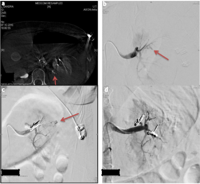
    Figure 5:  Extended intrarenal vascular imaging for localization of the bleeding
origin (red arrows) a) by cone beam CT and b) selective angiography for c),d)
successful selective coil embolization of two active bleedings in the upper and
the middle part of the left kidney (red arrows).
In the further course, the flank pain could be managed with analgesics. The patient still presented high blood pressure, in this setting most likely due to pain, and a temporary intravenous antihypertensive therapy was necessary. Heparin was stopped as well as the dual antiplatelet therapy, temporary. Only the Aspirin® therapy was continued. The next day the flank pain nearly resolved and the hemoglobin level dropped to minimal 8.0g/dl. During the day the antihypertensive therapy could be continued per os (Ramipril 2.5mg and Spironolactone) and the next day the patient was transferred to regular ward. Creatinine and urinary excretion was unobtrusively.
The patient was discharged a few days later with reduced antihypertensive therapy (Hydrochlorthiazide 12.5mg/d, Ramipril 5mg/d). Follow-ups revealed a blood pressure with RR 135/65mmHg, and a creatinine 1.1mg/dl (glomerular filtration rate 53.5ml/min/1.73m²). The antihypertensive medication consisted of Hydrochlorthiazide 12.5mg/d and Bisoprolol 2.5mg/d. Ramipril had to be stopped because of the suspicion of an angioedema as a severe side effect of angiotens in converting enzyme inhibitors in the longtime follow up.
Discussion
FMD is a non–inflammatory, non-atherosclerotic vasculopathy that can lead to arterial stenosis up to occlusion as well as aneurysms and dissection. Renal artery FMD is the most common nonatherosclerotic cause of renal artery stenosis, accounting up to 10% [1]. The stenosis is classified into four types: multifocal (multiple stenosis or string-of-beads appearance), unifocal (stenosis < 1cm in length), tubular (stenosis > 1cm in length) and mixed [2]. It can also be classified by its apparition site: ostial lesions are defined as lesions within 0.5cm of the renal artery orifice; proximal lesions are defined as lesions within 1 cm of the renal artery opening; middle lesions are defined as lesions not classified as ostial or proximal, but appearing before any branching of the main renal artery [3].
Loss of renal mass occurs in up to 63% of FMD patients and progression occurs in up to 37% of patients [6]. The cause of FMD remains unknown, despite the fact that even a variety of genetic, mechanical and humoral factors have been proposed. Smoking cessation is the only known preventable risk factor and FMD tends to affect women between 15 and 50 years [5,6]. Revascularization is recommended for those patients who have had a short duration of arterial hypertension; for those with resistant hypertension, severe stenosis, a loss of renal volume leading to ischemic nephropathy and for those with intolerance to antihypertensive medication [6]. Success correlates with an age of less than 50 years, the absence of associated coronary or carotid stenosis and the duration of arterial hypertension less than eight years [7]. Complications of rPTA occur up to 14% of patients and most commonly involve minor accessrelated problems, e.g. access site haematoma and pseudoaneurysm. Other complications with rare or minimal clinical significance are contrast related nephropathy, distal embolization or vasospasm. Rare are renal-artery perforation, dissection or segmental renal infarction. Technical success of rPTA is seen in 96% with about 80% immediate clinical benefit, cure of arterial hypertension in about 36%, improvement in hypertension and lower requirement for antihypertensive medication in about 44% [8] and free of restenosis in about 85%. A rare complication of rPTA is bleeding including perirenal, renal subcapsular or retroperitoneal haematoma. In the conducted literature search with the key words “ angioplasty or stent, renal artery stenosis and haematoma” we got 26 results on pubmed, including four relevant publications (guide wire perforation) and only two case report of a reperfusion injury after rPTA [9,10].
Reperfusion injury can be seen after endovascular treatment of peripheral arterial occlusive disease as well as high-grade carotid stenosis (0.67% of patients) [11]. The mechanism of cerebral hyperperfusion syndrome is described as a result of impaired or loss of auto regulation of cerebral blood flow. High-grade carotid stenosis causes a chronic low-flow state those results in a compensatory dilation of cerebral vessels distal to the stenosis as part of the normal auto regulatory response to maintain adequate cerebral blood flow. As the result of this chronic dilation, the vessels lose their ability to auto regulate vascular resistance in response to changes in blood pressure and this results in increased cerebral blood flow after recanalization [11].
The pathophysiology of perirenal or sub capsular haematoma seems similar and has mostly been explained as a transient hyperperfusion of renal tissue after revascularization. Analogous to cerebral loss of auto regulation, the persistent renal artery occlusive disease and hypoperfusion cause compensatory renal branch dilation and loss of auto regulation. Our patient presented signs of chronically reduced perfusion by MRI. After rPTA, hyperperfusion results from increased pressure into a distal bed fixed in maximum dilation. Hyperperfused renal artery branch vessels might rupture and result in haemorrhage as in our patient. Capsular arteries transversing the subcapsular space ruptured and, because of the anticoagulated and dual antiplatelet therapy of our patient, the haematoma worsened with an enlargement resulting in a persistent haemorrhage from multiple branches. An alternative possible mechanism of the bleeding is a guide wire perforation of a distal renal artery during renal artery stenting. In our case, a second look was performed during first angiography immediately with no evidence for perforation or periprocedural harm. Risk factors for haemorrhage complication after rPTA include advanced age (> 60 years), highgrade stenosis with poor collateral flow, pre-and post procedural arterial hypertension, evidence of chronic ipsilateral hypoperfusion, a small kidney, periprocedural anticoagulation and/or antiplatelet therapy, diabetes mellitus, generalized arteriosclerosis, coronary artery disease and obesity [10]. Choice of treatment depends on the patient’s hemodynamic and anticoagulated state, renal function and the feasibility of treatment modality. Perirenal haematoma due to other causes of renal bleeding most times has been managed conservatively. Nevertheless, embolotherapy or surgical intervention is occasionally required [12].
Conclusion
In conclusion, we describe a multilocular haemorrhage complication after rPTA in a woman with FMD mediated by reperfusion injury, which was successfully treated by embolotherapy. Long-standing renal artery stenosis might contribute to bleeding complication mediated by reperfusion.
References
- Safian RD and Textor SC. “Renal-artery stenosis.” N Engl J Med. 2001; 344: 431-442.
- Kincaid OW, Davis GD, Hallermann FJ and Hunt JC. “Fibromuscular dysplasia of the renal arteries. Arteriographic features, classification, and observations on natural history of the disease.” Am J Roentgenol Radium Ther Nucl Med. 1968; 104: 271-282.
- Kim HJ, Do YS, Shin SW, Park KB, Cho SK, et al. “Percutaneous transluminal angioplasty of renal artery fibro muscular dysplasia: mid-term results.” Korean J Radiol. 2008; 9: 38-44.
- Slovut DP and Olin JW. “Fibromuscular dysplasia.” N Engl J Med. 2004; 350: 1862-1871.
- Plouin PF, Fiquet B, Bobrie G and Jeunemaitre X. “[Fibromuscular dysplasia of renal arteries].” Nephrol Ther. 2016; S135-138.
- Sanidas EA, Seferou M, Papadopoulos DP, Makris A, Viniou NA, and et al. “Renal Fibromuscular Dysplasia: A Not So Common Entity of Secondary Hypertension.” J Clin Hypertens (Greenwich). 2016; 18: 240-246.
- Giavarini A, Savard S, Sapoval M, Plouin PF and Steichen O. “Clinical management of renal artery fibromuscular dysplasia: temporal trends and outcomes.” J Hypertens. 2014; 32: 2433-2438.
- Yang YK, Zhang Y, Meng X, Yang KQ, Jiang XJ, et al. “Clinical characteristics and treatment of renal artery fibro muscular dysplasia with percutaneous transluminal angioplasty: a long-term follow-up study.” Clin Res Cardiol. 2016; 105: 930-937.
- Kang KP, Lee S, Kim W, Han YM, Kang SK and Park SK. “Renal subcapsular hematoma: a consequence of reperfusion injury of long standing renal artery stenosis.” Electrolyte Blood Press. 2007; 5: 136-139.
- Xia D, Chen SW, Zhang HK and Wang S. “Renal subcapsular haematoma: an unusual Complication of renal artery stenting.” Chin Med J (Engl). 2011; 124: 1438-1440.
- Abou-Chebl A, Yadav JS, Reginelli JP, Bajzer C, Bhatt D and Krieger DW. “Intracranial hemorrhage and hyperperfusion syndrome following carotid artery stenting: risk factors, prevention, and treatment.” J Am Coll Cardiol. 2004; 43: 1596-1601.
- Morris CS, Bonnevie GJ and Najarian KE. “Nonsurgical treatment of acute iatrogenic renal artery injuries occurring after renal artery angioplasty and stenting.” AJR Am J Roentgenol. 2001; 177: 1353-1357.