Case Report
Austin J Cerebrovasc Dis & Stroke. 2014;1(2): 1010.
Resolution of Pulsatile Tinnitus after Coil Embolization of Sigmoid Sinus Diverticulum
Amans MR1*, Stout C2, Dowd CF1, Higashida RT1, Hetts SW1, Cooke DL1, Narvid J1 and Halbach VV1
1Department of Radiology and Biomedical Imaging, University of California San Francisco, USA
2Department of Radiology, University of Massachusetts, USA
*Corresponding author: Amans MR, Department of Radiology and Biomedical Imaging, University of California San Francisco, 505 Parnassus Ave, Room L352, San Francisco, CA, 94143-0628, USA
Received: July 10, 2014; Accepted: August 10, 2014; Published: August 11, 2014
Abstract
Venous sinus diverticulum is a rare vascular cause for pulsatile tinnitus characterized by an out pouching of the venous sinus into the calvarium, usually involving the sigmoid venous sinus. Sigmoid sinus diverticulum is often associated with upstream sinus stenosis. While the exact mechanism of sound generation from a sinus diverticulum is unclear, several case reports have suggested that pulsatile tinnitus can resolve after remodeling of venous blood flow such that the diverticulum is excluded from the circulation. Case reports have also suggested treatment of both the sigmoid sinus diverticulum and the often-associated upstream sinus stenosis may ameliorate pulsatile tinnitus. We report a case of trans-venous coil embolization of a sigmoid sinus diverticulum without treatment of an additionally identified upstream sinus stenosis, resulting in cure of the patient’s pulsatile tinnitus. A review of endovascular and open surgical treatment of sinus diverticula in the treatment of pulsatile tinnitus is also presented.
Keywords: Pulsatile tinnitus; Sigmoid sinus; Dural diverticulum; Venous sinus aneurysm
Abbreviations
PT: Pulsatile Tinnitus; CSF: Cerebrospinal Fluid; DSA: Digital Subtraction Angiography; DAVF: Dural Arteriovenous Fistula; IJ: Internal Jugular vein
Case Presentation
A 59-year-old postmenopausal woman presented with subjective right-sided pulse synchronous pulsatile tinnitus (PT) increasing in intensity over the previous 18 months. Her PT became increasingly bothersome resulting in both difficulties falling asleep and waking her from sleep as well as difficulty concentrating. Her PT abated with right neck compression and was exacerbated by strenuous activity, bending forward at the waist, and with Valsalva, suggestive of a venous etiology [1-4]. She denied changes in the pitch of the sound over time and also denied changes in hearing acuity, balance, swallowing, coordination, strength, or sensation.
A broad workup for causes of PT was performed. A complete history and physical were performed (including otoscopy to evaluate for middle ear mass, auscultation for a bruit, and ophthalmoscopy to search for papilledema) and were normal. Audiologic assessment of auditory acuity and discrimination (understanding of words) was also unremarkable. Lumbar puncture demonstrated bland cerebrospinal fluid (CSF) with a normal opening pressure.
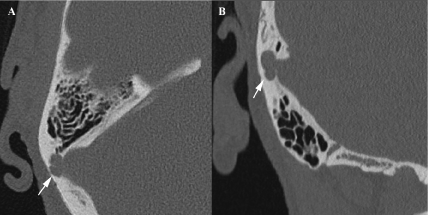
Noninvasive imaging, including contrast enhanced CT of the temporal bone, carotid ultrasound, and contrast enhanced MRI of the brain with MR venography and MR angiography were obtained. CT demonstrated a smoothly-marginated scalloping of the inner table of the sigmoid plate with narrow neck and extension of the sinus laterally into the sigmoid bone defect, characteristic of a sigmoid sinus diverticulum [5-9] (Figure 1). No other vascular neoplasm, vascular malformation, or vascular anomaly was identified.
Figure 1 : Axial (A) and coronal (B) images from noncontrast CT of the temporal bone demonstrating smoothly marginated scalloping of the inner table of right sigmoid plate consistent with sigmoid sinus aneurysm (white arrow).
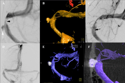
A conventional digital subtraction angiogram (DSA) excluded a dural arteriovenous fistula (DAVF) or other vascular pathology that might have been occult on noninvasive imaging. Venous phase images from the DSA (Figure 2) demonstrated a lobulated 6.8 x 8.0 x 4.2 mm right sigmoid sinus diverticulum with a 3.4 mm neck. In addition, a stenosis in the sigmoid sinus upstream from the diverticulum was lobular in appearance, suggestive of indentations in the sinus caused by arachnoid granulations.
Figure 2 : Venous phase images from a DSA demonstrate a lobulated right sigmoid sinus diverticulum (white arrow) in the AP projection prior to treatment (A) measuring6.8 x 8.0 x 4.2 mm with a 3.4 mm neck. 3-D reconstruction (B) outlines the narrow neck, as well as the upstream stenosis (black arrow head) in the sigmoid sinus. Mid coiling image (C) demonstrates the catheter in the diverticulum (black arrow), stenosis in the sigmoid sinus proximal to the diverticulum, and coils filling the diverticulum with partial filling of the neck of the diverticulum with contrast. Post coiling AP image (D) of the diverticulum demonstrates the diverticulum is no longer filling with contrast and the sigmoid sinus remains patent. 3-D reconstructions in the venous phase post coiling (E and F) demonstrate the relationship of the coil mass to the sinus, as well as sinus patency.
Due to the debilitating nature of the right-sided PT, the patient elected to undergo coil embolization of the sigmoid sinus diverticulum. Under general anesthesia, a 7F vascular sheath was placed in the common femoral vein and 5F vascular sheath in the contralateral common femoral artery. A 5000 unit Heparin bolus was given intravenously (appropriately increasing the activated clotting time from 134 seconds to 314 seconds). A coaxial 7F VBL guide catheter (Cordis Neurovascular, Miami Lakes, FL, USA) with a 5F Vert (Cook Medical, Bloomington, IN, USA) inner catheter was guided over a Bentson guide wire (Boston Scientific, Natick, MA, USA) from the common femoral vein, through the right atrium, and into the right internal jugular vein (IJ). With the 7 F guide catheter remaining in the high right IJ, and the 5 F catheter positioned at the right IJ sigmoid sinus junction, a 0.021 inches Prowler select plus (Cordis Neurovascular) microcatheter was guided over a Synchro 2 Standard (Stryker Neurovascular, Fremont, CA, USA) microwire into the sigmoid sinus diverticulum. Venous roadmap was obtained from venous phase of images of a DSA following injection of iodinated contrast into the contralateral left common carotid artery. Injection of the arteries on the contralateral side of the venous lesion allowed for visualization of the venous sinuses without ipsilateral arteries toconfound the images. The diverticulum was then embolized with 7 detachable coils. The upstream stenosis was not treated.
Upon recovery from general anesthetic, the patient reported complete resolution of her PT. She was admitted to the intensive care unit for observation overnight, and was discharged the following morning. She was placed on 81 mg ASA orally daily for two weeks to allow for endothelialization of the coil-sinus interface and minimize the risk of sinus thrombosis. At 6 month follow up, the patient reported complete resolution of right-sided PT. Follow up angiogram demonstrated complete obliteration of the sigmoid sinus diverticulum and preserved patency of the dominant right sigmoid sinus.
Discussion/Conclusion
Pulsatile tinnitus (PT) is the auditory perception of a rhythmic sound in the absence of external source. The differential diagnosis of PT can be classified into vascular and nonvascular etiologies. Vascular etiologies can be divided into arterial causes (e.g. carotid artery dissection, fibromuscular dysplasia, aberrant internal carotid artery, glomus tumor, contralateral carotid artery stenosis resulting in ipsilateral carotid high-flow state), and venous causes (e.g. stenosis, dural arteriovenous fistula, sinus diverticulum, high jugular bulb, intracranial hypertension). Sigmoid sinus diverticulum, also referred to as sigmoid sinus “aneurysm”, is a rare vascular etiology for PT characterized by an out pouching of the venous sinus into the calvarium usually involving the sigmoid sinus. The exact mechanism of sound generation from a sinus diverticulum is unclear. Previous authors suggest that sinus diverticulum-induced PT may be a result of vibration of the venous sinus wall (caused by turbulence in the sigmoid sinus diverticulum) that is sensed by the cochlea [6,7]. An upstream stenosis of the venous sinuses is often noted in association with a sigmoid sinus diverticulum, and it has been speculated to play an additive role in sound generation [6,10,11], or even a causative role in sinus diverticulum generation [6,7,10-13]. As such, several authors suggest the treatment of both lesions simultaneously to treat venous PT [10,11]. We report a case of coil embolization of a venous sinus diverticulum without treatment of an associated venous sinus stenosis resulting in cure of PT without complication.
Several authors have proposed the coincidence of venous sinus stenosis and sigmoid sinus diverticulum as a cause of PT [6,10,11]. Since each lesion individually or the two together may be the causative factor in a patient with PT, a diagnostic and therapeutic dilemma is incurred. Eisenman approached this dilemma by performing surgical reconstruction of the sinus to treat both lesions simultaneously [10]. While his method was successful in treating PT in 13 patients, he also reports two major post operative complications including one patient with venous sinus thrombosis and another with signs and symptoms of venous sinus thrombosis without “radiologic evidence of dural sinus thrombosis”. Signorelli and colleagues treated both conditions simultaneously by stent placement across both the stenosis and sigmoid sinus diverticulum with subsequent balloon dilatation of the stent in the area of stenosis. Antiplatelet therapy was subsequently used to minimize the risk of sinus thrombosis [11].
The incidence of dural venous sinus stent thrombosis, which could result in complete dural venous sinus thrombosis, is unknown. A retrospective review of venous sinus stent placement for intracranial hypertension did not report a single case of in stent stenosis or thrombosis out of 52 stents placed [14]. A recently published multicenter analysis of cerebral arterial stent placement to assist aneurysm coil embolization found a 0.7% incidence of in-stent thrombosis (1 in 142), which resolved with intraprocedural administration of abciximab [15]. Signorelli’s decision to treat the venous stenosis was in a large part attributed to measuring 10 mmHg pressure gradient across the stenosis suggesting turbulent flow downstream from the stenosis [11]. When approaching our case, we considered the large number of patients who have a stenosis in the dominant and non-dominant transverse or sigmoid sinus due to arachnoid granulations with very few of these patients presenting with PT. As such, we hypothesized the stenosis may be an incidental finding, or possibly contributory in the pathogenesis of the sigmoid sinus diverticulum, and not the main factor in sound generation. In treating the diverticulum only, we cured the PT without having to remodel the venous sinus in the region of the stenosis, thereby decreasing the complexity of the procedure, reducing the amount of implanted hardware and decreasing the complication risk of the procedure. We suggest the use of a stent be reserved for cases in which the sigmoid sinus diverticulum cannot otherwise be occluded endovascularly, and treatment of sinus stenosis only in cases of PT refractory to treatment of the sigmoid sinus diverticulum.
Endovascular treatment of sigmoid sinus diverticulum using detachable coils for treatment of PT was initially described by Houdart [5]. Since that publication, 4 additional endovascular case reports have been published using a combination of detachable coils, detachable coils in conjunction with vascular stenting, as well as vascular stenting alone [2,12,13,16,17]. All cases involve female subjects (age range 31 to 59), involving the dominant sinus (either directly stated by the authors or ascertained from published images using previously proposed standards [6]), with resolution of symptoms following treatment, and no reported complications. Many of the authors utilized medications with platelet inhibitory properties to mitigate the risk of thrombus formation on the implanted coils and stents, presumably allowing for endothelialization as occurs in intra-arterial stent placement.
Several surgical methods to treat sigmoid sinus diverticula have also been described. In addition to Eisenman, Gologorsky et al reported surgical treatment of venous sinus diverticula using self-tying U clips, as an alternative to stent-assisted coiling, in a case where primary coil embolization was technically unsuccessful; this approach resulted in resolution of symptoms (3 month follow up) [1]. A series by Otto et al described transmastoid sigmoid sinus reconstruction to treat PT in 3 patients with sigmoid sinus diverticula. In each of these cases, the wall of the sinus was reconstructed with extraluminal placement of bone wax or temporalis muscle and fascia resulting in complete resolution of PT (average 16.3 months follow up) [18]. Surgical remodeling of venous diverticula appears to carry a significant risk of morbidity including venous bleeding, hemotympanum, visual loss, embolization of bone wax, intracranial hypertension, and sinus thrombosis [1,18,19]; however, the incidence of complication remains unknown. None of the published operative cases have reported the length of stay. The relative merits of open surgical sinus remodeling versus endovascular embolization require larger patient cohorts.
The potential risks of endovascular treatment of sigmoid sinus diverticulum include coil migration, thrombus formation on coils and, in cases requiring stent placement, stent migration, and in-stent thrombosis [1,2,12]. In order to minimize procedural risk, stents should be limited to cases requiring their use to adequately position coils in the venous diverticulum. Similar to treatment of arterial aneurysms, fastidious technique, appropriate coil selection, and mid embolization angiography to identify potential coil encroachment on the sinus or thrombus formation on the coils are required to minimize procedural risks. Use of a stent will likely require extended (at least six months) use of dual platelet inhibitor therapy with aspirin and clopidogrel to minimize risks of stent thrombosis and subsequent sinus occlusion [2,11]. As per Mehanna et al, we chose to place our patient on 325 mg aspirin orally for two weeks to minimize the risks of thrombus formation on the coils [13].
In summary, we present a case of PT in a 59-year-old woman with two potential venous etiologies, sigmoid sinus diverticulum and upstream sinus stenosis. In contrast to previous case reports, we elected to coil embolize the diverticulum only. The patient’s PT immediately resolved without treatment of the ipsilateral sinus stenosis.
References
- Gologorsky Y, Meyer SA, Post AF, Winn HR, Patel AB, Bederson JB. Novel surgical treatment of a transverse-sigmoid sinus aneurysm presenting as pulsatile tinnitus: technical case report. Neurosurgery. 2009; 64: E393-394.
- Zenteno M, Murillo-Bonilla L, Martínez S, Arauz A, Pane C, Lee A. Endovascular treatment of a transverse-sigmoid sinus aneurysm presenting as pulsatile tinnitus. Case report. J Neurosurg. 2004; 100: 120-122.
- Mattox DE, Hudgins P. Algorithm for evaluation of pulsatile tinnitus. Acta Otolaryngol. 2008; 128: 427-431.
- Sonmez G, Basekim CC, Ozturk E, Gungor A, Kizilkaya E. Imaging of pulsatile tinnitus: a review of 74 patients. Clin Imaging. 2007; 31: 102-108.
- Houdart E, Chapot R, Merland JJ. Aneurysm of a dural sigmoid sinus: a novel vascular cause of pulsatile tinnitus. Ann Neurol. 2000; 48: 669-671.
- Krishnan A, Mattox DE, Fountain AJ, Hudgins PA. CT arteriography and venography in pulsatile tinnitus: preliminary results. AJNR Am J Neuroradiol. 2006; 27: 1635-1638.
- Liu Z, Chen C, Wang Z, Gong S, Xian J, Wang Y, et al. Sigmoid sinus diverticulum and pulsatile tinnitus: analysis of CT scans from 15 cases. Acta Radiol. 2013; 54: 812-816.
- Weissman JL, Hirsch BE. Imaging of tinnitus: a review. Radiology. 2000; 216: 342-349.
- Xue J, Li T, Sun X, Liu Y. Focal defect of mastoid bone shell in the region of the transverse-sigmoid junction: a new cause of pulsatile tinnitus. J Laryngol Otol. 2012; 126: 409-413.
- Eisenman DJ. Sinus wall reconstruction for sigmoid sinus diverticulum and dehiscence: a standardized surgical procedure for a range of radiographic findings. Otol Neurotol. 2011; 32: 1116-1119.
- Signorelli F, Mahla K, Turjman F. Endovascular treatment of two concomitant causes of pulsatile tinnitus: sigmoid sinus stenosis and ipsilateral jugular bulb diverticulum. Case report and literature review. Acta Neurochir (Wien). 2012; 154: 89-92.
- Gard AP, Klopper HB, Thorell WE. Successful endovascular treatment of pulsatile tinnitus caused by a sigmoid sinus aneurysm. A case report and review of the literature. Interventional neuroradiology. 2009; 15: 425-428.
- Mehanna R, Shaltoni H, Morsi H, Mawad M. Endovascular treatment of sigmoid sinus aneurysm presenting as devastating pulsatile tinnitus. A case report and review of literature. Interventional neuroradiology. 2010; 16: 451-454.
- Ahmed RM, Wilkinson M, Parker GD, Thurtell MJ, Macdonald J, McCluskey PJ, et al. Transverse sinus stenting for idiopathic intracranial hypertension: a review of 52 patients and of model predictions. AJNR Am J Neuroradiol. 2011; 32: 1408-1414.
- Mocco J, Snyder KV, Albuquerque FC, Bendok BR, Alan S B, Carpenter JS, et al. Treatment of intracranial aneurysms with the Enterprise stent: a multicenter registry. J Neurosurg. 2009; 110: 35-39.
- Park YH, Kwon HJ. Awake embolization of sigmoid sinus diverticulum causing pulsatile tinnitus: simultaneous confirmative diagnosis and treatment. Interventional neuroradiology. 2011; 17: 376-379.
- Santos-Franco JA, Lee A, Nava-Salgado G, Zenteno M, Vega-Montesinos S, Pane-Pianese C, et al. Hybrid carotid stent for the management of a venous aneurysm of the sigmoid sinus treated by sole stenting. Vasc Endovascular Surg. 2012; 46: 342-346.
- Otto KJ, Hudgins PA, Abdelkafy W, Mattox DE. Sigmoid sinus diverticulum: a new surgical approach to the correction of pulsatile tinnitus. Otol Neurotol. 2007; 28: 48-53.
- Hou ZQ, Han DY. Pulsatile tinnitus in perimenopausal period. Acta Otolaryngol. 2011; 131: 896-904.

