Editorial
Austin J Cerebrovasc Dis & Stroke. 2014;1(5): 1025.
Aortic Arch Plaque and Ischemic Stroke Detection Modalities and Statin Effects
Kazuyoshi Kaneko1*
Department of Cardiology, Kitamurayama Municipal Hospital, Japan
*Corresponding author: Kazuyoshi Kaneko, Department of Cardiology, Kitamurayama Municipal Hospital, 2-15-1, Onsen-machi, Higashine, Yamagata 999-3792, Japan
Received: August 18, 2014; Accepted: September 26, 2014; Published: September 29, 2014
Keywords
Aortic Arch Plaque; Ischemic Stroke; Transesophageal Echocardiography; Statin
Abbreviations
CAP: Complicated Aortic Arch Plaque; TEE: Transesophageal Echocardiography; LDL-C: Low-Density Lipoprotein-Cholesterol; HDL-C: High-Density Lipoprotein-Cholesterol; Apoa-1: Apolipoprotein A-1; Apob: Apolipoprotein B
What should we do when severe aortic arch atheroma is detected on Transesophageal Echocardiography (TEE) in patients with ischemic stroke? Aortic arch atheroma is associated with atherosclerotic risk factors such as advanced age, hypertension, hypercholesterolemia, diabetes mellitus, and smoking [1,2]. In addition the importance of apolipoprotein A-1 (ApoA-1) and the ratio apolipoprotein B/A-1 on lipid profiles in thoracic aortic plaque detected by TEE has also been described in a recent clinical report [3]. Accordingly, aggressive and strict control of hypertension, hypercholesterolemia, diabetes mellitus and smoking cessation may be required.
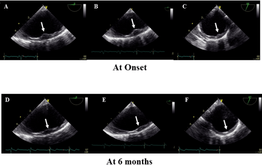
What is the rationale for treating severe aortic arch atheroma? The answer has been shown over the past 20 years in many previous clinical reports concerning the correlation between aortic atheroma and ischemic stroke recurrence and death [1,4,5]. In these reports, aortic arch intima media thickness ≥4 mm increased ischemic stroke relative risk 4-9 fold, and aortic arch intima media thickness ≥5 mm increased all cause mortality [6]. Furthermore, characteristics of aortic arch atheroma such as low echoic status, lack of calcification, or presence of ulcerated and mobile plaques increased risk further [4]. Progression of aortic arch atheroma assessed by TEE over a 12 month period was associated with recurrent vascular events, suggesting the need for medical intervention such as use of statins, which may improve prognosis [7]. Recently new recommendations for treatment for aortic arch atheroma in patients with stroke and transient ischemic attacks published in the AHA/ASA guidelines have received widespread recognition [8]. According to theguidelines, prophylactic endarterectomy, aortic arch stenting, and anticoagulation with warfarin for purposes of stroke prevention are not recommended, whereas antiplatelet and statin therapies arerecommended with assignment to high grade evidence levels and recommendation class. Nevertheless, no randomized clinical trials have yet been reported which specifically examine the effectiveness of statin therapy for reducing the risk of first or recurrent stroke among patients with complex aortic plaque. However, observational studies among patients with a recent embolic event including stroke or transit ischemic attack suggest that statins may be effective in preventing recurrent events [9]. Although there are clinical reports showing that statins may reduce plaque volume and morphology based on intravascular ultrasound study of coronary arteries [10] or magnetic resonance imaging study of descending aortic plaque [11], few reports concerning aortic arch plaque regression have been published. Recently, we have reported that rosuvastatin improves aortic arch plaque morphology concomitant with improving lipid profiles in cerebral embolism patients with normal low-density Lipoprotein-Cholesterol (LDL-c) levels [12]. In this paper we studied Complicated Aortic Arch Plaque (CAP) morphology, defined as presence of atheromatous plaque with ≥4 mm wall thickness or presence of ulcerated or mobile plaque in the aortic arch. In a group of 56 acute cerebral embolism patients without hypercholesterolemia, defined as LDL-c below 140 mg/dl, CAP was detected in 24 patients (43%) despite their non-hypercholesterolemia status. However they were older and had lower serum levels of ApoA-1 and High- Density Lipoprotein-Cholesterol (HDL-c) than the group of 32 patients without CAP. Rosuvastatin was given to these 24 patients for 6 months, with a follow-up TEE in 18 of them. The results showed that aortic arch plaque surfaces were improved (Figure) in 11 of the 18 patients (61%) with no change in the other 6 of the 18 (33%). CAP diameter was significantly decreased, and was accompanied by an increase in ApoA-1 and HDL-c. No patients had recurrent ischemic stroke or died during the 6-month treatment period. The data suggest that rosuvastatin therapy may lead to CAP regression and inhibit progression to acute ischemic stroke in patients without hypercholesterolemia, and that rosuvastatin therapy may represent a beneficial option for treating CAP. Therefore when severe aortic arch atheroma is detected, statin therapy may be considered as additional antithrombotic therapy in not only dyslipidemic but also non-dyslipidemic ischemic stroke patients [9,12,13,14].
Figure 1 : A representative case showing improved aortic arch plaque morphology on TEE. A-C show longitudinal (A and B) and transverse (C) images on admission. CAP diameter was 9.2 mm with a protruding, ulcerating, and irregular surface (arrows). D-F show corresponding sectorsfollowing administration of 5 mg of rosuvastatin for 6 months. CAP diameter had decreased to 5.7 mm with smoothing of the plaque surface (arrows). Abbreviations: TEE: Tran thoracic Echocardiography; CAP: Complicated Aortic Arch Plaque.
What is the best method for searching for severe aortic arch atheroma? Although TEE is regarded as the gold standard for determining plaque diameter and morphology, it is often difficult to perform this semi-invasive procedure in patients who are in an unstable condition, and may also require the participation of a cardiologist trained in the procedure. For this reason another more non-invasive modality for evaluating cerebral embolism patients in the acute phase to detect or estimate aortic arch atheroma may be required. Enhanced 3D-CT may also be able to detect presence of aortic arch and aortic wall atheroma [15,16], but is not superior to TEE, and furthermore enhanced contrast media sometimes induce critical allergic responses. Although detection of aortic arch atheroma using magnetic resonance angiography has been attempted as well [17,18,19], motion artifact is an important problem, with attempts currently being made to overcome this shortcoming with ECG and respiratory adjustments. At present all these additional approaches for detecting aortic arch plaque must be regarded as preliminary and in the pilot study phase. An alternative approach to estimating the likelihood of CAP existence may involve consideration of the combined 3 parameters of Ankle Brachial Index (ABI) <0.9, present smoking, and age >70 years old, which can predict CAP with high specificity [20]. Carotid artery Intima Media Thickness (IMT) =2.0 mm also predicts CAP with high sensitivity and negative predictive value [21]. In another clinical report from Tessitore et al. prediction of CAP using only IMT at the carotid bifurcation had high sensitivity and negative predictive value, albeit with low specificity and positive predictive value [22]. It follows that ABI and carotid artery IMT examinations may be useful for initial screening, with decisions about subsequent use of TEE based on these results.
What is needed to advance our studies of CAP further? First would be to establish clearly the overall value of statin therapy in treating CAP in patients with ischemic stroke. Second would be to develop improved non-invasive methods of detecting CAP. Lastly would be to determine objectively that statins cause aortic arch plaque regression and improve the prognosis of patients. Accomplishment of the second of these aims may provide options for the neurologist caring for embolic stroke patients to evaluate likelihood of CAPs non-invasively, thereby avoiding the semi-invasive TEE procedure, and making it possible to reach a decision whether statin therapy is indicated.
References
- Amarenco P, Cohen A, Tzourio C, Bertrand B, Hommel M, Besson G, et al. Atherosclerotic disease of the aortic arch and the risk of ischemic stroke. N Engl J Med. 1994; 331: 1474-1479.
- Jones EF, Kalman JM, Calafiore P, Tonkin AM, Donnan GA. Proximal aortic atheroma: an independent risk factor for cerebral ischemia. Stroke. 1995; 26: 218-224.
- Kohsaka S, Jin Z, Rundek T, Homma S, Sacco RL, Di Tullio MR. Relationship between serum lipid values and atherosclerotic burden in the proximal thoracic aorta. Int J Stroke. 2010; 5: 257-263.
- Cohen A, Tzourio C, Bertrand B, Chauvel C, Bousser MG, Amarenco P. Aortic plaque morphology and vascular events: a follow-up study in patients with ischemic stroke. FAPS Investigators. French Study of Aortic Plaques in Stroke. Circulation. 1997; 96: 3838-3841.
- Di Tullio MR1, Russo C, Jin Z, Sacco RL, Mohr JP, Homma S, et al. Patent Foramen Ovale in Cryptogenic Stroke Study Investigators. Aortic arch plaques and risk of recurrent stroke and death. Circulation. 2009; 119: 2376-2382.
- Izumi C, Takahashi S, Miyake M, Sakamoto J, Hanazawa K, Yoshitani K, et al. Impact of aortic plaque morphology on survival rate and incidence of a subsequent embolic event – long-term follow-up data –. Circ J. 2010; 74: 2152-2157.
- Sen S, Hinderliter A, Sen PK, Simmons J, Beck J, Offenbacher S, et al. Aortic arch atheroma progression and recurrent vascular events in patients with stroke or transient ischemic attack. Circulation. 2007; 116: 928-935.
- Kernan WN, Ovbiagele B, Black HR, Bravata DM, Chimowitz MI, Ezekowitz MD, et al. Guidelines for the prevention of stroke in patients with stroke and transient ischemic attack: a guideline for healthcare professionals from the American Heart Association/American Stroke Association. Stroke. 2014; 45: 2160-2236.
- Tunick PA, Nayar AC, Goodkin GM, Mirchandani S, Francescone S, Rosenzweig BP, et al. Effect of treatment on the incidence of stroke and other emboli in 519 patients with severe thoracic aortic plaque. Am J Cardiol. 2002; 90: 1320-1325.
- Takayama T, Hiro T, Yamagishi M, Daida H, Hirayama A, Saito S, et al. Effect of rosuvastatin on coronary atheroma in stable coronary artery disease: multicenter coronary atherosclerosis study measuring effects of rosuvastatin using intravascular ultrasound in Japanese subjects (COSMOS). Circ J. 2009; 73: 2110-2117.
- Yonemura A, Momiyama Y, Fayad ZA, Ayaori M, Ohmori R, Higashi K, et al. Effect of lipid-lowering therapy with atorvastatin on atherosclerotic aortic plaques detected by noninvasive magnetic resonance imaging. J Am Coll Cardiol. 2005; 45: 733-742.
- Kaneko K, Saito H, Takahashi T, Kiribayashi N, Omi K, Sasaki T, et al. Rosuvastatin improves plaque morphology in cerebral embolism patients with normal low-density lipoprotein and severe aortic arch plaque. J Stroke Cerebrovasc Dis. 2014; 23: 1682-1689.
- Everett BM, Glynn RJ, MacFadyen JG, Ridker PM. Rosuvastatin in the prevention of stroke among men and women with elevated levels of C-reactive protein: justification for the Use of Statins in Prevention: an Intervention Trial Evaluating Rosuvastatin (JUPITER). Circulation. 2010; 121: 143-150.
- Amarenco P, Bogousslavsky J, Callahan A 3rd, Goldstein LB, Hennerici M, Rudolph AE, et al. High-dose atorvastatin after stroke or transient ischemic attack. N Engl J Med. 2006; 355: 549-559.
- Kim SJ, Ryoo S, Hwang J, Noh HJ, Park JH, Choe YH, et al. Characterization of the infarct pattern caused by vulnerable aortic arch atheroma: DWI and multidetector row CT study. Cerebrovasc Dis. 2012; 33: 549-557.
- Boussel L, Cakmak S, Wintermark M, Nighoghossian N, Loffroy R, Coulon P, et al. Ischemic stroke: etiologic work-up with multidetector CT of heart and extra- and intracranial arteries. Radiology. 2011; 258: 206-212.
- Bitar R, Moody AR, Leung G, Kiss A, Gladstone D, Sahlas DJ, et al. In vivo identification of complicated upper thoracic aorta and arch vessel plaque by MR direct thrombus imaging in patients investigated for cerebrovascular disease. AJR Am J Roentgenol. 2006; 187: 228-234.
- Fayad ZA, Nahar T, Fallon JT, Goldman M, Aguinaldo JG, Badimon JJ, et al. In vivo magnetic resonance evaluation of atherosclerotic plaques in the human thoracic aorta: a comparison with transesophageal echocardiography. Circulation. 2000; 101: 2503-2509.
- Faber T, Rippy A, Hyslop WB, Hinderliter A, Sen S. Cardiovascular MRI in Detection and Measurement of Aortic Atheroma in Stroke/TIA patients. J Neurol Disord. 2013; 1: 139.
- Terasawa Y, Kimura K, Iguchi Y, Shibazaki K, Okada Y, Matsumoto N. Predictors of aortic complicated lesions in stroke patients. Hypertens Res. 2009; 32: 462-465.
- Fasseas P, Brilakis ES, Leybishkis B, Cohen M, Sokil AB, Wolf N, et al. Association of carotid artery intima-media thickness with complex aortic atherosclerosis in patients with recent stroke. Angiology. 2002; 53: 185-189.
- Tessitore E, Rundek T, Jin Z, Homma S, Sacco RL, Di Tullio MR. Association between carotid intima-media thickness and aortic arch plaques. J Am Soc Echocardiogr. 2010; 23: 772-777.
