
Department of Neurology, Fudan University, China
*Corresponding author: Wu Guofeng, Emergency Department, Affiliated Hospital of Guiyang Medical College, and Department of Neurology, Affiliated Huashan Hospital of Fudan University, Guiyang City, Guizhou Province, PRC 550004, China
Received :June 21, 2014; Accepted: October 20, 2014; Published: October 22, 2014
Citation: Guofeng W, Likun W, Yuanhong M and Lan X. Diffusion Tensor Imaging and Motor-Evoked Potential Changes in 2 Patients with Locked-In Syndrome Due To Brainstem Stroke. Austin J Cerebrovasc Dis & Stroke. 2014;1(6): 1028. ISSN: 2381-9103.
Objective: Locked-in syndrome is a devastating state that occurs after brainstem stroke. The combined use of Diffusion Tensor Imaging (DTI) and Motor-Evoked Potential (MEP) monitoring has the potential to confirm and evaluate concisely the damage to the pyramidal pathway in the brainstem, and it is possible that DTI and MEP changes during the clinical course could reflect the clinical outcome.
Clinical Presentation: Two patients were admitted to the hospital because of sudden weakness affecting the four extremities and an inability to talk. The patients presented with quadriplegia, anarthria, and preserved consciousness and intellectual functioning, but they could not speak and could only communicate with the medical staff by blinking their eyes. Brain CTs showed hypo density in the ventral part of the upper brainstem in one case and hyper density in the same area in the other.
Intervention: Positive-pressure ventilation assistance was performed after admission to the hospital, and other treatments were also administered, including antiplatelet aggregation in response to brainstem infarction and hemostasis in response to brainstem hemorrhage. DTI and MEP monitoring were conducted during their hospital stays.
Conclusion: DTI demonstrated that FA values were decreased and that the white matter fibers descending through the brainstem were significantly reduced in number on admission. Both FA values and the number of the white matter fibers were increased two weeks after admission. Cortical MEPs were absent on admission but were observed two weeks later as FA values increased. The changes observed by DTI were associated with appearance of the MEPs, and both were positively correlated with motor function.
Keywords: Locked-in syndrome; Motor-evoked potential; Diffusion tensor imaging; Motor function; Corticospinal tract
Locked-in syndrome remains a rare but devastating state after brainstem stroke. It is most commonly associated with upper ventral brainstem infarction caused by an anterior pontine vascular lesion involving the perforating pontine vessels of the basilar artery and is characterized by quadriplegia and anarthria with preserved consciousness and intellectual functioning [1]. While MEP findings have been reported for the syndrome, to the authors’ knowledge, few studies have investigated both MEP and diffusion tensor imaging findings in patients with locked-in syndrome. Here, we report MEP and DTI findings in two patients presenting with locked-in syndrome. One patient suffered from brainstem infarction, and the other suffered from brainstem hemorrhage.
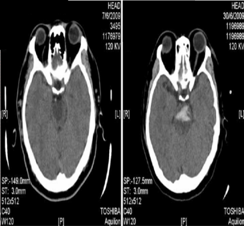
Case 1 is a 42-year old male patient who was admitted to the hospital because of sudden weakness affecting all four extremities and an inability to talk for the previous 3 days. He had a history of hypertension for 20 years. A physical examination on admission showed the following: T 37.5°C, P 88/min, R 20/min, BP 130/90 mmHg, poor general condition, an acutely and critically sick appearance, bilateral pupils of the same large diameter (D=3.0mm) and sensitive responsiveness to light. No pathological signs were detected. No abnormalities were found when examining the heart and the lungs. The patient’s consciousness was preserved; he was able to understand and communicate with the medical staff by opening and closing his eyes, but he was unable to move his extremities. Brain CT showed hypo density in the ventral part of the upper brainstem (Figure 1); the ventral pons and part of the ventral midbrain were involved. T2-weighted MRI showed high signal intensity in the same area of the brainstem.
Case 2 is a 54-year old female patient who was brought to the emergency department because of sudden weakness of the left extremities; her right extremities became involved twenty minutes after the initial symptoms began. She had a history of hypertension for 5 years and had taken antihypertensive medication irregularly. A neurological examination showed that the patient had a clear mind, but the muscle powers of the four extremities were grade 0, and the patient could not speak and swallow. She could only look up and down and could only communicate with others by blinking her eyes. A sensory disturbance was not found. Brain CT showed a region of hyperdensity in the ventral part of the pons (Figure 1).
Positive-pressure ventilation assistance was administered after admission to the hospital, and other treatment measures were also performed, including maintenance of the airway, dehydration, neuroprotective efforts for both patients, anticoagulation and antiplatelet aggregation for the brainstem infarction, and hemostasis for the brainstem hemorrhage. However, both patients developed complications. Both developed severe ventilator-associated pneumonia, for which powerful antibiotics were administered intravenously. Diffusion Tensor Imaging (DTI) and Motor-Evoked Potentials (MEPs) were conducted during their hospital stays.
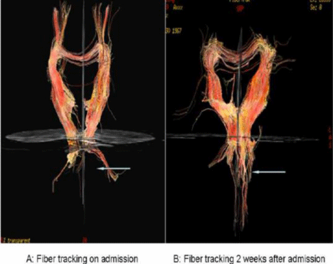
Diffusion tensor imaging was performed using a single-shot spin echo-planar imaging sequence (GE 1.5T Singa Horizon equipped with a standard head coil). We evaluated diffusion tensor images using mean Fractional Anisotropy (FA) in Regions Of Interest (ROIs). Typical ROI size was set between 10 and 15 voxels to include only the pyramidal pathway. The FA values were measured using fiber-tracking software (the Tensor module of FONCTOOL3.1.22 software). We also evaluated fiber connectivity using fiber assignment by continuous tracking. A normal subject was examined as a control. The results demonstrated that FA values in the ventral part of the brainstem were 0.389 and 0.385, respectively, which represented a significant decrease compared with the normal control (0.588). DTI-based fiber tracking showed that white matter fibers through the pons were significantly reduced in number or had disappeared in both patients. These findings suggested that the Corticospinal tract and the cortico-brainstem tract were destroyed at the level of the brainstem. Two weeks after admission, DTI showed partial recovery of FA values and of the pyramidal pathway in the two patients (Figure 2).
A CCS-1 DC chopping cerebral cortical electric nerve stimulator was connected to an NDI-400 EMG/evoked potential instrument. A stimulating electrode was placed on the scalp 7 cm lateral to the CZ location, corresponding to the area of the cortex representing the upper extremities, to record cortical potentials. The stimulating electrode was placed between C6 and C7 to record cervical spinal cord potentials and between L1 and L2 for lumbar spinal cord potentials. The recording electrodes were placed in the thenar muscle site of the upper limb and in the anterior tibial muscle. The reference electrode was in the distal muscle tendon and the ground line was in the wrist. The intensity of stimulation ranged from 800 V to 1500 V. Cortical MEPs could not be recorded from the thenar or anterior tibial muscles of both the two patients when stimulating the motor cortex at an intensity of 1500 V. When the stimulating electrode was placed between C6 and C7, an intensity of 800 V produced a clear waveform in the thenar muscles; the latency of the cervical spinal potential was 11.17 ms (left) and 10.97 ms (right) in case 1 and 10.47 ms (left) and 11.17 ms (right) in case 2. The lumbar spinal cord potential was also evoked successfully, and the latency of this potential was in normal range. These findings suggest that while the motor pathway had been nearly obliterated in the central part of the nervous system, nerve conduction from the cervical spinal cord to the thenar muscles and from the lumbar spinal cord to the anterior tibial muscle was normal. Two weeks after admission, cortical MEPs were recorded from the upper and lower extremities; however, the latency of the MEPs was prolonged, and the amplitude was significantly less (Figure 3) than that observed in normal subjects. Both DTI and MEP in case 1 demonstrated that the patient experienced a partial recovery; the muscle power of the upper extremities and of the left extremity returned to grade 3, while that of the right lower extremity returned only to grade 2. Fiber tracking based on DTI showed that the white matter passing through the brainstem increased significantly, and the FA values were also increased compared with the first examination. Similar improvements were observed for case 2.
Poorly differentiated waveforms for waves I, III, and VI were observed; however, the latencies and amplitudes were in the normal range. BAEP wave V was recorded successfully; the latencies of BAEPs on both sides were in the normal range, but the amplitudes were decreased.
In our report, the patients presented with sudden respiratory difficulties, quadriplegia, and anarthria; intubation and ventilatory assistance were required. The patients’ consciousness was preserved, and they were able to understand and communicate with medical staff members by opening and closing their eyes. Brain CT showed hypodensity in the ventral part of the upper brainstem in one patient and hyperdensity in the ventral pons and part of the midbrain in the other patient. T2-weighted MRI showed abnormal signal intensity in the same areas of the brainstem. The clinical features of the two patients were consistent with the diagnosis of locked-in syndrome. The Corticospinal and cortical medullary tracts dominate motor function. The two pathways descend in the ventral part of the brainstem. When they are damaged or disrupted completely, patients lose all motor function and present with quadriplegia and anarthria. Diffusion Tensor Imaging (DTI) has been introduced as a newly developed technique used to evaluate structural abnormalities in white matter and to visualize white matter fibers in the human brain; it has become a powerful tool for predicting clinical outcomes [2]. In the present report, DTI showed disruption or severe destruction of white matter in the ventral pons; the FA values in the ventral part of the pons were significantly lower compared with those in normal subjects. These findings were consistent with the only previous report of DTI findings for a patient with locked-in syndrome; in this patient, with locked-in syndrome due to a surgical procedure, increased mean diffusivity and decreased fractional anisotropy were observed in the ventral brainstem [3]. The Motor-Evoked Potential (MEP) is the key indicator for evaluating the function of the Corticospinal tract and has been used by some authors to measure the extent of motor function injury [4,5,7,8]. Bruno et al. reported that MEPs from the right upper limb in a patient with locked-in syndrome were delayed and low-voltage in the acute stage, and MEPs from the left upper limb were absent; however, Somatosensory Evoked Potentials (SEPs) from the median nerve and Auditory Brainstem Responses (ABR) were normal, suggesting that the Corticospinal tract was damaged incompletely and that the somatosensory and auditory conduction pathways were preserved. These findings differed from our observations that cortical MEPs from both the right and left limbs were absent on admission and that BAEPs showed poorly differentiated waveforms for waves I, III and VI and decreased amplitude and slightly prolonged latency for wave V; our observations suggested that the pyramidal pathways were damaged severely and that the lesion extended into the auditory pathway in the brainstem. Smart also observed a similar phenomenon [6] in one case of locked-in syndrome following ventral pontine hemorrhage, together with central deafness secondary to extension of the lesion to the inferior colliculus.
In our report, both MEPs and DTI in the two patients showed severe abnormalities associated with motor function. MEP allowed the level of neurophysiological function of the pyramidal pathway to be determined, and DTI allowed the detection of damage to the white matter in the brain using FA values and, by tracking fibers, observations of the extent of white matter connectivity and continuity. DTI abnormalities were closely correlated with MEP changes. MEP and DTI have the potential to be used in combination to confirm and efficiently evaluate the brainstem damage involved in the pyramidal pathway, and DTI and MEP changes during the clinical course appear to reflect the clinical outcome.
Brain CT of patients with locked-in syndrome.
A: Brain CT showed hypo density in the ventral pons, suggesting brainstem infarction. B: Brain CT showed ventral pontine hyper density, suggesting brainstem hemorrhage.
Fiber tracking based on diffusion tensor imaging.
A: Diffusion tensor imaging showed that the number of white fibers descending through the brainstem was significantly decreased and that some fibers were disrupted. B: Diffusion tensor imaging showed that the number of images of white fibers descending through the brainstem increased significantly.
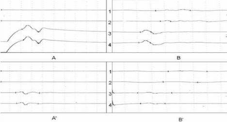
Motor-evoked potential.
A: A cortical MEP was not recorded in the thenar and anterior tibial muscles while stimulating the motor cortex, suggesting that the connection from the motor cortex to the spinal cord was disrupted (lines 1 and 2 in figure 3 A and A’). When a stimulation electrode was placed between C6-C7 and L1-L2, a cervical spinal cord MEP could be recorded in the thenar muscles, and a lumbar spinal cord potential could be recorded in the anterior tibial muscle; the latencies of these recordings were slightly prolonged (lines 3 and 4 in figure 3 A and A’). B: Cortical MEPs were recorded in the thenar and anterior tibial muscles two weeks after admission, but the latencies were prolonged and the amplitudes decreased (lines 1 and 2 in figure 3 B and B’).
Austin Publishing Group is an emerging open access publisher specialising in Science, Technology and Medicine is dedicated to serve the biomedical community through its initiatives. Austin Publishing Group is an academic publisher with 100+ peer reviewed open access journals in various subjects such as biomedical, Pharma, Life Sciences, Environmental, Engineering and Management. Austin Publishing Group publishes Open Access eBooks providing free access to vast scientific literature.