Case Report
Austin J Cerebrovasc Dis & Stroke. 2015;2(1): 1035.
A Pure Dissociated Sensory Loss of Face-Arm-Trunk: An Exceptional Presentation of Midbrain Hemorrhagic Stroke
Rajendra Singh Jain, Sunil Kumar*
Department of Neurology, SMS Medical College, Jaipur,Rajasthan, India
*Corresponding author: Sunil Kumar, Department of Neurology, SMS Medical College, Jaipur, Rajasthan, India.
Received: April 02, 2015; Accepted: May 19, 2015; Published: May 30, 2015
Abstract
A pure sensory syndrome is characterized by impairment of sensations on one side of body, without any other associated neurological features. The most common cause is lacunar ischemic stroke involving the pons, midbrain, thalamus, posterior part of internal capsule and cortex. Lacunar syndromes due to intraparenchymal hemorrhage are rarely described in literature. Hemorrhagic lacunar stroke presenting with pure dissociated sensory loss is exceptional. Herein, we describe a case of 72-year-old man, who presented with sudden onset pure dissociated sensory loss (loss of pain and temperature sensations with intact joint position and vibration senses) of face-arm-trunk on left side. The computed tomography and magnetic resonance imaging with gradient echo sequences of brain showed a small hemorrhage in the right dorsolateral region of midbrain. The patient was managed symptomatically and improved within six days. Our case highlights that a small hemorrhage in dorsal spinothalamic sensory pathway before terminating to the thalamus, can produce pure dissociated sensory syndrome. The topography of dorsal spinothalamic tract may explain the restricted dissociated sensory loss on left half of face, arm and trunk.
Keywords: Hemorrhagic Lacunar Stroke; Midbrain Hemorrhagic Stroke; Dissociated Sensory Loss; Dorsal Spinothalamic Tract
Introduction
A pure sensory stroke syndrome is characterized by sensory loss involving the face, arm, trunk and leg on one side, without any associated neurological features [1]. This is due to selective lesion of ascending sensory pathways. The most common cause is lacunar infarct involving pons, midbrain, thalamus, posterior part of internal capsule and cortex [2]. Rarely, a small hemorrhage can also cause this syndrome. A pure dissociated sensory loss as a presenting feature of hemorrhagic midbrain stroke is a rare clinical feature.
Case Presentation
A 72-year-old male with history of chronic smoking and uncontrolled hypertension, presented with sudden onset numbness of left half of face, arm and trunk. There were no associated pain, paresthesias, headache, vomiting, vertigo, and change in voice, limb weakness, facial deviation, seizure or altered sensorium. There was no history of diabetes mellitus, transient ischemic attack (TIA), dyslipidemia or similar illness in family.
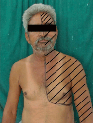
On examination, pulse was 84 per minute, regular and blood pressure was 180/90 mm Hg. The pain and temperature sensations were absent on left half of the face, upper limb and trunk up-to thoracic T5 vertebral level [Figure 1]. The posterior column sensations including light touch, joint position and vibration sensations were intact. Rest of the neurological examination including higher mental functions, visual acuity, field of vision, ocular motility, fundi, pupillary size and reactions, motor system, deep tendon reflexes, plantar response and cerebellar functions were unremarkable.

    Figure 1:  Photograph of patient depicting left sided sensory loss (pain and
temperature) in face-arm-trunk distribution.
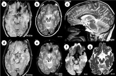
Hemogram, coagulation profile and serum biochemistry including lipid profile, serum vitamin B12 level and thyroid function tests were normal. Enzyme-linked immunoassay (ELISA) test for human immunodeficiency virus (HIV) was negative. X-ray chest, electrocardiography (ECG), 2D-echocardiography, somatosensory evoked potentials (SSEP) and brainstem evoked potentials (visual and auditory) were normal. The non-contrast computed tomography (CT) revealed a well-defined rounded hyperdensity in right dorsolateral midbrain region. Magnetic resonance imaging (MRI) with gradient echo sequence (GRE) of brain showed a well-defined hypointensity on T1-weighted [Figure 2.a], T2-weighted [Figure 2.b,c] and fluid attenuation inversion recovery (FLAIR) [Figure 2.d] images in the right dorsolateral region (tegmentum) of midbrain. There was no diffusion restriction [Figure 2.f,g]. The GRE images (Figure 2.e) showed single discrete “blooming of black” in tegmentum of midbrain on right side. The magnetic resonance angiography of brain and neck vessels was normal.

    Figure 2:  Magnetic resonance imaging (MRI) of brain showing hypointense signal on TI-weighted and T2-weighted images in right dorsolateral (tegmentum)
midbrain. There is no restriction on diffusion-weighted image. Gradient echo (GRE) sequence imaging showing a single discrete “blooming of black” in tegmentum
of midbrain on right side. A small perilesional edema is seen.
In view of pure dissociated sensory loss (loss of pain and temperature sensations with intact joint position and vibration senses) of left half of face-arm-trunk and findings on brain imaging, the patient was diagnosed with acute hemorrhagic stroke involving dorsolateral midbrain. He was managed symptomatically with target systolic blood pressure less than 140 mm Hg. He started showing improvement in sensory symptoms after six days. On six-month follow up, patient does not have any sensory symptoms.
Discussion
The pure sensory stroke syndrome is a rare phenomenon, accounting for about seventeen percent of all lacunar stroke and approximately five percent of acute ischemic stroke [3]. The pure sensory syndrome is mostly associated with ischemic lacunar stroke [3]. Lacunar syndromes due to intracerebral hemorrhage are rarely described in literature [4-6]. Hemorrhagic lacunar stroke presenting with pure sensory syndrome is exceptional. The common sites for pure sensory syndrome are pons [7], thalamus [8], posterior part of internal capsule [9] and cortex. Rarely, it may be due to hemorrhagic stroke of midbrain [10,11].
Our patient presented with sudden onset of left hemisensory loss of pain and temperature restricted to face-arm-trunk with intact posterior column sensations (light touch, joint position and vibration sense) without any other neurological features. A clinical diagnosis of pure dissociated sensory stroke was made. The magnetic resonance imaging showed acute hemorrhage in right dorsolateral part of midbrain (tegmentum). The hemorrhage in our patient was due to chronic uncontrolled hypertension, however other causes like bleeding diathesis, platelets dysfunction and vascular malformation were ruled out by appropriate investigations.
The lateral spinothalamic tract (LST) is located dorsal to the lateral part of the substantia nigra in midbrain and has somatotopic organization, however, the medial lemniscal fibers are located anteriorly. There is segmental organization of sacral, lumbar, thoracic and cervical components, which is arranged from lateral to medial, respectively [12,13]. The LST terminate at ventrolateral posterior nucleus of thalamus. The contralateral dissociated sensory loss on face-arm-trunk distribution observed in our patient was due to this somatotopic organization of the spinothalamic tract in midbrain and sparing of anteriorly located medial lemniscal fibers [14]. The ventral trigeminothalamic tract carries facial pain and temperature sensations from opposite side. It is located medially in spinothalamic tract. Rarely, reverse dissociated sensory loss (loss of joint position or vibration sensations and normal pain and temperature sensations) is also described [7,15] in literature. Our case highlights that a small hemorrhage in dorsal spinothalamic sensory pathway before terminating to the thalamus, can produce pure dissociated sensory syndrome.
Conclusion
In our patient, the small hemorrhage involved the sensory pathway limited to right dorsal spinothalamic tract in midbrain without hampering the medial lemniscus functions, resulted in dissociated sensory loss. The hemorrhagic lacunar stroke presenting with such dissociated sensory loss is exceptional. The topography and location of dorsal spinothalamic tract in midbrain may explain the dissociated sensory loss of face-arm-trunk on left side.
References
- Fisher CM. Pure Sensory Stroke Involving Face, Arm, and Leg. Neurology. 1965; 15: 76-80.
- Derouesné C, Mas JL, Bolgert F, Castaigne P. Pure sensory stroke caused by a small cortical infarct in the middle cerebral artery territory. Stroke. 1984; 15: 660-662.
- Arboix A, García-Plata C, García-Eroles L, Massons J, Comes E, Oliveres M, et al. Clinical study of 99 patients with pure sensory stroke. J Neurol. 2005; 252: 156-162.
- Arboix A, García-Eroles L, Massons J, Oliveres M, Targa C. Hemorrhagic lacunar stroke. Cerebrovasc Dis. 2000; 10: 229-234.
- Milandre L, Donnet A, Graziani N, Grisoli F, Khalil R. [Lacunar syndromes due to intracerebral hemorrhage]. Acta Neurol Belg. 1992; 92: 125-137.
- Mori E, Tabuchi M, Yamadori A. Lacunar syndrome due to intracerebral hemorrhage. Stroke. 1985; 16: 454-459.
- Araga S, Fukada M, Kagimoto H, Takahashi K. Pure sensory stroke due to pontine haemorrhage. J Neurol. 1987; 235: 116-117.
- . Azouvi P, Pappata S, Baron JC, Bousser MG, LaPlane D. [Pure sensory stroke caused by thalamic hematoma]. Rev Neurol (Paris). 1988; 144: 212-214.
- Groothuis DR, Duncan GW, Fisher CM. The human thalamocortical sensory path in the internal capsule: evidence from a small capsular hemorrhage causing a pure sensory stroke. Ann Neurol. 1977; 2: 328-331.
- Azouvi P, Tougeron A, Hussonois C, Schouman-Claeys E, Bussel B, Held JP. Pure sensory stroke due to midbrain haemorrhage limited to the spinothalamic pathway. J Neurol Neurosurg Psychiatry. 1989; 52: 1427-1428.
- Tuttle PV, Reinmuth OM. Midbrain hemorrhage producing pure sensory stroke. Arch Neurol. 1984; 41: 794-795.
- Brodal A. Neurological anatomy in relation to clinical medicine. Oxford University Press, Oxford. 1981: 141-143.
- Heines DE. Neuroanatomy and Atlas of Structures, Sections and Systems. 2nd ed. Berlin, Germany: Urban & Schwartzenberg. 1987:158-163.
- Kim JS. Pure sensory stroke. Clinical-radiological correlates of 21 cases. Stroke. 1992; 23: 983-987.
- Graveleau P, Decroix JP, Samson Y, Masson M, Cambier J. [Isolated sensory deficit of 1 side of the body as a result of a hematoma of the pons]. Rev Neurol (Paris). 1986; 142: 788-790.