Case Report
Austin J Cerebrovasc Dis & Stroke. 2016; 3(1): 1041.
Deterioration of Pre-Existing Hemiparesis Following an Ipsilateral Corona Radiata Infarct: A Rare Entity
Jain RS, Sisodiya MS*, Kookna JC, Bhana I and Verma YK
Department of Neurology, SMS Medical College, India
*Corresponding author: Sisodiya MS, Department of Neurology, SMS Medical College, Jaipur, Rajasthan 302004, India
Received: May 12, 2016; Accepted: June 25, 2016; Published: June 27, 2016
Abstract
Stroke presenting as contralateral hemiparesis is predominantly related to contralateral projection of the corticospinal tract. While most corticospinal fibers decussate at the level of the medulla, some tracts continue to descend as ipsilateral anterior corticospinal fibers. The anterior corticospinal tract (CST) has been suggested as one of the ipsilateral motor pathways which contribute to motor recovery following stroke. Few case reports in literature show ipsilateral hemiparesis due to involvement of anterior corticospinal fibers. We are reporting a case who showed deterioration of pre-existing hemiparesis due to involvement of the ipsilateral anterior CST following a corona radiata infarct.
Keywords: Hemiparesis; Ipsilateral; Anterior corticospinal tract
Introduction
Supratentorial stroke commonly results in neurological weakness on the contralateral side of the body. The reason for this is the predominance of contralateral corticospinal projections which arise from the cortical regions of the brain and decussate in the caudal medulla [1]. While not all fibers decussate, 70–90% of them cross, resulting in this ‘crossed’ hemiparesis [2]. The CST is generally divided into the crossed lateral CST and the uncrossed anterior CST. The anterior CST is considered to be one of the ipsilateral motor pathways from the unaffected motor cortex to the affected extremities, which contribute to motor recovery following stroke incidents [3]. There are few case reports of ipsilateral hemiparesis due to affection of uncrossed fibers. We are reporting a case who showed deterioration of pre-existing hemiparesis due to involvement of the ipsilateral anterior CST following a corona radiata infarct.
Case Presentation
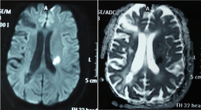
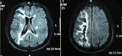
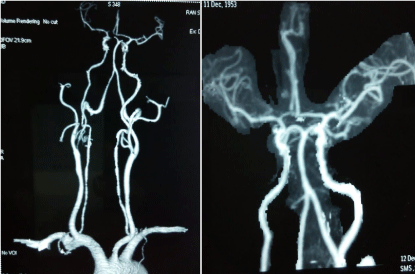
A 58-year-old right handed male, known case of hypertension, diabetes mellitus and old stroke- 3 years back in the form of left hemiparesis with residual deficit (MRC grade power 3/5) and residual left facial weakness, developed sudden deterioration in left hemiparesis including facial weakness. On neurological examination, he had left upper and lower limb weakness with MRC grade 0/5. DTR were brisk on left side with extensor plantar.MRI brain DWI showed an area of restricted diffusion in the left corona radiata representing acute infarct (Figure 1). There was an old infarct in the right frontoparietal region (Figure 2). A 2-D echocardiogram and carotid Doppler study was normal. CT angiography brain and neck vessels were normal (Figure 3). Diffusion tensor tractography (DTT) could not be done due to non-availability. Patient was discharged on conservative treatment as well as physiotherapy and recovering in follow up.

Figure 1: MRI DWI & corresponding ADC image showing acute infarct in left corona radiata and remote infarct in right fronto - parietal cortex.

Figure 2: MRI T2 FLAIR image showing remote infarct in right fronto-parietal cortex and acute infarct in left corona radiata.

Figure 3: Normal CT Angiography of brain & neck vessels.
Discussion
Ipsilateral hemiparesis after a supratentorial cerebral stroke has rarely been reported. There are only few case reports in literature in which ipsilateral hemiparesis occurred due to stroke involving uncrossed anterior CST (Table 1).
    Authors
    No. of cases
    Age
    Sex
    Hemiparesis
    Dominant    hemisphere
    Type of stroke
    Location
    Confirmatory    imaging
    DTT


    [4]
    1
    55
    M
    R
    L
    IS
    CR
    MRI
    -


    [5]
    1
    62
    M
    R
    L
    ICH
    Putamen
    CT
    -


    [6]
    1
    -
    -
    R
    L
    ICH
    IC and thalamus
    CT
    -


    [7]
    2
    62
    F
    L
    L
    IS
    CR
    MRI,Fmri
    -


    41
    M
    R
    L
    IS
    CR
    MRI


    [8]
    1
    59
    M
    L
    L
    IS
    CR
    fMRI
    -


    [9]
    1
    55
    M
    L
    L
    IS
    CR and putamen
    MRI
    Yes


    [10]
    1
    35
    M
    R
    L
    IS
    IC
    MRI
    -


    [11]
    2
    57
    M
    L
    L
    IS
    O,P,F
    MRI
    -


    67
    F
    R
    L
    IS
    F
    MRI


    L = Left,    R=Right, IS= Ischemic Stroke, ICH= Intracerebral Hemorrhage, IC= Internal    Capsule, CR=Corona Radiate, DTT= Diffusion Tensor Imaging Tractography, O=Occipital,    P= Parietal, F=Frontal
Table 1: Summary of previous case reports.
We evaluated a left hemiparetic patient with a new left corona radiata infarct and an old right middle cerebral artery infarct. Before the onset of the new left corona radiata infarct, the patient was able to do his daily routine activities with mild discomfort. However, after the onset of the new infarct in the left corona radiata, the motor function of his left extremities deteriorated to complete weakness (MRC grade 0/5). We presumed that some neural tract other than the right whole CST had been responsible for the partial motor function (MRC grade 3/5) of the left extremities before the onset of new left corona radiata infarct. Considering that the new infarct involved anterior CST area of the left corona radiata, the left anterior CST appeared to be the most plausible motor tract for deterioration of pre-existing left hemiparesis.
Diffusion tensor tractography (DTT) can visualize the neural tracts in three dimensions, so DTT should be used for confirmation of anterior CST as used by Ng, et al. [9]. Absence of proof by DTT is the limitation of our case report as this modality is not available to us.
Association of ipsilateral hemiparesis with cerebral malformations, such as posterior fossa malformations, occipital encephalocele, Dandy-Walker malformation, Joubert syndrome, and Moebius syndrome was not found in our case as described in literature [12,13], however findings in our case are consistent with higher incidence of ipsilateral hemiparesis in right handed person and in those with past history of stroke as in literature [6,11] .
Conclusion
Our case report suggests that the new infarct in the left corona radiata damaged the uncrossed ipsilateral motor pathway, which had been responsible for the motor function of the left extremities after remote infarct, causing further deterioration of the pre-existing hemiparesis. Our observation favours previous case reports that ipsilateral hemiparesis is common in right handed males with past history of stroke; however malformation of the brain was not found in our case which were detected earlier in the literature.
References
- Davidoff RA. The pyramidal tract. Neurology. 1990; 40: 332-339.
- York DH. Review of descending motor pathways involved with transcranial stimulation. Neurosurgery. 1987; 20: 70-73.
- Jang SH. A review of the ipsilateral motor pathway as a recovery mechanism in patients with stroke. Neuro Rehabilitation. 2009; 24: 315-320.
- Alurkar A, Karanam L, Atre P, Nirhale A, Nayak S, Oak S. Ipsilateral stroke with uncrossed pyramidal tracts and underlying right internal carotid artery stenosis treated with percutaneous transluminal angioplasty and stenting: a rare case report and review of the literature. Neuroradiol J. 2012; 25: 237–242.
- Hosokawa S, Tsuji S, Uozumi T, Matsunaga K, Toda K, Ota S. Ipsilateral hemiplegia caused by right internal capsule and thalamic hemorrhage: demonstration of predominant ipsilateral innervation of motor and sensory systems by MRI, MEP, and SEP. Neurology. 1996; 46: 1146-1149.
- Song YM, Lee JY, Park JM, Yoon BW, Roh JK. Ipsilateral hemiparesis caused by a corona radiata infarct after a previous stroke on the opposite side. Arch Neurol. 2005; 62: 809-811.
- Ago T, Kitazono T, Ooboshi H, Takada J, Yoshiura T, Mihara F, et al. Deterioration of pre-existing hemiparesis brought about by subsequent ipsilateral lacunar infarction. J Neurol Neurosurg Psychiatry. 2003; 74: 1152- 1153.
- Terakawa H, Abe K, Nakamura M, Okazaki T, Obashi J, Yanagihara T. Ipsilateral hemiparesis after putaminal hemorrhage due to uncrossed pyramidal tract. Neurology. 2000; 54: 1801-1805.
- Ng AS, Sitoh YY, Zhao Y, Teng EW, Tan EK, Tan LC. Ipsilateral stroke in a patient with horizontal gaze palsy with progressive scoliosis and a subcortical infarct. Stroke. 2011; 42: e1-3.
- Kang K, Choi NC. Ipsilateral hemiparesis and spontaneous horizontal nystagmus caused by middle cerebral artery territory infarct in a patient with agenesis of the corpus callosum. Neurol Sci. 2012; 33: 1165-1168.
- Saada F, Antonios N. Existence of ipsilateral hemiparesis in ischemic and hemorrhagic stroke: two case reports and review of the literature. Eur Neurol. 2014; 71: 25-31.
- Lagger RL. Failure of pyramidal tract decussation in the Dandy-Walker syndrome. Report of two cases. J Neurosurg. 1979; 50: 382-387.
- ten Donkelaar HJ, Lammens M, Wesseling P, Hori A, Keyser A, Rotteveel J. Development and malformations of the human pyramidal tract. J Neurol. 2004; 251: 1429-1442.