Case Report
Austin J Cerebrovasc Dis & Stroke. 2017; 4(3): 1061.
Cervical Spinal Arteriovenous Malformation with Intramedullar Hemathoma
Salihović D¹*, Smajlović D², Čičkušić A² and Moranjkić M³
1Department of Neurology, University Clinical Centre Tuzla, Bosnia and Herzegovina
2Department of Physical Medicine and Rehabilitation, University Clinical Centre Tuzla, Bosnia and Herzegovina
3Department of Neurosurgery, University Clinical Centre Tuzla, Bosnia and Herzegovina
*Corresponding author: Salihović D, Department of Neurology, University Clinical Centre Tuzla, 75000 Tuzla, Bosnia and Herzegovina
Received: May 04, 2017; Accepted: May 30, 2017; Published: June 08, 2017
Abstract
Intramedullar arterial-venous malformation (AVM) in the cervical region is rare, but they are important clinical entities (1). We would like to present one our patient with AVM of spinal arteries and intramedullar hemathoma.
Keywords: Intramedullar hemathoma; Arterial-venous malformation
Case Presentation
Female patient 28 years old, was hospitalized at our Department with sudden quadriplegia. Information that she was treated at our department six years ago due to weakness in the arms and legs was obtained after admission. Computed tomography (CT) of the brain, which was done at first hospitalization, showed a subarachnoid haemorrhage (SAH), while spinal digital subtraction angiography (DSA) has showed AV-malformation of spinal arteries at the level C4- C5. Afterwards, endovascular treatment has been done and patient recovered fully. The current hospitalization, on admission patient was in poor general somatic condition with dyspnoea, tachyarrhythmia and urinary retention. She was quadriplegic with extinguished deep tendon reflexes. Superficial sensibility was damaged for all qualities to shoulder level, and pathological reflexes were not present.
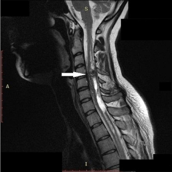
During the current hospitalization CT of the brain and cervical spine were performed, and both of them were normal. The next day lumbar puncture was done and we found uniformly haemorrhagic liquor. Magnetic resonance imaging (MRI) of brain was normal, while the cervical spine MRI showed intramedullar/intradural lesion at the level C4/C5 corresponding to intra and juxtamedullar AV malformation with acute intramedullar hemathoma (from C3 to Th1 vertebra) and subarachnoid haemorrhage (Figure1).

    Figure 1:  Intramedullar/intradural AV malformation in level C4/C5 segment
with intra medullar hemathoma (from C3 to Th1) and subarachnoid
haemorrhage.
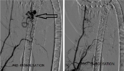
Patient was treated at our Department with pharmacological and physical procedures during one month. Initially and before rehabilitation, she had Rankin Scale 5 and Barthel index 0 (total dependence). Two months after the onset she was treated with endovascular embolization (Figure 2) at Department of Neurosurgery. Using “road-map” technique, marathon micro catheter cannulised nidus of AVM. Afterwards, using micro catheter DMSO solution was placed, which lead to placing of “Onyx”, embolization agent in feeder of AVM, with technique of blank road-map. Neurosurgeon plans were to do another endovascular treatment which will be on the left side to fulfil AVM with embolization agent and totally excluded it from circulation. After endovascular treatment patient has resumed rehabilitation. Currently she has a light weakness of right limb, plegic left hand and moderate paresis of the left leg, and she walks with a cane (Rankin Scale 3, and Barthel index 62 - moderate dependence).

    Figure 2:  AVM of spinal arteries before and after the Onyx endovascular
embolisation.
Discussion
AVM of the spinal cord is a rare malformation characterised by arterial-venous shunt with or without vascular network between feeder arteries and drainage veins. Incidence of AVM range is between 20 to 30% of all spinal vascular shunts [1]. According to our knowledge and available literature, AVM in our region are rare. We did not find any publications on this topic in Bosnia and Herzegovina, and because of that we decided to present this case. Rangel-Castilla, et al. were described 110 patients with AVM and AV fistulas during 13 years [2]. We do not have such a sample of patients in our region, and we could not find any publication on this topic in our country.
There are four types of AVM [3]. It is difficult to determine precisely the shunt using MRI. Angiography remains the gold standard for analysing anatomical, morphological and architectural features necessary for therapeutic decisions in both pediatrics and adult population [1]. The experience of one Polish study suggested that endovascular treatment is effective and safe method for the treatment of AVM located in the cervical region of spinal cord [4]. Kring, et al. point out that hyper-acute treatment is rarely indicated, because acute re-bleeding rarely occurs. After bleeding in spinal AVM occurs, it is custom to wait for approximately six weeks for potential vasospasm to be resolved or the haemorrhage to resorbed [5].
Our patient had a spinal AVM type II with multiple feeders with origin predominantly on the right vertebral and costocervical arteries, as well as right thyrocervical truncus. Treatment was done according to the criteria indicated in the above studies, and endovascular treatment was not carried out in acute phase, but was done when hemathoma was resolved. Our neurosurgeons have better results and experience with endovascular embolization and because of that we prefer to endovascular than surgical treatment.
Conclusion
Arterial-venous malformations of the spinal artery are rare and usually are not in the focus of differential diagnostic interest of clinicians. When we see symptoms and signs of spinal cord dysfunction we should give a thought about arterial-venous malformation in that region.
References
- Lorenzoni PJ, Scola RH, Kamoi Kay CS, de Queiroz E, Cardoso J, Leite Neto MP, et al. Spinal cord arteriovenous malformation: a pediatric presentation. Arq. Neuro-Psiquiatr. 2009; 67: 2b.
- Rangel-Castilla L, Russin JJ, Zaidi HA, Martinez-Del-Campo E, Park MS, Albuquerque FC, et al. Contemporary management of spinal AVFs and AVMs: lessons learned from 110 cases. Neurosurg Focus. 2014; 37: E14.
- Ferch RD, Morgan MK, Sears WR. Spinal arteriovenous malformations: a review with case illustrations. J Clin Neurosci. 2001; 8: 299-304.
- Acewicz A, Richter P, Tykocki T, Czepiel W, Ryglewicz D, Dowzenko A. Endovascular treatment of cervical intramedullary arteriovenous malformation. Neurol Neurochir Pol 2014; 48: 223-227.
- Krings T, Thron AK, Geibprasert S, Agid R, Hans FJ, Lasjaunias PL, et al. Endovascular management of spinal vascular malformations. Neurosurg Rev. 2010; 33: 1-9.