
Research Article
Austin J Cerebrovasc Dis & Stroke. 2018; 5(1): 1076.
Vertebral Artery Dissection: Can we Afford to Miss it?
Ojha PT, Shashank N*, Patil S, Chheda A, Kadam NS and Ansari A
Department of Neurology, Grant medical College and Sir JJ Group of Hospitals, India
*Corresponding author: Shashank N, Department of Neurology, Grant medical College and Sir JJ Group of Hospitals, Byculla, Mumbai, India
Received: January 26, 2018; Accepted: March 12, 2018; Published: March 30, 2018
Abstract
Vertebral artery dissection (VAD) is an infrequent occurrence but is a leading cause of stroke in young and otherwise healthy patients. CTA, MRI and catheter angiography can all be used to detect vertebral artery dissection, and each has pros and cons. Here we discuss two cases of stroke in young where the presence of double lumen sign on axial sequences of GRE provided the clue to intracranial arterial dissection as the etiology of stroke. This helped avoid unnecessary and expensive etiological evaluation of stroke in young.
Keywords: Vertebral artery dissection; Double lumen sign; Ipsilateral appendicular ataxia
Abbreviations
VAD: Vertebral Artery Dissection; CTA: CT Angiography; MRI: Magnetic Resonance Imaging; GRE: Gradient ECHO; ER: Emergency Room; LMWH: Low Molecular Weight Heparin
Case 1
A 43 year old male developed sudden weakness of left side of body, along with imbalance and dizziness after strenuous exercise in gymnasium. On presentation in ER after 1 hour of the onset of symptoms, his blood pressure was found elevated to 180/110 mm Hg. Though, he was fully conscious, left side limb power was reduced to 3-4/5 MRC grade with marked ipsilateral appendicular ataxia.
Urgent MRI Brain imaging showed Left cerebellar infarct. MRI Brain Angiography showed poor visualisation of left vertebral artery (possibilities being: hypoplastic artery or affected by dissection, stenosis or thrombosis). None of the commonly described signs of dissection were observed, but there was a strong suspicion of vertebral artery dissection. On close observation, GRE brain axial sequences showed presence of both true and false lumen (double lumen sign), confirming our suspicion of dissection of the intracranial segment of left vertebral artery. Patient was thrombolysed with intravenous alteplase and improved nearly completely within next 24 hours. He was administered LMWH for then next 5 days and discharged on antiplatelets. Follow up MRI brain angiography, showed complete recanalisation of the left vertebral artery with resolution of double lumen sign (Figure 1,2 and 3).
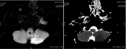
Figure 1: MRI Brain showing left cerebellar acute infarct.
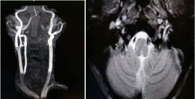
    Figure 2:  Left vertebral artery poorly visualised and Double Lumen sign of
left vertebral artery.
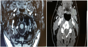
    Figure 3:  Follow up angiogram of the patient showing recanalisation of left
vertebral artery.
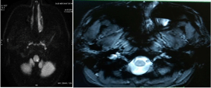
Figure 4: MRI Brain showing left medullary infarct.
Case 2
A 39 year old male had been experiencing shooting pain in the neck along with transient difficulty in swallowing and tingling in left side of the body for the last one week. He was admitted in hospital when he further developed for left upper limb weakness and imbalance occuring for over 12 hours. On examination his Blood pressure was 160/100 mm Hg. Clinical examination revealed left lateropulsion of stance, left upper limb incoordination and gait ataxia. This was accompanied by dysarthria, dysphagia to solids and left hemianesthesia.
MRI Brain showed Left medullary acute infarct and MRI Brain Angiography showed poor visualisation of the left vertebral artery. GRE axial brain sequences revealed double lumen sign in intracranial segment of left Patient was started on antiplatelets with LMWH. As he was out of time window for IV thrombolysis, it was withheld. He improved rather quickly in a matter of few days. Follow up MRI brain angiography showed complete recanalisation of the intracranial left vertebral artery with resolution of double lumen sign (Figure 4 and 5).
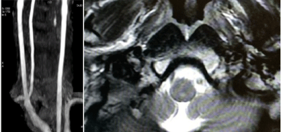
    Figure 5:  MRI Angiography images and Double Lumen sign of left vertebral
artery.
Discussion
Intracranial arterial dissection (ICAD), especially vertebral artery dissection (VAD) is an increasingly recognized cause of stroke in patients younger than 45 years., Advances in neuro-imaging have contributed to growing awareness of this entity.
Typical patients with VAD present with posterior neck pain or headache, followed by ischemia of the vertebrobasilar system. Initial manifestations of VAD however, are less distinct than those of carotid artery dissection and usually interpreted as musculoskeletal pain [1,2]. Therefore, an accurate neurologic examination and detailed historytaking are mandatory, particularly in young adults, in order to search for symptoms or focal signs of brainstem stroke. Ischemic symptoms occur in more than 90% of VAD patients and may involve the brain stem, especially the lateral medulla (Wallenberg’s syndrome)
Though Catheter angiography is the gold standard for diagnosis of VAD, it is not routinely available at many centers. Characteristic features are vessel irregularity and/or stenosis, string sign, double lumen, pseudoaneurysm formation, or complete occlusion. Magnetic resonance techniques [3,4] are now replacing conventional angiography in the diagnosis of most strokes, especially dissections of intracranial arteries. The resolution of magnetic resonance angiography now approaches that of conventional angiography, and magnetic resonance imaging very often shows the intramural hematoma on routine sequences. The intramural hematoma characteristically has a crescentic shape adjacent to the vessel lumen and often spirals along the length of the artery. Gradient Echo/ SWI axial images of MRI brain have the capability to show blood/ thrombus in brain, hence can be used in early detection of ICAD/ VAD.
As MRI is being widely used, recognition of this “DOUBLE LUMEN SIGN on GRE or SWI” is helpful in detecting intracranial dissection. Detection of double lumen sign in intracranial VAD helped in accurate etiologic diagnosis and adequate treatment in both of these patients.
To prevent thromboembolic complications, anticoagulation with intravenous heparin followed by oral warfarin has been recommended for all patients with acute dissections of the vertebral artery, regardless of the type of symptoms, unless there are contraindications such as the presence of a large infarct with associated mass effect, hemorrhagic transformation of the infarcted area, an intracranial aneurysm, and intracranial extension of the dissection [5]. Although antithrombotic treatment has been advocated since the 1970s [3], no randomized trials have been reported, and the validity of such treatment has never been proved and use of antiplatelets have been shown comparable to the use of anticoagulation. Anticoagulation with a target international normalized ratio (INR) of 2.0 to 3.0 is generally used for three to six months.
While thrombolysis [6,7] in the setting of dissection may theoretically cause enlargement of the intramural hematoma, accumulating evidence suggests that the effectiveness and safety of thrombolysis for patients with ischemic stroke related to cervical artery dissection are similar to its effectiveness and safety for patients with ischemic stroke from other causes.
Most VADs heal spontaneously [8]. However, an urgent surgical intervention may be required in patients presenting with SAH. Symptomatic aneurysmal dilatation of the artery may also warrant surgery. Chronic VADs have also been treated by surgical reconstruction to prevent further ischemic or thromboembolic complications, if medical treatment with six month anticoagulation fails or if the dissecting aneurysms and/or high grade stenosis persist. Surgical interventions include endovascular treatment and the arterial repair.
In conclusion, axial GRE/SWI sequences of MRI brain should be examined carefully for presence of double lumen sign related to intracranial artery dissection. Arterial dissection remains and important cause of stroke in young. It might help save a lot of unnecessary etiologic investigations in such patients. The further management of such cases might also differ on the basis of type/ severity of dissection.
References
- Silbert PL, Mokri B, Schievink WI. Headache and neck pain in spontaneous internal carotid and vertebral artery dissections. Neurology. 1995; 45: 1517-1522.
- Lanfranchi S, Di Falco M, Perini M, Zarcone D. Posterior headache as a warning symptom of vertebral dissection: a case report. J Headache Pain. 2005; 6: 478-479.
- Schievink WI. Spontaneous dissection of the carotid and vertebral arteries. N Engl J Med. 2001; 344 : 898-906.
- Rizzo L, Crasto SG, Savio D, Veglia S, Davini O, Giraudo M, et al. Dissection of cervico-cephalic arteries: early diagnosis and follow-up with magnetic resonance imaging. Emerg Radiol. 2006; 12: 254-265.
- Schievink WI. The treatment of spontaneous carotid and vertebral artery dissections. Curr Opin Cardiol. 2000; 15: 316-321.
- Engelter ST, Rutgers MP, Hatz F, Georgiadis D, Fluri F, Sekoranja L, et al. Intravenous thrombolysis in stroke attributable to cervical artery dissection. Stroke. 2009; 40: 3772-3776.
- Georgiadis D, Baumgartner RW. Thrombolysis in cervical artery dissection. Front Neurol Neurosci 2005; 20: 140-146.
- Muller BT, Luther B, Hort W, Neumann-Haefelin T, Aulich A, Sandmann W. Surgical treatment of 50 carotid dissections :indications and results. J Vasc Surg. 2000; 31: 980-988.