Editorial
Austin J Clin Cardiolog. 2014;1(2): 1013.
Lacrosse NSE Balloon using the “Leopard-Crawl” Technique is Efficacious for Predilation of Severely Calcified Lesions
Kazuhiro Ashida*, Taichiro Hayase, Takayuki Shinmura, Kei Kawai and Masayuki Orimo
Department of Cardiology, Yokohama Shintoshi Neurosurgical Hospital, Yokohama, Japan
*Corresponding author: Kazuhiro Ashida, Department of Cardiology, Yokohama Shintoshi Neurosurgical Hospital, Yokohama, Japan
Received: February 14, 2014; Accepted: February 20, 2014; Published: February 24, 2014
Editorial
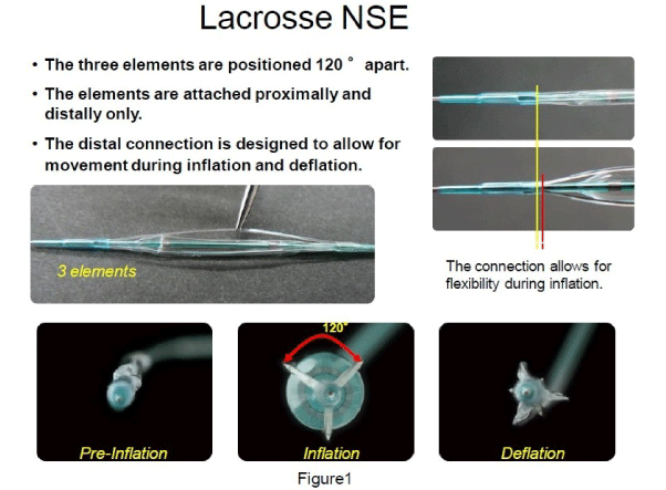
Difficulties associated with stent delivery and under expansions [1–5] are often encountered with calcified lesions. Lesion preparation of calcified lesions prior to stent implantation is important to facilitate stent delivery and provide concentric stent expansion [6,7]. Recently, the novel Lacrosse NSE catheter (Goodman Co.,Ltd. ) has become commercially available. The catheter contains three triangular, nylon elements (width, 0.014”, height, 0.015”) that are free–floating on the outside of the balloon surface, and attached proximal and distal to a 13 mm balloon length (Figure1). Dilatation using a Lacrosse NSE creates a scoring effect into calcified tissue through a focused transmission of force through the elements.
Figure 1: Lactose NSE.
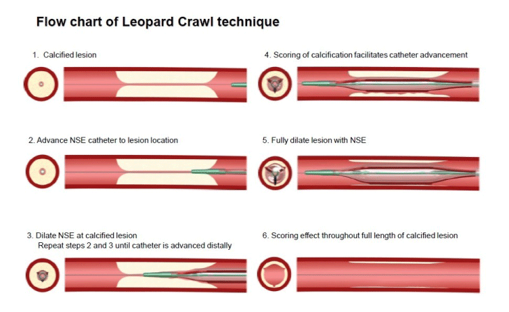
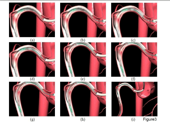
Unfortunately, current designs of scoring balloons result in reduced functionality in regard to delivery in comparison to conventional balloons, and difficulties associated with delivery and lesion cross ability of scoring catheters occur in a clinical setting. One method for overcoming the obstacles faced by difficult delivery is use of the “leopard–crawl” technique. This technique uses allow inflation pressure to create a wedge into the calcification and then subsequently advances the catheter during balloon deflation to facilitate catheter delivery across the stenosis (Figure 2, 3). The Lacrosse NSE elements are attached distal to the balloon location, and for instances whereby the catheter is unable to cross the lesion location, a “leopard–crawl” technique can assist in facilitating device delivery. We reported the initial clinical use of the leopard–crawl technique for facilitating catheter delivery in cases of severely calcified lesions in which standard delivery was unsuccessful, while creating an efficacious scoring effect into the calcified lesion that reflects the results of bench testing [8].
Figure 2: Flow chart of Leopard Crawl technique.
Figure 3: At the time of advancing the Lacrosse NSE (a) into the coronary artery there is a reverse reaction in force applied that disengages the guiding catheter (b). Lacrosse NSE is then inflated at low pressure (3˜6atm) (c), with the slack in guide wire then removed while ensuring the guide catheter positioning is corrected (d).Following deflation of the Lacrosse NSE, further insertion of the catheter results in the balloon advancing distally (e). Further repeating this process (f, g, h) results in the catheter advancing closer toward the target lesion (i).
Experimental and clinical studies with the Lacrosse NSE (NSE) balloon for predilatation of calcified lesions were conducted. Methods and results: Six artificial, circumferentially calcified lesions were prepared in silicon tubes by using plaster. Balloon inflation was performed with the same sized NSE or non–compliant balloon (NC) until cracks were observed. The luminal area and eccentricity (short⁄ long diameter) of the stent, as indices of expansion, were measured by intravascular ultrasound (IVUS) after the same stent was placed at rated burst pressure. Clinical study: Balloon delivery to target lesion location using standard delivery techniques for severely calcified lesions is typically more problematic. The “leopard–crawl” technique was utilized to overcome the obstacles faced by difficult delivery. Predilation was performed with the NSE using the “Leopard–Crawl” technique or NC for lesions with >270 degree calcification on IVUS before stenting between May, 2011 and August, 2012. Eccentricity in minimum stent area (MSA) was measured retrospectively in 47 consecutive patients stented after NSE expansion (NSEgroup) and 23 consecutive patients stented after NC expansion (NCgroup). In the experimental results, the NSE caused more multiple cracks (NSE 2.3 vs. NC 1.3) across the entire lesion at a lower pressure (9.3atm vs. 20.3atm) and the expanded stent was more concentric (eccentricity; 0.88 vs. 0.76). Clinical results: Stent eccentricity was 0.83±0.11 vs. 0.71±0.18 for NSE vs. NC (p<0.05). The number of cracks was higher in the NSE–group than in the NC–group (1.6±0.4 vs. 0.9±0.2, p<0.05). Conclusions: Experimental NSE caused multiple full–length cracks at lower pressure than NC and the stent was more concentric in nature. Clinical results were similar, suggesting that good stent expansion can be achieved by predilation of severely calcified lesions with NSE.
References
- Albrecht D, Kaspers S, Füssl R, Höpp HW, Sechtem U. Coronary plaque morphology affects stent deployment: assessment by intracoronary ultrasound. Cathet Cardiovasc Diagn. 1996; 38: 229-235.
- Sonoda S, Morino Y, Ako J, et al. SIRIUS Investigators. Impact of inal stent dimensionson long-term results following sirolimus-eluting stent implantation: serial intravascularultrasoundanalysis from SIRIUS trial. J Am Coll Cardiol. 2004; 43: 1959-1963.
- Park DW, Park SW, Park KH, Lee BK, Kim YH, Lee CW, et al. Frequency of and risk factors for stent thrombosis after drug-eluting stent implantation during long-term follow-up. Am J Cardiol. 2006; 98: 352-356.
- Fujii K, Carlier SG, Mintz GS, Yang YM, Moussa I, Weisz G, et al. Stent underexpansion and residual reference segment stenosis are related to stent thrombosis after sirolimus-eluting stent implantation: an intravascular ultrasound study. J Am Coll Cardiol. 2005; 45: 995-998.
- Hong MK, Mintz GS, Lee CW, Park DW, Choi BR, Park KH, et al. Intravascular ultrasound predictors of angiographic restenosis after sirolimus-eluting stent implantation. Eur Heart J. 2006; 27: 1305-1310.
- Asakura Y, Furukawa Y, Ishikawa S, Asakura K, Sueyoshi K, Sakamoto M, et al. Successful predilation of a resistant, heavily calciied lesion with cutting balloon for coronary stenting: a case report. Cathet Cardiovasc Diagn. 1998; 44: 420-422.
- Urban P, Gershlick AH, Guagliumi G, Guyon P, Lotan C, Schofer J, et al. Safety of coronary sirolimus-eluting stents in daily clinical practice: one-year follow-up of the e-Cypher registry. Circulation. 2006; 113: 1434-1441.
- Ashida K, Hayase T, Shinmura T. Eficacy of lacrosse NSE using the “leopard-crawl” technique on severely calciied lesions. J Invasive Cardiol. 2013; 25: 555-564.


