Case Report
Austin J Clin Cardiolog. 2014;1(3): 1021.
An Unusual Etiology for an Acute Coronary Syndrome
Claudiu Ungureanu1*, Christophe Laruelle1, Denis Pieters1, Eleonor Ponlot1, Philippe Blouard1, Patrick Timmermans1, Pierre-Yves Etienne2
1Departments of Cardiology, Cliniques Universities Saint- Luc, Belgium
2Department of Cardiovascular Surgery, Cliniques Universities Saint-Luc, Belgium
*Corresponding author: Ungureanu Claudiu, Department of Cardiology, University Hospital Saint Pierre Brussels, rue Haute, nr 322, 1000, Belgium
Received: April 28, 2014; Accepted: May 20, 2014; Published: May 22, 2014
Keywords
Tirone david procedure; Heparin induced thrombocytopenia syndrome
Case Report
A 46–years–old man was admitted in our hospital for the treatment of an aneurysm of the ascending aorta and aortic root.
As planned, a valve sparing operation (Tirone David procedure) was performed. The intervention and the postoperative period were uneventful.
However the day before discharge the patient developed severe chest pain accompanied by diaphoresis and nausea. The ECG showed ST segment elevation of 2mm in the inferior leads.
The patient underwent urgent coronary angiography that revealed proximal occlusion of the right coronary artery by a large thrombotic mass (figure 1). This was managed by thromboaspiration and a subsequent stenting for an ostial residual thrombotic image (figure 2).
Figure 1: Selective injection in the right coronary artery.( OAD 90). An massive and subocclusive thrombus is visualised in the proximal part of the artery. The distal part of this mass is floating.
Figure 2: Selective injection in the right coronary artery. We can suspecte that the thrombotic mass protruding from ascending aorta in the coronary artery.
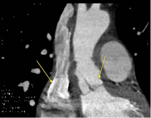
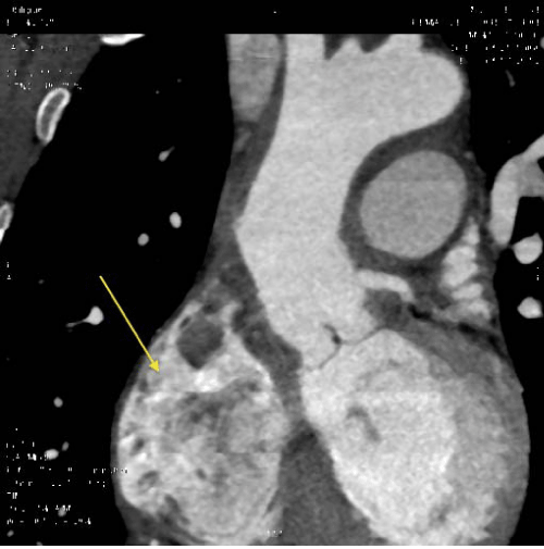
A CT scan confirmed the presence of the thrombotic mass in the ascending aorta (figure 3), but surprisingly also revealed the presence of multiple thrombi located in the right pulmonary artery, the right atrium (figure 4) and the superior vena cava.
Figure 3: Aortic CT scan. Presence of a thrombus in the aortic sinus of Valsalva with extension at the level of the aortic cusps and a large thrombus in the superior vena cava.
Figure 4: Aortic CT scan. Large thrombotic mass inside the anterior part of the right atrium.
Our first hypothesis, that the aetiology of this inferior STEMI was related to the surgical intervention, was challenged by the presence of thrombotic charge both in the arterial and venous system.
A diagnosis of heparin induced thrombocytopenia syndrome was sustained by a halving fall of platelet count and thrombosis (venous and arterial) five days following heparin administration. The ELISA test for antibodies against heparin–PF4 was positive and diagnosis of HIT was confirmed.
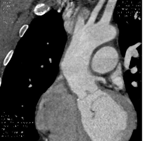
Despite an extensive aortic, intracardiac, intracoronary and venous thrombosis the evolution of our patient was favorable (figure 5) after the percutaneous revascularisation of the right coronary artery and replacement of LMWH by fondaparinux, without any other complications.
Figure 5: Aortic CT scan. Two months later the thrombotic mass inside right atrium and superior vena cava disappeared. Partial regression of the thrombus in the proximal ascending aorta.


