
Case Report
Austin J Clin Cardiolog. 2016; 3(1): 1046.
Subacute Anterior Myocardial Infarction due to the Discontinuation of Warfarin in a Patient with Aortic Valve Prosthesis; What is the Best Treatment
Ozturk C*, Celik T, Demir M, Yildirim AO, Balta S and Iyisoy A
Department of Cardiology, Gulhane Military Medical Academy, School of Medicine, Turkey
*Corresponding author: Cengiz Ozturk, Department of Cardiology, Gulhane School of Medicine, Tevfik Saglam St., 06018 Etlik, Ankara, Turkey
Received: June 10, 2016; Accepted: July 19, 2016; Published: July 20, 2016
Case Report
Acute Myocardial Infarction (MI) is a common entity in patients due to the atherosclerotic plaque rupture. But there are other suspicious etiologic reasons that lead to coronary embolism, such as intracardiac prosthesis, infective endocarditis, mural thrombus or a cardiac tumor [1-8]. Even if the patient did not use warfarin, the real reason for acute myocardial infarction whether embolism or
atherosclerotic plaque rupture should be revealed in order to decide optimal treatment option. Intravascular ultrasonography and optical coherence computed tomography are very useful for diagnosis so which treatment is the best; thrombolysis, angioplasty or stenting. Previous cases of coronary emboli in association with prosthetic mechanical valves have been reported previously, but the treatment is controversial [9-18]. We present a patient who had a mechanical aortic valve and was admitted to the hospital for a subacute anterior ST elevation myocardial infarction due to the discontinuation of warfarin.
A 58-year- old male presented with severe retrosternal chest and back pain radiates to his left arm for 20 hours was admitted to our coronary care unit. His symptoms were resolved on admission. He had previous mechanical aortic valve implantation nine years earlier because of a rheumatic aortic valve. The patient did not have a history of traditional cardiovascular risk factors except smoking (1 pack a day) and hypertension. No other cormobidities are present. His familial medical history is otherwise unremarkable. Pulse rate was 68/bpm, regular, arterial blood pressure was 110/70mmHg, lungs were clear,
breath rate was 18/pm, no murmur on physical examination. Systolic ejection murmur grade 2/6 on the second right parasternal space and a mechanical valve click was heard with the cardiac auscultation. The Electrocardiography (ECG) revealed ST-segment elevation in the precordial leads consistent with subacute anterior Myocardial Infarction (MI); ST segment elevation in V1-6, T inversions and pathological Q waves on admission (Figure 1). There was significant elevation of the cardiac markers (troponin T level of 8022 ng/ml and creatinine kinase-MB level of 340 U/L). The patient’s INR level was 1.34 reflecting an unprotected and prothrombotic state. Physical findings were not remarkable. The patient was diagnosed to have a subacute anterior ST-Elevation Myocardial Infarction (STEMI). LVEF was 30% and severe akinesia of the anterior and apical wall of the left ventricle was detected on transthoracic echocardiography so we started the dual anti-platelet therapy (aspirin 300 mg +180 mg ticagrelor as for loading dosage), and unfractionated heparin infusion. After that, the patient was transferred to the catheter laboratory and Coronary Angiography (CAG) showed total occlusion of the proximal part of the Left Anterior Descending Artery (LAD) (Figure 2). There were no significant stenosis on the right coronary artery and circumflex artery. We advanced a 0.014 inch floppy guidewire and passed through the lesion, we attempted to aspirate the thrombus three times by using intracoronary catheter aspiration was but was not successful. The occlusion was inflated with 1.5x15 mm and 2.5x15 mm balloons (Figure 3) but they were also unsuccessful due to the severe residue coronary thrombus (Figures 4-7). A drug eluting stent (3.0x15mm) was inserted and TIMI flow was so good but distal embolism developed, which completely not obstructed the lumen (Figures 8-10). The patient was transferred to the coronary care unit and intravenous glycoprotein II b III a inhibitor (abciximab) plus heparin were also started for 24 hours. Control CAG showed the flow was good but there was no thrombus formation in the LAD (Figures 11-12). The ECG was improved (Figure 13). The patient was discharged 7 days later on acetylsalicylic acid, and beta-blockers, and angiotensin-converting enzyme inhibitor therapy under maintenance of warfarin (with an INR value of 3-3.5).
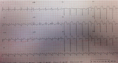
Figure 1: The ECG during admission.
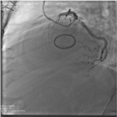
    Figure 2:  The angiographic images of the patient with total occlusion of the
LAD.
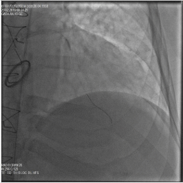
Figure 3: The angiographic images of the patient during balloon inflation.
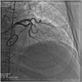
Figure 4: The angiographic images of the patient after first balloon inflation.
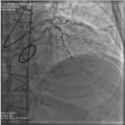
    Figure 5:  The angiographic images of the patient after second balloon
inflation with cranial projection.
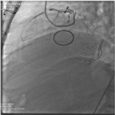
    Figure 6:  The angiographic images of the patient after second balloon
inflation and residue thrombus with cranial projection.
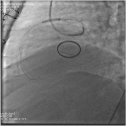
    Figure 7:  The angiographic images of the patient after second balloon
inflation with lateral projection.
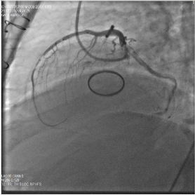
Figure 8: The angiographic images of the patient during stent implantation.
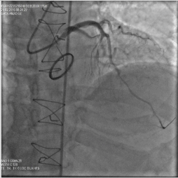
    Figure 9:  The angiographic images of the patient after stent implantation with
lateral projection.
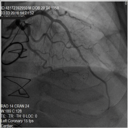
    Figure 10:  The angiographic images of the patient after stent implantation
and residue thrombus in the distal LAD with cranial projection.
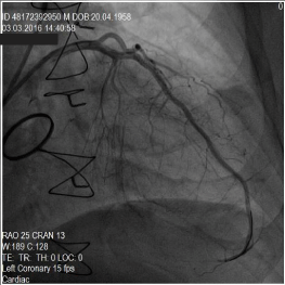
    Figure 11:  The angiographic images of the patient after stent implantation
and no residue thrombus in the distal LAD with cranial projection.
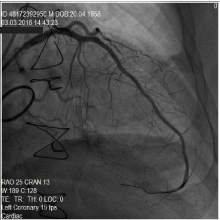
Figure 12: The final result of the distal LAD with cranial projection.
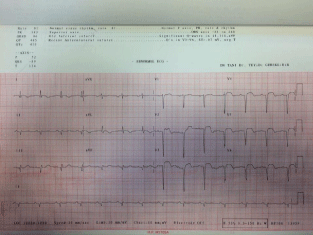
Figure 13: The ECG after stent implantation to the proximal LAD.
In conclusion, in case of a high thrombus burden causes total occlusion of the coronary vessels, aggressive antithrombotic treatment is feasible in case of low bleeding risk as in our case. So, in case of persistant thrombus formation despite aggresive antitrombotic and anticoagulant therapy, thrombus aspiration, angioplasty and stenting should be preferred rather than conservative approach. Prosthetic mechanical valves requires the use of long-term conventional anticoagulation therapy with warfarin and it is important for the doctors to be lack of awareness of medication of patient who carries significant thromboembolic risk.
References
- Patel M, Bhangoo M, Prasad A. Successful percutaneous treatment of suspected embolic left main thrombosis in a patient with a mechanical aortic valve. J Invasive Cardiol. 2011; 23: 263-266.
- Eguchi K, Ohtaki E, Misu K, Aikawa M, Sumiyoshi T, Hosoda S, et al. Acute myocardial infarction caused by embolism of thrombus in the right coronary sinus of Valsalva: a case report and review of the literature. J Am Soc Echocardiogr. 2004; 17: 173-177.
- Kamishirado H, Yamanaka T, Morooka S, Takayanagi K, Sasaki T, Koshikawa K, et al. A case of coronary artery embolism associated with combined valvular heart disease. Kokyu To Junkan. 1993; 41: 81-84.
- Härle T, Reimers J, Hertting K, Kuck KH. Successful trapping of an organized thrombus in a coronary artery aneurysm in myocardial infarction: case report and literature review. Cardiovasc Revasc Med. 2008; 9: 52-55.
- Kiernan TJ, Flynn AM, Kearney P. Coronary embolism causing myocardial infarction in a patient with mechanical aortic valve prosthesis. 2006; 112: 14-16.
- Nakazone MA, Tavares BG, Machado MN, Maia LN. Acute Myocardial Infarction due to Coronary Artery Embolism in a Patient with Mechanical Aortic Valve Prosthesis. Case Rep Med. 2010.
- Ozturk C, Demirkol S, Demir M, Yildirim AO, Balta S, Celik T, et al. Mobile mass lesion in the aorta after transcatheter aortic valve implantation: Thrombus or residue calcification. Int J Cardiol. 2015; 198: 45-46.
- Ali Buturak, Egemen Duygu, Ekrem Aksu, Orhan Alper Güngördük, Sami Özgül. Embolic acute myocardial infarction treated by intracoronary catheter aspiration embolectomy in a patient with mechanical aortic valve prosthesis. Anatol J Cardiol. 2011; 11: 461-462.
- Ozturk C, Yildirim AO, Demir M, Balta S. Coronary Obstruction During Transcatheter Aortic Valve Replacement: Related to Calcification or Thrombus? Rev Esp Cardiol (Engl Ed). 2016; 69: 456-457.
- Ozturk C, Celik T, Ozturk A, Balta S, Iyisoy A. Duration of antiplatelet or anticoagulant therapy after transcatheter aortic valve implantation in high risk patients; longer or shorter. Int J Cardiol. 2016; 210: 14-15.
- Ozturk C, Celik T, Demirkol S, Demir M, Balta S, Unlu M, et al. The healing of spontaneous coronary artery dissection with conservative treatment:When to stop. Int J Cardiol. 2015; 189: 249-251.
- Öztürk C, Çelik T, Balta S, Iyisoy A. Do spontaneous coronary artery dissections always need intervention in patients with no atherosclerosis? Anatol J Cardiol. 2016; 16: 365-366.
- Ozturk C, Yildirim AO, Demir M, Haqmal H, Balta S, Unlu M, et al. The spontaneous coronary artery dissection may need intervention in the proximal segment of the arteries. Int J Cardiol. 2016; 202: 943-944.
- Iyisoy A, Kursaklioglu H, Kose S, Yesilova Z, Ozturk C, Saglam K, et al. Acute myocardial infarction and left subclavian artery occlusion in Behçet's disease: a case report. Mt Sinai J Med. 2004; 71: 330-334.
- Iyisoy A, Kursaklioglu H, Kose S, Ozturk C, Amasyali B, Demirtas E. Spontaneous intimal dissection in a patient with post-infarct angina: identification with intravascular ultrasound and treatment with coronary stenting. Jpn Heart J. 2003; 44: 557-564.
- Celik T, Balta S, Ozturk C, Iyisoy A. Survival of the young patients with acute ST segment elevation myocardial infarction treated with primary percutaneous coronary intervention: Does gender matters? Int J Cardiol. 2016; 210: 54-55.
- Celik T, Balta S, Ozturk C, Kaya MG, Aparci M, Yildirim OA, et al. Predictors of No-Reflow Phenomenon in Young Patients With Acute ST-Segment Elevation Myocardial Infarction Undergoing Primary Percutaneous Coronary Intervention. Angiology. 2015.
- Ozturk C, Celik T, Iyisoy A, Haqmal H, Demir M, Yildirim AO, et al. Acute anterior myocardial infarction after heavy exercise in a young sportsman: Importance of intravascular ultrasonography on differential diagnosis. Int J Cardiol. 2016; 215: 169-172.