Case Report
Austin J Clin Cardiolog. 2016; 3(2): 1048.
Rapid Diagnosis of Left Circumflex Coronary Artery Occlusion as a Complication in the Immediate Postoperative Period of Cardiac Surgery
Fiol M¹*, Carrillo A², Riera M³ and Rodríguez A2
¹Palma Institute of Health Research, Hospital Son Espases, Spain
²Coronary Care Unit, Hospital Son Espases, Spain
³Cardiac Surgery Post Operative Unit, Hospital Son Espases, Spain
*Corresponding author: Miguel Fiol, Palma Institute of Health Research, Hospital Son Espases, Ctra. Valldemossa 79, 07120 Palma, Balearic Islands, Spain
Received: June 20, 2016; Accepted: August 11, 2016; Published: August 12, 2016
Abstract
A case of acute myocardial infarction due to inadvertent circumflex artery ligation in the immediate postoperative valve, cardiac surgery, diagnosed using the algorithms of localization of culprit artery.
Keywords: Acute myocardial infarction; Postoperative cardiac surgery; Left atrial appendage; Postsurgical complication
Case Report
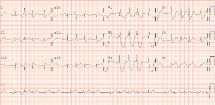
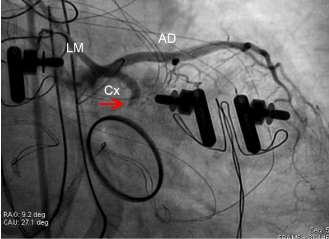
A 70-year-old woman with a previous history of hypertension, diabetes, stroke and chronic atrial fibrillation was admitted for increased effort dyspnea. Echocardiogram showed double mitral and aortic stenosis. Preoperative coronary angiography was normal. A mitral mechanical prosthesis replacement (Saint Jude 27 mm), aortic mechanical prosthesis replacement (Saint Jude Regent 17 mm) and Cox-Maze procedure with ligation of left atrial appendage was indicated. She arrived at the cardiac surgery post-operative unit with support vasoactive with dobutamine 5μg/kg/min and noradrenaline 0.25μg/Kg/min. The ECG showed a pattern compatible with occlusion of Left Circumflex Coronary Artery (LCx) (Figure 1). Transthoracic echocardiogram showed segmental inferobasalmobility disorder. Requested emergent coronary angiography showed a stop from a complete LCx occlusion (Figure 2) that we unsuccessfully tried to revascularize with PCI, which made us suspect that the occlusion was due to an LCx ligature. Thus, the patient was moved directly to the operating room where the left atrial appendage was reopened, normalizing ECG alterations.

    Figure 1:  The ECG shows atrial fibrillation, right bundle branch block and a
pattern of circumflex artery occlusion: ST, measured at 60 m sec from j point,
isoelectric in lead I, ST elevated 1 mm from baseline in leads II and III and
marked depression of ST in V1-V3 leads.

    Figure 2:  Coronary angiogram shows a total occlusion of the left circunflex
coronary artery (Cx) (red arrow); AD: Anterior Descending Coronary Artery;
LM: Left Main Coronary Artery Left.
Discussion
Hilltrop et al. [1], identified 44 cases of mitral valve repair/ replacement associated myocardial ischemia. They provide a comprehensive review of current knowledge regarding mitral valve surgery-related LCx. Electrocardiography showed regional STsegment elevations in 68%, especially in inferior and lateral leads. Registration of a full 12-lead ECG at the moment of intensive care unit arrival after cardiac surgery should be routine clinical practice. However, ST-segment changes are often observed in the operating room during mitral valve surgery, are often transient and attributed to intracardiac air causing intracoronary air embolization. This often affects the right coronary artery due to the high position in the surgical field. Coronary air embolism is a recognized complication after cardiac operations performed with cardiopulmonary bypass, especially open heart operations [2]. It has been shown that after coronary artery bypass grafts, air emboli occur in the period between cross-clamp removal and termination of cardiopulmonary bypass [3- 5].
We think that some of the ECG signs are pathognomonic for LCx injury. From our experience [6,7] the following electrocardiographic criteria are useful in the diagnosis of right coronary artery versus LCx occlusion when included in a 3-step algorithm: (a) ST changes in lead I, (b) the ratio of ST elevation in lead III to that in lead II, and (c) the ratio of the sum of ST depression in precordial leads to the sum of ST elevation in inferior. Therefore, neither the first step, ST isoelectric in lead I, nor the second step, ST-segment elevated 1 mm in II and III leads is conclusive, but the third step, ST-depression in V1-V3 higher than ST-segment elevation in inferior leads, is a diagnosis of LCx occlusion. Thus, the observer might think that they have accidentally ligaged the circumflex artery on closing the left atrial appendage [8], as this is the normal technique to prevent the formation of clots and peripheral embolism. Nevertheless, to our knowledge, acute myocardial infarction due to the ligation of left atrial appendage has never been described. On the other hand, injury to the LCx during mitral valve surgery is a recognized complication, because the LCx courses near the anterolateral commissure of the mitral valve [9-12].
When coronary artery occlusion occurs after mitral valve surgery, one of the main concerns is to determine whether to return the patient to the operating room or to perform emergent cardiac catheterization. If the underlying cause is the inadvertent suturing of the vessel, the result is unpredictable and the potential for revascularization by PCI is less [12-15].
Conclusion
This case illustrates the usefulness of knowing the culprit artery location algorithms, in patients with narrow QRS in the ECG, for a timely diagnosis and treatment. A correct and careful study of the relationship between the injury dipole with hemifield positive and negative different leads allows us to better understand which is the area of myocardium at risk as a consequence of both the occluded responsible artery and occlusion point. It can also help in the most likely differential diagnosis in case of the ST-segment elevation in the inferior territory in the postoperative cardiac surgery mechanism involved.
References
- Hiltrop N, Bennett J, Desmet W. Circumflex coronary artery injury after mitral valve surgery: a report of four cases and comprehensive review of the literature. CardiovascInterv. 2016.
- Justice C, Leach J, Edwards WS. The harmful effects and treatment of coronary air embolism during open-heart surgery. Ann ThoracSurg. 1972; 14: 47-53.
- Tingleff J, Joyce FS, Pettersson G. Intraoperative echocardiographic study of air embolism during cardiac operations. Ann ThoracSurg. 1995; 60: 673-677.
- Suastika LO, Oktaviono YH. Multiple air embolismduring coronary angiography: how do we deal with It? Clin Med Insights Cardiol. 2016; 10: 67-70.
- Chaudhuri N, Hickey MJ. A simple method of treating coronary air embolism after cardiopulmonary bypass. Ann ThoracSurg. 1999; 68: 1867-1868.
- Fiol M, Cygankiewicz I, Carrillo A, Bayés-Genis A, Santoyo O, Gómez A, et al. Value of electrocardiographic algorithm based on "ups and downs" of ST in assessment of a culprit artery in evolving inferior wall acute myocardial infarction. Am J Cardiol. 2004; 94: 709-714.
- Fiol M, Carrillo A. Culprit artery in evolving inferior wall acute myocardial infarction: RCA vs LCx. Europace. 2010; 12: 758.
- Su P, McCarthy KP, Ho SY. Occluding the left atrial appendage: anatomical considerations. Heart. 2008; 94: 1166-1170.
- Somekh NN, Haider A, Makaryus AN, Katz S, Bello S, Hartman A. Left circumflex coronary artery occlusion after mitral valve annuloplasty: "a stitch in time". Tex Heart Inst J. 2012; 39: 104-107.
- Tavilla G, Pacini D. Damage to the circumflex coronary artery during mitral valve repair with sliding leaflet technique. Ann ThoracSurg. 1998; 66: 2091-2093.
- Virmani R, Chun PK, Parker J, McAllister HA Jr. Suture obliteration of the circumflex coronary artery in three patients undergoing mitral valve operation. Role of left dominant coronary artery. J ThoracCardiovascSurg 1982; 84: 773-778.
- Vaquerizo B, Serra A, García-Picart J. Perioperative ST-segment elevation myocardial infarction during mitral valve annuloplasty: role of earlyangiography. J Clinic Experiment Cardiol. 2011, 2: 5.
- Mantilla R, Legarra JJ, Pradas G, Bravo M, Sanmartin M, Goicolea J. Percutaneous coronary intervention for iatrogenic occlusion of the circumflex artery after mitral annuloplasty. Rev EspCardiol. 2004; 57: 702-704.
- Raza JA, Rodriguez E, Miller MJ. Successful percutaneous revascularization of circumflex artery injury after minimally invasive mitral valve repair and left atrial cryo-MAZE. J Invasive Cardiol. 2006; 18: 285-287.
- Grande AM, Fiore A, Massetti M, Vigano M. Iatrogenic circumflex coronary lesion in mitral valve surgery: Case report and review of the literature. Tex Heart Inst J. 2008; 35: 179-183.