
Case Report
Austin J Clin Cardiolog. 2016; 3(2): 1050.
A Tumour Thrombus due to Hepatocellular Carcinoma Presenting as Right Atrial Mass
Hasan A*, Anwar MR and Amir SH
Centre of Cardiology, Jawaharlal Nehru Medical College, India
*Corresponding author: Asif Hasan, Professor of Cardiology, Centre of Cardiology, Jawaharlal Nehru Medical College, Aligarh Muslim University, Aligarh, India
Received: August 11, 2016; Accepted: October 13, 2016; Published: October 14, 2016
Abstract
Inferior Vena Cava Tumour Thrombus (IVC-TT) is a common complication of abdominal malignancies such as renal cell carcinoma, hepatocellular carcinoma and adrenocortical carcinoma. Extension of IVC thrombus into Right Atrium (RA) presenting as RA mass is a rare manifestation of hepatocellular carcinoma. Here we present a case of hepatocellular carcinoma, which was diagnosed to have right atrial mass due to IVC tumour thrombus extending to RA detected in echocardiography.
Keywords: tumor thrombus; Hepatocellular carcinoma; Inferior Vena Cava; Right atrium
Introduction
Extension of IVC thrombus into Right Atrium (RA) can present as RA mass [1]. These are often detected during tumour work up or during evaluation of cardiorespiratory symptoms. Here we present a case of hepatocellular carcinoma which was diagnosed to have IVC thrombus extending to RA.
Case Report
A 62 Year old male presented to OPD with the complaints of breathlessness on exertion (NYHA class 2-3), loss of appetite for the last 2 months, swelling over feet from the last 2 weeks. Patient was a chronic smoker for the last 30 years. There was no history of orthopnoea and paroxysmal nocturnal dyspnoea. On physical examination patient had fair general condition, normal built and nutrition. His vital parameters were within normal limits. There was 1+ bilateral pitting type pedal oedema present. A hard, nodular mass in right hypochondrium was found in per abdominal examination which was moving with respiration. His blood investigations were positive for hepatitis B surface antigen. Rest other parameters were within normal limits. CECT abdomen revealed a well-defined mass in segment 8 of liver with invasion of portal vein which was dilated and enhancing tumour thrombus within its lumen. Right and middle hepatic veins were also distended with enhancing heterogenous content which were extending into intrahepatic segment of IVC and further superiorly into suprahepatic IVC and floor of RA. In chest CT there was enhancing tumour thrombus in RA along with hypodense non-enhancing bland thrombus and pulmonary thrombo-embolism involving both lower lobe pulmonary arteries. FNAC of the lesion showed malignant cytology suggestive of Hepatocellular Carcinoma (HCC). In 2D Echo examination a 30.4x20.3 mm irregular RA mass was present which had echogenic shadows within. The mass did not prolapse in right ventricle. Distal end of IVC also showed a mass leading to almost total obstruction of IVC which was dilated with decreased inspiratory collapse. Rest other echo parameters were unremarkable. Gastroenterology and surgery opinions were sought and transarterial chemo-embolization with or without radiofrequency ablation was planned. Besides this, he was managed conservatively with anticoagulation using enoxaparin and acenocoumarol and followed up with echocardiographic evaluation of IVC and RA thrombus and INR monitoring. Despite two weeks of antithrombotic therapy the IVC and RA mass did not show significant change in size (Figures 1-3).
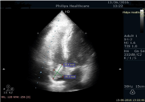
    Figure 1:  Heterogenous Right atrial mass as seen in apical four chamber
view.
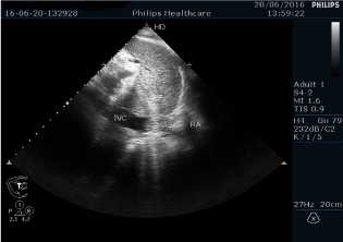
    Figure 2:  Tumour thrombus with IVC dilatation seen at the junction of IVC
and RA in subcostal view.
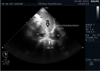
    Figure 3:  Tumour thrombus visible in hepatic vein showing continuity with
IVC-TT in subcostal view.
Discussion
IVC tumour thrombus (IVC-TT) extending to RA is an uncommon complication of hepatocellular carcinoma (HCC). IVC involvement occurs in about 1-4% cases of HCC [2]. However renal cell carcinoma is by far the most common malignancy to extend into IVC in up to 10% of cases and to RA in 1% of patients [3,4]. Other tumours that have a tendency for IVC invasion are adrenocortical carcinoma, Wilm’s tumour, primary leiomyoma or leiomyosarcoma of the IVC. There can be two types of IVC thrombus, bland thrombus and tumour thrombus. Bland thrombus is an isolated thrombus, which commonly arise from DVT of lower extremities. However it can also form due to hypercoagulable states, venous stasis, compression by neoplastic lesions, lymph nodes, retroperitoneal masses/ fibrosis or haemorrhage, foreign bodies such as vena cava filter or catheters, extension from benign tumours like angiomyolipoma, IVC leiomyoma, adrenalpheochromocytoma [5,6]. Tumour thrombus contains both tumour and thrombotic components and may not be adherent and usually extends in the direction of blood flow towards right atrium. The presence of tumour thrombus affects staging of neoplasms and also the prognosis. Only biopsy can differentiate these two types of thrombus with certainty. However imaging modalities such as CT, MRI and abdominal ultrasonography are useful in rapid detection and follow-up of such tumour thrombus. Echocardiogram can also help in differentiating the two types of thrombus. Features suggestive of tumour thrombus in 2D Echo are expansion of IVC by the tumour thrombus and tumour thrombus will often show continuity with primary tumour which can be seen by IVC echocardiography especially in case of HCC. In contrast a bland thrombus either results from external compression of the IVC by a neoplastic lesion (i.e. no direct invasion), so the IVC is usually narrowed at the site of thrombosis. Enhancement on Contrast CT is also suggestive of tumour thrombus while bland thrombus appears as hypodense nonenhancing mass [5-7]. In our case 2D Echo and CECT features were suggestive of tumour thrombus in IVC and RA. Presence of IVC-TT and RA thrombus is a poor prognostic sign and is also a risk factor for pulmonary embolism. Tumour thrombus which are completely or near-completely occluding IVC, embolic event, or bland thrombus in addition to tumor thrombus are indication for preoperative therapeutic anticoagulation with Low Molecular Weight Heparin (LMWH) which can be started in the outpatient setting in such cases. For patients with a contraindication to LMWH or who are unable to obtain long-term LMWH, warfarin may be started with appropriate bridging therapy with target INR of 2-3. As our patient also had pulmonary thromboembolism therefore was started on LMWH and acenocooumarol along with other treatment [8].
Conclusion
IVC and RA involvement by the abdominal malignancies is a common phenomenon. Timely and accurate identification of these lesion is now possible with the use of different imaging modalities which is very important in staging, prognostication, therapeutic decision making and follow up.
References
- Panidis IP, Kotler MN, Mintz GS, Ross J. Clinical and echocardiographic features of right atrial masses. Am Heart J. 1984; 107: 745-758.
- Vallakati A, Chandra PA, Frankel R, Shani J. Intra-atrial tumor thrombi secondary to hepatocellular carcinoma responding to chemotherapy. North American Journal of Medical Sciences. 2011; 3: 435-437.
- Keamey GP, Waters WB, Klein LA, et al. Results of IVC resection for renal cell carcinoma. J Urol. 1981; 125: 769-773.
- Pritchett TR, Lieskovsky G, Skinner DG. Extension of renal cell carcinoma into the vena cava: clinical review and surgical approach. J Urol. 1986; 135: 460-464.
- Sheth S, Fishman EK. Imaging of the inferior vena cava with MDCT. AJR Am J Roentgenol. 2007; 189: 1243-1251.
- Kaufman LB, Yeh BM, Breiman RS, Joe BN, Qayyum A, Coakley FV. Inferior vena cava filling defects on CT and MRI. AJR Am J Roentgenol. 2005; 185: 717-726.
- Smillie RP, Shetty M, Boyer AC, Madrazo M, Jafri RVT. Imaging evaluation of the inferior vena cava. Radiographics. 2015; 35: 578-592.
- Woodruff D, Van Veldhuizen P, Muehlebach G, Johnson P, Williamson, Holzbeierlein J. The perioperative management of an inferior vena caval tumor thrombus in patients with renal cell carcinoma. Urol Oncol. 2013; 31: 517-521.