Case Report
Austin J Clin Cardiolog. 2016; 3(2): 1052.
Cerebrogenic Sudden Cardiac Death
Ali Raza Ghani*, Saddaf Mirrani, Hafeez Virk, Khawar Maqsood and Ghazi Mirrani
Thomas Jefferson University Hospital, USA
*Corresponding author: Ali Raza Ghani, Thomas Jefferson University Hospital, Philadelphia, USA
Received: August 10, 2016; Accepted: November 04, 2016; Published: November 08, 2016
Abstract
An ST segment elevation is a life threatening condition requiring immediate intervention in many cases. The presentation can be very misleading, specially the presence of neurological symptoms warrants to an alternate diagnosis. We are presenting an interesting case with ST elevation on EKG, in a patient with acute insular infarction.
Introduction
Various EKG patterns have been well documented with cerebral infarction and particularly insular infarct has been associated with ST elevation on the EKG. The Mechanism of this finding is not clearly understood but insular infarct seems to activate sympathetic - adrenal system because of decreased inhibitory insular activity.
Case Report
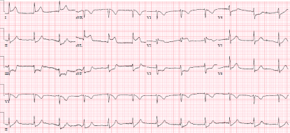
A 43-year-old female with endometrial cancer admitted for hysterectomy had a syncopal episode on the second postoperative day with a small amount of vaginal bleeding. Patient was hypotensive, diaphoretic and delirious. She was noticed to have dense left hemi paresis. Her ECG (Figure 1) showed ST elevation in leads I, a VL and ST depression in lead II, III, a VF and V3-V6. The obvious diagnosis was acute inferior ST elevation myocardial infarction but patient also developed dense left hemi paresis and was very confused. Her CT scan of brain revealed acute right middle cerebral artery stroke. Pt was not a t-PA candidate due to risk of hemorrhagic conversion of infarct. Patient had Pulseless Electrical Activity (PEA) cardiac arrest in radiology department and she re-gained her pulse and blood pressure after 2 cycles and was taken to catheterization lab. Her coronary angiogram did not show any obstructive disease. She again had PEA arrest in catheterization lab and died despite resuscitative measures. Autopsy showed patent foramen ovale raising concern for paradoxical embolism.

Figure 1: ECG showing elevation and depression.
ST Elevation on Electrocardiogram (EKG) is a serious and life threatening finding that warrants an immediate action to prevent life threatening complications and death. There are several causes of ST Elevation on EKG apart from Myocardial Infarction (MI) and studies have shown an incidence of 2.3% where an alternate diagnosis was established in suspected ST elevation MI patient [1]. Although majority of patients with ST elevation suffer from true ST Elevation MI (STEM I) but up to 2.6 % patients had normal coronary arteries and the ST elevation in these patients could be coronary vasospasm, thrombus or any non cardiac causes of this alarming EKG finding [2].
Alternate diagnosis for ST elevation on EKG include but not limited to Non obstructive Coronary artery disease, Pericarditis, myocarditis [3-6], Pulmonary elbolism, aortic dissection [7,8], acute cholecystitis or pancreatitis [9,10], subarachnoid hemorrhage [11], and cerebrovascular infarcts [12]. Cerebrovascular Accident (CVA) can result in various cardiovascular complications including cerebrogenic pulmonary edema, electrocardiographic changes, cardiac arrhythmias, elevation of cardiac enzymes and altered blood pressure regulation [13].
EKG changes have been reported in Subarachnoid Hemorrhage (SAH) (40-70%), Ischemic stroke (15-40%) and intra cerebral hemorrhage (60-70%) and these patterns include, Prolonged QT, ST depression, Inverted and Flat T waves, ST elevation, Notched T waves, Q waves [14,15].
Patients with abnormal EKG has higher mortaliry (95%) as compared to 86% in those with normal EKG though this finding was not statistically significant. Those patients who died had 2-5 fold higher echocardiographic incidence of recent MI, Atrial Fibrillation and conduction defects as compared to those who survived [16].
Brain controls cardiovascular system in different ways including increased sympathetic tone, enhanced catecholamines secretions and decreased parasympathetic tone [14,15].
It has been well established that various centers including hypothalamus, brainstem cardiovascular centers, and spinal autonomic centers control cardiovascular response. Recent studies suggest that these complex controls are actually managed by higher cortex and subcortex including insula and amygdale [17]. Parasympathetic nervous system controls Sinoatrial (SA) node and Atrioventricular (AV) node and ventricular muscles are mainly controlled by Sympathetic nervous system [15].
Stimulation of Insular cortex in rats, cats, dogs, monkeys and human, seems to change arterial pressure, heart rate, respiration, and adrenaline stimulation [18]. Insular role in cerebrogenic cardiovascular and autonomic disturbance was initially observed in a cat and was later on confirmed in a rat [19].
Right insular stroke was associated with significant lower sympathetic and parasympathetic activities than other patients with stroke. There were 5 sudden deaths in right insular infarction as compared to 2 sudden deaths in left insular infarction [20].
Discussion
EKG changes in the absence of coronary artery disease have been reported with head injury, intracranial hemorrhage, brain tumors, meningitis, hydrocephalus and ischemic stroke. Increased sympathetic tone and augmentation of intracardiac sympathetic nerve activity have been proposed as etiology. Cerebrogenic sudden death has been described in literature and involves infarction of insular cortex, which controls the brain-heart interactions. Insula is supplied by the middle cerebral artery and probably is the reason for circulatory collapse seen in our patient.
Conclusion
EKG changes like this are very rare but treatment can be totally different in setting of neurological compromise and urgent brain imaging can be life saving.
References
- YL Gu, T Svilaas, I.CC van der Horst, F Zijlstra. Conditions mimicking acute ST-segment elevation myocardial infarction in patients referred for primary percutaneous coronary intervention. 2008; 16: 325-331.
- Widimsky P, Stellova B, Groch L. Prevalence of normal coronary angiography in the acute phase of suspected ST-elevation myocardial infarction: experience from the PRAGUE studies. Can J Cardiol. 2006; 22: 1147-1152.
- Bertrand ME, Simoons ML, Fox KA. Management of acute coronary syndromes in patients presenting without persistent STsegment elevation. Eur Heart J. 2002; 23:1809-1840.
- Wang K, Asinger RW, Marriott HJ. ST-segment elevation in conditions other than acute myocardial infarction. N Engl J Med. 2003; 349: 2128-2135.
- Angelini A, Calzolari V, Calabrese F, Boffa G, Maddalena F, Chioin R, et al. Myocarditis mimicking acute myocardial infarction: role of endomyocardial biopsy in the differential diagnosis. Heart. 2000; 84: 245-250.
- Surawicz B, Lasseter KC. Electrocardiogram in pericarditis. Am J Cardiol.1970; 26: 471-474.
- Livaditis IG, Paraschos M, Dimopoulos K. Massive pulmonary embolism with ST elevation in leads V1-V3 and successful thrombolysis with tenecteplase. Heart. 2004; 90: 41.
- Spittell PC, Spittell JA Jr, Joyce JW, Tajik AJ, Edwards WD, Schaff HV, et al. Clinical features and differential diagnosis of aortic dissection: experience with 236 cases (1980 through 1990). Mayo Clin Proc 1993; 68: 642-651.
- Ryan ET, Pak PH, DeSanctis RW. Myocardial infarction mimicked by acute cholecystitis. Ann Intern Med. 1992; 116: 218-220.
- Fulton MC, Marriott HJ. Acute pancreatitis simulating myocardial infarction in the electrocardiogram. Ann Intern Med. 1963; 59: 730-732.
- Beard EF, Robertson JW, Robertson RCL. Spontaneous subarachnoid hemorrhage simulating acute myocardial infarction. Am Heart J. 1959; 58: 755-759.
- Christensen H, Boysen G, Christensen AF, Johannesen HH. Insular lesions, ECG abnormalities and outcome in acute stroke. J NeurolNeurosurg Psychiatry. 2005; 76: 269-271.
- Cheung RT, Hachinski V. The insula and cerebrogenic sudden death. Arch Neurol. 2000; 57: 1685-1688.
- Oppenheimer SM, Cechetto DF, Hachinski VC. Cerebrogenic cardiac arrhythmias: cerebral electrocardiographic influences and their role in sudden death. Arch Neurol. 1990; 47: 513-519.
- Cheung RTF, Hachinski V. Cardiology. In: Samuels MA, Editor Hospitalist Neurology. Woburn, Mass: Butterworth-Heinemann. 1999: 305-330.
- Cerebrovascular accident Electrocardiographic changes and myocardial damage in patients with acute, J Dimant and D Grob. Stroke. 1977; 8: 448- 455.
- Raymond TF, Cheung, Vladimir Hachinski. The Insula and Cerebrogenic Sudden Death. Arch Neurol. 2000; 57: 1685-1688.
- Oppenheimer SM, Cechetto DF. Cardiac chronotropic organization of the rat insular cortex. Brain Res. 1990; 533: 66-72.
- Cechetto DF, Wilson JX, Smith KE, Wolski D, Silver MD, Hachinski VC. Autonomic and myocardial changes in middle cerebral artery occlusion: stroke models in the rat. Brain Res. 1989; 502: 296-305.
- Tokgözoglu SL, Batur MK, Topçuoglu MA, Saribas O, Kes S, Oto A. Effects of stroke localization on cardiac autonomic balance and sudden death. Stroke. 1999; 30: 1307-1311.