
Research Article
Austin J Clin Cardiolog. 2019; 5(1): 1061.
Pulse Pressure as a Risk Factor for Cardiovascular Events: Myth or Reality?
Braite MS1*, Santos MA1, Wilson PG1, França
JCQ1, Carvalho IG1 and Godoy MF
¹Department of Hemodynamics and Interventional Cardiology Service, Regional Medical School, Brazil
²Department of Cardiology and Cardiovascular Surgery, Sao Jose do Rio Preto Medical School, Brazil
*Corresponding author: Braite MS, Department of Hemodynamics and Interventional Cardiology Service, Regional Medical School - Funfarme, Rua Gastão Vidigal, Cruzeiro do Oeste, Brazil
Received: March 12, 2019; Accepted: May 22, 2019; Published: May 29, 2019
Abstract
Increased SBP and/or decreased DBP leads to an increase in systolic load, concomitant with a decrease in coronary perfusion pressure. Previous research has suggested a relationship between increases in PP and morbidity / mortality due to cardiovascular events.
Objective: This study aimed to demonstrate whether pulse pressure is in fact a predisposing factor for coronary heart disease and / or an aggravating risk in patients with CAD.
Methods: A total of 5,027 exams were studied. Of these, 3,052 (60.7%) were males. Age ranged from 20 to 92 years (59.0 {plus minus} 11.0 years). PP was determined invasively in the ascending aorta. Coronary artery disease was diagnosed if at least one of the major branches had obstructive lesion with a reduction of 50% or more in the diameter of its lumen. Statistical analyzes were performed with unpaired t-test or Mann-Whitney test for intergroup comparisons, as indicated. Categorical variables were compared using the chi-square test. Long-term survival was assessed using Kaplan-Meier curves. Values of P less than or equal to 0.05 were considered statistically significant.
Results: Pulse pressure varied from 20.0 to 160.0 mmHg, with a mean and standard deviation of 68.4 {plus minus} 22.3 (median of 66.0mmHg, 75th percentile of 82mmHg).
Conclusion: pulse pressure was not shown to be a risk factor for coronary heart disease in the comparison between patients with and without obstructive coronary disease. In addition, it was not independently relevant in the group with CAD patients with more advanced degrees of coronary disease and the pulse pressure showed no association with the patients age, being the only relevant independent variable. In other words, the influence of wrist pressure has confirmed to be a myth!
Keywords: Pulse pressure; Cardiovascular events; Atherosclerotic disease
Introduction
Pulse Pressure (PP) is the difference between Systolic Blood Pressure (SBP) and Diastolic Blood Pressure (DBP) and it may be elevated due to increased SBP and decreased DBP [1].
Increased SBP and decreased DBP lead to elevated systolic load, concomitantly with decreased coronary perfusion pressure [2]. Importantly, increases in SBP cause a disproportionate increase in the end-systolic stress, which is the major hemodynamic factor that promotes cardiac hypertrophy, increased ventricular oxygen consumption and Left Ventricular (LV) hypertrophy, strongly compromising coronary perfusion.
Prior researches have suggested a relationship between increases in PP and morbidity/mortality due to cardiovascular events, which would even have a physiopathological explanation for Coronary Artery Diseases (CAD), based on the arguments described above [2- 6].
However, although cross-sectional analyses usually support the hypothesis that widened pulse pressure is an independent risk factor for cardiovascular diseases in general and more specifically CAD, prospective analyzes do not confirm this hypothesis [7].
Thus, based on a considerably large population and longer follow-up periods, this study aimed to demonstrate if pulse pressure is in fact a predisposing factor for coronary artery diseases and/or an aggravating risk in CAD patients, or if it fails to have an impact on the disease. Therefore, this is an existing conflict between Myth and Reality.
Methods
The examinations of 12,997 consecutive patients who underwent cardiac catheterization in the Hemodynamic Service of a University Hospital were assessed. Patients with valvopathy, congenital heart diseases, heart transplantation, hemodynamic instability or those who had already undergone myocardial revascularization procedures were excluded. Hence, a total of 5,027 examinations were used in the present study.
Of this total, 3,052 (60.7%) were males. Age ranged from 20 to 92 years (59.0 ± 11.0 years). PP was determined in an invasive manner by the absolute difference between the systolic and diastolic pressures in the ascending aorta at the beginning of the hemodynamic study. Coronary artery disease was diagnosed if at least one of the main branches (right coronary artery, interventricular anterior artery or diagonal artery, circumflex artery or left marginal artery) or Left Main Coronary Artery (LMCA) showed obstructive injury with a reduction of 50% or more in diameter of its light. The obstructive injury was considered advanced when at least 3 of the main branches, associated or not with the concomitant obstructive injury in the left main coronary artery, were involved.
Cut-off values for pulse pressure as a risk factor were established based on the median and 75% percentile of the selected sample. The digital records of each patient were analyzed prospectively, and death event was selected for analysis. To evaluate long-term mortality, 3 cut-off ranges were empirically established: pulse pressure greater than 40.0mmHg, pulse pressure greater than 60.0mmHg, and pulse pressure greater than 80.0mmHg. The statistical analyses were performed with unpaired t-test or Mann-Whitney test for the intergroup comparisons, depending on the Gaussian distribution or not of the variables. Categorical variables were compared using the Chi-Square test. Long-term survival was assessed using Kaplan-Meier curves. P values less than or equal to 0.05 were considered statistically significant.
Results
Pulse pressure ranged from 20.0 to 160.0 mmHg, with mean and standard deviation of 68.4 ± 22.3 (median of 66.0mmHg, 75th percentile of 82mmHg). Considering the median as a cut-off point, there were 2,453 (48.8%) patients with pulse pressure above that value. Of these, 1,481 had at least 1 major vessel with =50% obstruction. On the other hand, of the 2,574 patients with pulse pressure up to 66mmHg, 1,549 had obstructive CAD. The Chi-square analysis revealed P=0.9047 (OR = 1.00, 95% CI = 0.900 to 1.128).
The advanced degree of obstructive impairment was used as an analysis criterion. In the group with pulse pressure above median level, 411 patients showed tri-arterial impairment and 27 of them had compromised left main coronary artery. In the group with pulse pressure up to the median value, 368 patients presented with tri-arterial impairment and 20 of them had impaired left main coronary artery. The Chi-square statistical test compared tri-arterial impairment versus no obstruction and revealed P=0.053 (therefore not significant), with OR of 1.18 (95% CI 0.998 to 1.390).
Considering 75th percentile value (82mm Hg) as cut-off point, there were 1,217 (24.2%) patients with pulse pressure above that value. Of these, 750 had at least 1 major vessel with =50% obstruction. On the other hand, of the 3,810 patients with pulse pressure up to 82mmHg, 2280 had obstructive CAD. The Chi-square analysis revealed P=0.2829 (OR 1.08, 95% CI 0.944 to 1.230).
Using the advanced degree of obstructive impairment, the group with pulse pressure above the 75th percentile showed 208 patients with tri-arterial impairment, and 8 of them also had compromised left main coronary artery. In the group with pulse pressure below the 75th percentile, there were 571 patients with tri-arterial impairment, and 39 of them also had compromised left main coronary artery. Statistical analysis using the Chi-square test compared tri-arterial impairment and no obstruction and yielded P=0.075, OR of 1.19 and 95% CI 0.987 to 1.442). Thus, the analysis of significant numbers of cases (5,027) gives evidence that the elevated pulse pressure was not statistically more frequent in patients with obstructive coronary artery diseases than in non-coronary artery diseases, even when comparing a subgroup with advanced and normal coronary impairment.
Since pulse pressure was not confirmed as a risk marker for the incidence of coronary artery diseases, there was an attempt to verify whether pulse pressure exerted some effect related to the prognosis of the disease within a subgroup of CAD patients. According to the theory, the higher oxygen consumption due to a greater systolic work and due to the lower supply of oxygen in diastole would be logical pathophysiological mechanisms of greater myocardial damage. 922 patients with at least one compromised major branch (loss of vessel diameter greater than 50%) were prospectively evaluated. These patients were followed up during up to 225 months (mean, 142.7 ± 35.2 months, median 140 months). Comparisons were made based on different cut-off values.
The group with pulse pressure greater than 40mmHg included 821 patients, with 112 deaths. In the group of 101 patients with pulse pressure of 40mmHg or less, 11 deaths were observed (Hazard Ratio 1.134239, 95% CI 0.575462 to 2.233591, P=0.73). Survival at the end of follow-up in the group with pulse pressure greater than 40mmHg was 69.5 ± 5.6%; whereas survival was 81.3 ± 7.3% in the group with pulse pressure of 40mmHg or less.
The group with pulse pressure greater than 60mmHg comprised 488 patients, with 61 deaths. The group of 434 patients with pulse pressure of 60mmHg or less showed 62 deaths (Hazard Ratio 0.989931; 95% IC0.696182 to1.407626; P=0.955). Survival at the end of follow-up in the group with pulse pressure greater than 60mmHg was 75.4 ± 5.2%; and yet 68.6 ± 7.3% in the group with pulse pressure of 60mmHg or less.
The group with pulse pressure greater than 80 mmHg consisted of 220 patients with 27 deaths. In the group of 702 patients with pulse pressure of 80mmHg or less, 96 deaths could be observed (Hazard Ratio 1.109008; 95% CI 0.718917 to1.710764; P=0.629). Survival at the end of follow-up in the group with pulse pressure greater than 80mmHg was 80.9 ± 3.5%, and 69.6 ± 6.0 in the group with pulse pressure of 80mmHg or less. (Figures 1-3) illustrate death-free survival with Kaplan-Meier curves.
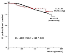
    Figure 1: Kaplan-Meier curves for comparison of death-free survival in
groups with pulse pressure above 40mmHg versus 40mmHg or less.
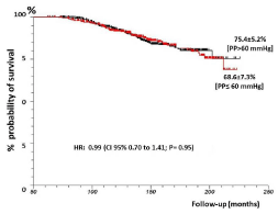
    Figure 2: Kaplan-Meier curves for comparison of death-free survival in
groups with pulse pressure above 60mmHg versus 60mmHg or less.
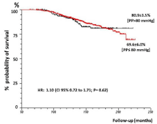
    Figure 3: Kaplan-Meier curves for comparison of death-free survival in
groups with pulse pressure above 80mmHg versus 80mmHg or less.
Notably, statistically significant differences between the groups could not be detected in any of the combinations tested, in relation to the death events (Figure 4).
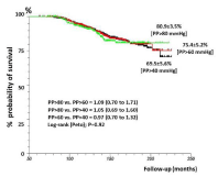
    Figure 4: Demonstrates the comparative evolution of late mortality, and the
Log-Rank statistical evaluation, in relation to the three cut-off levels for pulse
pressure, using Kaplan-Meier Curves.
Finally, a logistic regression analysis was performed including sex, age, BMI, systolic arterial pressure, diastolic arterial pressure, coronary obstruction (RCA, IVA, CX and LMCA) and pulse to the potential independent variables.
The first step of the logistic regression included all mentioned variables and cut-off level of P ‹0.20 was set for the evaluation of the relevance of the variable as risk independent factor. In this regression, only age, BMI left main coronary artery injury and interventricular anterior artery injury remained independently significant. Pulse pressure was not significant (P=0.8451) (Table 1).
![]()
    Intercept
    b0 = -3.956348
    z = -3.583425
    P = 0.0003


    SEX F-0 M-1
    b1 = 0.015906
    z = 0.072467
    P = 0.9422


    AGE
    b2 = 0.042862
    z = 4.014626
    P < 0.0001


    BMI
    b3 = 0.034181
    z = 1.577063
    P = 0.1148


    SYST AP
    b4 = -0.087796
    z = -0.196154
    P = 0.8445


    DIAST AP
    b5 = 0.069564
    z = 0.155378
    P = 0.8765


    PULSE P
    b6 = 0.087446
    z = 0.195406
    P = 0.8451


    RCA
    b7 = 0.145648
    z = 0.717201
    P = 0.4732


    LMCA
    b8 = -1.174959
    z = -1.585078
    P = 0.1129


    IVA
    b9 = -0.301691
    z = -1.334102
    P = 0.1822


    Parameter
    Estimate
    OR
    95% CI


    Constant
    -3.956348


    SEX F-0 M-1
    0.015906
    1.016033
    0.660813 to 1.562201


    AGE
    0.042862
    1.043794
    1.022179 to 1.065866


    BMI
    0.034181
    1.034772
    0.991735 to 1.079676


    SYST AP
    -0.087796
    0.915948
    0.380963 to 2.202208


    DIAST AP
    0.069564
    1.072041
    0.445782 to 2.578102


    PULSE P
    0.087446
    1.091383
    0.454002 to 2.623598


    RCA
    0.145648
    1.156789
    0.776950 to 1.722325


    LMCA
    -1.174959
    0.308832
    0.072237 to 1.320339


    IVA
    -0.301691
    0.739567
    0.474776 to 1.152036
Table 1: Kaplan-Meier curves for comparison of death-free survival in
groups with pulse pressure above 40mmHg versus 40mmHg or less.
Assuming that pulse pressure could be relevant only in the presence of severe coronary impairment, logistic regression was repeated only with pulse pressure and LMCA impairment. Nevertheless, pulse pressure was not independently significant (P=0.4956) (Table 2).
![]()
    Intercept
    b0 = -2.031908
    z = -6.721222
    P < 0.0001


    PULSE P
    b1 = 0.002859
    z = 0.681445
    P = 0.4956


    LMCA
    b2 = -0.950075
    z = -1.296531
    P = 0.1948
Table 2: Kaplan-Meier curves for comparison of death-free survival in
groups with pulse pressure above 60mmHg versus 60mmHg or less.
As age appeared as an independent variable significantly related to the cases of death, it was assessed whether the relationship between pulse pressure and age could become relevant. Once more, age remained relevant; however, pulse pressure was not independently significant (P=0.7261) (Table 3).
![]()
    Intercept
    b0 = -4.187173
    z = -6.685964
    P < 0.0001


    AGE
    b1 = 0.040713
    z = 4.062871
    P < 0.0001


    PULSE P
    b2 = -0.001525
    z = -0.350289
    P = 0.7261


    Parameter
    Estimate
    OR
    95% CI


    Constant
    -4.187173


    AGE
    0.040713
    1.041553
    1.021296 to 1.062212


    PULSE P
    -0.001525
    0.998476
    0.989991 to 1.007033
Table 3: Pulse pressure was not independently significant (P=0.7261).
Discussion
In addition to damaging the vascular wall, increased pulse pressure is associated with increased left ventricular stress, which may result in ventricular hypertrophy and Heart Failure (HF). Increased pressure during systole increases the need for oxygen in the myocardium and reduced diastolic value may become a limiting factor for coronary perfusion, resulting in ischemia. The end result of these combined effects is that increased pulse pressure could be a predictor of a variety of adverse cardiovascular outcomes.
The discussion about the importance of blood pressure components (systolic pressure, diastolic pressure and pulse pressure) on cardiovascular risks still persists [8].
With increasing age, pulse pressure correlates more closely with systolic pressure than with diastolic pressure, and therefore, it has also been reported as a good predictor of cardiovascular disease among the elderly. In some cases, this measure has higher predictive power than that observed with isolated systolic pressure [9,10].
Elevated pulse pressure has been considered an independent predictor of cardiovascular events in the elderly [11].
These data provide strong evidence of a relationship between pulse pressure, which is directly related to vessels stiffness, and subsequent cardiovascular events following myocardial infarction in patients with left ventricular dysfunction [2].
In the Framingham Heart Study, for example, each 10mmHg increase in pulse pressure was associated with a 23% increased risk of developing coronary arterial diseases [1].
Vaccarino et al [11] followed-up on 2,152 elderly patients (>65 years), with no cardiovascular events at the beginning, during a ten-year period. There were 328 cases of coronary artery diseases, 224 cases of heart failure, and 1,046 individuals died due to other causes of death. The authors concluded that PP had a strong linear correlation with each specific event. This correlation was evident in both normotensive and in patients with isolated systolic hypertension. These findings could not be confirmed by our study, at least relating to the death events and occurrence of coronary artery diseases.
Other studies, such as the National Health and Nutrition Examination Survey (NHANES), reported that pulse pressure added little predictive value compared to systolic or diastolic pressure, since there were increased, reduced or unchanged risks, depending on the systolic and diastolic blood pressure values. They considered PP not relevant as a prognostic element or for therapeutic decision [8].
This study sought to evaluate all possible aspects to consider pulse pressure as a marker of cardiovascular death.
Thus, in 5,027 cases, we studied the association between the presence of coronary artery diseases and increased pulse pressure. There were no differences in the rate of coronary artery diseases using the cut-off level at the median (66mmHg) or at the 75th percentile (82mmHg). Furthermore, there were no cases of more advanced coronary artery diseases with higher pulse pressure. As no association was detected, we investigated whether, at least in the group with confirmed diagnosis of CAD, pulse pressure would be an event marker. Using three cut-off levels (40, 60 and 80 mmHg), there was no difference in mortality, not even considering the group with a more advanced disease, despite the suggestion of the pathophysiological mechanism (greater oxygen consumption and lower supply).
At last, the logistic regression analysis definitively excluded pulse pressure as an independent variable with only age remaining as a marker, which is consistent.
Conclusively, pulse pressure has not proved to be as risk factor for coronary artery diseases in the comparison between patients with and without obstructive coronary diseases. Furthermore, it was not independently relevant in the group with CAD patients with more advanced degrees of coronary artery diseases and the pulse pressure showed no association with the patients’ age, being the only relevant independent variable.
In other words, considering the association of PP as a marker for risk or prognosis of coronary artery diseases, in a considerably large group of patients with long-term follow-up, the influence of pulse pressure has confirmed to be a Myth!
References
- Franklin SS, Khan SA, Wong ND, Larson MG, Levy D. Is pulse pressure useful in predicting risk for coronary heart Disease? The Framingham heart study. Circulation. 1999; 27: 354-360.
- Mitchell GF, Moyé LA, Braunwald E, Rouleau JL, Bernstein V, Geltman EM, et al. Sphygnomanometrically Determined Pulse Pressure Is a Powerful Independent Predictor of Recurrent Events After Myocardial Infarction in Patients With Impaired Left Ventricular Function. Circulation. 1997; 96: 4254- 4260.
- Chen G, Bliden KP, Chaudhary R, Liu F, Kaza H, Navarese EP, et al. Central aortic pulse pressure, thrombogenicity and cardiovascular risk. J Thromb Thrombolysis. 2017; 44: 223-233.
- Mascha EJ, Yang D, Weiss S, Sessler DI. Intraoperative Mean Arterial Pressure Variability and 30-day Mortality in Patients Having Noncardiac Surgery. Anesthesiology. 2015; 123: 79-91.
- Benetos A, Rudnichi A, Safar M, Guize L. Pulse Pressure and Cardiovascular Mortality in Normotensive and Hypertensive Subject. Hypertension. 1998; 32: 560-564.
- Pinto E. Blood pressure and ageing. Postgrad Med J. 2007; 83: 109-114.
- Dyer AR, Stamler J, Shekelle RB, Schoenberger JA, Stamler R, Shekelle S, et al. Pulse pressure-III. Prognostic significance in four Chicago epidemiologic studies. J Chronic Dis. 1982; 35: 283-294.
- Pastor-Barriuso R, Banegas JR, Damián J, Appel LJ, Guallar E. Systolic blood pressure, diastolic blood pressure, and pulse pressure: an evaluation of their joint effect on mortality. Ann Intern Med. 2003; 139: 731-739.
- Farasat SM, Morrell CH, Scuteri A, Ting CT, Yin FC, Spurgeon HA, et al. Pulse pressure is inversely related to aortic root diameter implications for the pathogenesis of systolic hypertension. Hypertension. 2008; 51: 196-202.
- O’Rourke M, Frohlich ED. Pulse pressure: Is this a clinically useful risk factor? Hypertension. 1999; 34: 372-374.
- Vaccarino V, Holford TR, Krumholz HM. Pulse Pressure and Risk for Myocardial Infarction and Heart Failure in the Elderly. J Am Coll Cardiol. 2000; 36: 130-138.