Clinical Image
Austin J Clin Cardiolog. 2019; 5(1): 1063.
A Myocardial Infarction with “Normal Arteries”
Benjamin BM* and McCann G
Honorary Consultant Cardiologist, Department of Cardiovascular Sciences, University of Leicester, UK
*Corresponding author: Benjamin BM, Honorary Consultant Cardiologist, Department of Cardiovascular Sciences, University of Leicester, UK
Received: July 22, 2019; Accepted: July 29, 2019; Published: August 05, 2019 /p>
Clinical Image
A 46 year old male with a history of 28 pack year history of smoking, hypertension and polycystic kidney disease was admitted with typical cardiac chest pain his ECG demonstrated 1mm ST elevation in AVR and anterior leads. He was taken for primary PCI and no infarction was seen but his troponin I rose from 36.8 to 18877 (ng/l). He was referred for a cardiac magnetic resonance (CMR) imaging to determine the cause of his chest pain and troponin rise.

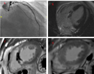
    Figure 1: RAO view of LAD during emergency coronary angiography; 4c
view demonstrating basal to mid infer septal LGE; Mid-level short axis LGE
demonstrating septal microvascular obstruction; Apical level short axis LGE
demonstrating no infarction.
The CMR images demonstrate infarction in the basal and mid anteroseptum and inferoseptum with microvascular obstruction but do not descend into the apical segments. This pattern suggests a septal perforator infarct. If the coronary angiogram is reviewed closely there is no obstruction of the left anterior descending artery but the first and second septal perforators are not visualised. This is a rare example of infarction of the septal arteries.