Special Article - Cardiac Pacing
Austin J Clin Cardiolog. 2022; 8(1): 1090.
Non Selective his Bundle Pacing: As Physiological as S-HBP but Simpler and Faster
Ortega DF*, Logarzo E, Paolucci A and Mangani N
Arrhythmias and Pacemakers Unit, Clínica San Camilo, Buenos Aires, Argentina
*Corresponding author: Daniel Felipe Ortega, Arrhythmias and Pacemakers Unit, Clínica San Camilo, Buenos Aires, Argentina
Received: March 04, 2022; Accepted: March 23, 2022; Published: March 30, 2022
Editorial
We have read with interest Dr. Burri’s Electrocardiographic Analysis for His Bundle Pacing at Implantation and Follow up [1]. A comprehensive guide to understand electrocardiographic (ECG) nuances of His Bundle Pacing (HBP) is presented in this review. The authors, with outstanding didactics, describe a well-organized analysis of challenging HBP electrocardiographic characteristics to confirm conduction tissue (selective or non-selective HBP) at implant and follow-up.
Caught our interest, in particular, the paragraph where practical pacing characteristics and benefits of non-selective HBP pacing (NSHBP) are emphasized. In this context we would like to point out the statement that pacing “ventricular myocardium adjacent to the atrioventricular septum, near the His bundle can also result in NSHBP”. Above all, of paramount clinical importance, when Dr. Burri et al. conclude that “No significant differences in cardiac mechanical synchrony or clinical outcome have been found between SHBP and NSHBP”.
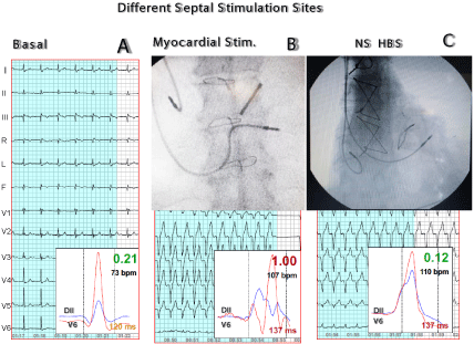
Our group is in total agreement with these essential features, having abandoned SHBP many years ago by developing a very simple technique to place standard pacing leads in the para-Hisian area (Figure 1). Concordant with the authors, we understand that “With non-selective His bundle pacing (NS-HBP), the lead is usually positioned in the ventricle at a site where the His bundle (HB) is surrounded by or at proximity to myocardial tissue”. That is why we named “para-Hisian pacing”. In our daily practice, at the same time, to verify and prove physiological capture of the conduction system we monitor synchrony online by analyzing the ECG signal variance [8] (Synchromax®) [2].

    Figure 1: A) Pre-implant basal ECG showing signal averaging of right septum (DII) and LV lateral wall (V6) with excellent agreement in the electrical flow direction,

QRS duration and coincidence of both intrinsic deflections. B) Myocardial Stimulation. Far from the conduction system (not reaching His-Purkinje capture) high

degree of electrical dyssynchrony is shown by the superposition of DII and V6 leads, due to the different direction of electrical flow and lack of concordance with

the intrinsic deflections. C) Pacing near the His-Purkinje, capturing it, NS HBP, shows excellent agreement between DII and V6 (flow direction base to apex)

and coincidence in the intrinsic deflection despite a QRS slightly wider due to a brief myocardial transit of the stimulus until capturing the conduction tissue (nonselective

stimulation).
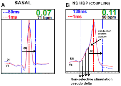
Briefly, Synchromax® provides supplementary information to the conventional 12-lead ECG by averaging the signals from standard ECG leads DII and V6 (right septum and lateral wall of the left ventricle). By these means our method allows to measure the direction of intrinsic cardiac activation, electrical flow and coupling of the intrinsic deflections. Verification of capture of the conduction system and thus physiological pacing is simplified [3]. Synchromax® ECG analysis allows us to confirm His-Purkinje system capture despite eventually a wider QRS. Effectively, the QRS shows a pseudodelta wave (His Bundle capture after a brief myocardial stimulation configuring NSHBP). We observed that the interval between the end of the delta wave and end of the QRS is the same as in native QRS (Figure 2). The QRS polarity is in most leads similar to basal as well. The pseudo-delta wave size will change depending on the distance needed to transit between the pacing site and the His-Purkinje system.

    Figure 2: A) Basal tracing, QRS duration 80msec; B) NSHBP: QRS is wider due to a delta wave corresponding to non-selective pacing. However, His-Purkinje

capture can be demonstrated (arrow) by having the same interval, 80 ms, between end of “pseudo delta” and end of QRS, having similar QRS polarity in most

leads. Pseudo delta size and duration will depend on the distance between pacing site and His-Purkinje system.
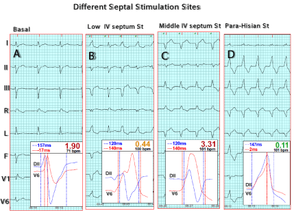
In fact, the Synchromax® technique discussed here was born from the thorough research of para-Hisian pacing (therefore NSHBP, but we adopted the former terminology) and the resulting ECG patterns. By performing parallel comparative analysis with tissue echo and intracardiac pressures we have confirmed that correlation with acute hemodynamic parameters is excellent [4]. Having 10- year experience with the Synchromax® method, we are sure that we have simplified para-Hisian pacing (and therefore physiological pacing), reaching over 500 such implants [5] (Figure 3). Of note, with our technique we use standard screw-in leads of all manufacturers, not being dependent upon special leads or sheaths for success. In addition, we advanced to include screw-in ICD leads in para-Hisian position. We also have normalized electrocardiographic Brugada pattern through para-Hisian pacing with remarkable results [6]. Furthermore, through analysis of cardiac electrical synchrony by using Synchromax®, in patients with or without pacemakers we have observed several findings that lead us to better understanding of interventricular coupling also in patients who were CRT candidates. Nowadays, Synchromax® allows verifying and estimating beforehand if such patients would be potential “electrical responders” [7].

    Figure 3: A) Basal ECG showing RBBB + LAFB. The intrinsic deflections of DII and V6 are not coincident due to the late LV lateral wall activation (uncoupling); B)

Low IV septum stimulation; strong discrepancy between intrinsic deflections and differences of electrical flow directions of DII and V6 (uncoupling) without capture

of His-Purkinje system; C) Middle IV septum stimulation, discrepancy between intrinsic deflections; D) NS HBP agreement in flow direction of both DII and V6,

coincident intrinsic deflections, disappearance of RBBB + LAFB. Both complexes are synchronous and there is His-Purkinje capture.
Finally, besides the practical advantages shown by the authors, on how to best use and troubleshoot the 12-lead ECG for HBP in daily practice, we congratulate Dr. Burri et al. for this comprehensive and critical review, that enlightens about the many controversial aspects of physiological pacing. Certainly simplicity and low index of complications (“higher R wave sensing amplitudes compared with selective (SHBP)), altogether with efficiency, safety (namely the importance of “back up of myocardial capture in case of loss of His capture or exit block”), association with, low implantation times, use of conventional leads, no requirement for special sheaths, and so forth, (all met with Synchromax®) will place non-selective His Bundle pacing / para-Hisian pacing as the future of physiological pacing.
We are convinced that the future of physiological pacing will be non-selective His pacing due to its simplicity and low index of complications8, in addition to the advantages clearly shown by the authors of the paper that we are commenting.
We congratulate them for this comprehensive and critical paper that enlightens about the controversial aspects of physiological pacing in these days.
References
- Burrri et al. JACC Clinical Electrophisiology. 2020; 6.
- Bonomini MP, et al. ECG Parameters to Predict Left Ventricular Electrical Delay. J Electrocard. 2018; 51: 844-850.
- Bonomini MP, et al. Depolarization Spatial Variance as a Cardiac Dyssynchrony Descriptor. Biomedical Signal Processing and Control. 2019; 49: 540-545.
- Ortega et al. Non-selective His bundle pacing with a biphasic waveform: enhancing septal resynchronization. Europace. 2017: 1-7.
- Logarzo E, et al. Para-Hisian pacemaker implantation technique guided by Synchromax method. Poster ESC. 2018 Munich.
- Logarzo E, et al. Novel Brugada syndrome treatment using para-Hisian stimulation. Poster HRS. 2019.
- De Zuloaga C, et al. Intraventricular dyssynchrony and electrical uncoupling: a disturbance unique to LBBB? Electrofisiología y Arritmias. 2018; 10: 48-55.
- Ortega DF, et al. Is traditional resynchronization therapy obsolete? Is para- Hisian pacing the new paradigm? Electrofisiología y Arritmias. 2019; 11: 38- 40.