
Research Article
Austin J Clin Cardiolog. 2022; 8(2): 1093.
Right Ventricular Involvement in Takotsubo Syndrome
Li Z#, Hao H#, Qiao S#, Chen J, Li G, Qi Y, Kang L* and Xu B*
¹Department of Cardiology, Nanjing University Medical School Affiliated Nanjing Drum Tower Hospital, P.R. China
#These authors contributed equally to this article
*Corresponding author: Lina Kang &, Biao Xu, Department of cardiology, Nanjing University Medical School Affiliated Nanjing Drum Tower Hospital, Nanjing, 210008, P.R. China
Received: June 21, 2022; Accepted: July 25, 2022; Published: August 01, 2022
Abstract
Objective: Patients with Right Ventricular (RV) involvement Takotsubo Syndrome (TTS), which leads to the higher risk of potential clinical comorbidities, higher mortality and extended recovery should be treated appropriately in the early stage, however, currently large-scale clinical data are lacking. Our article reviews epidemiological, clinical and prognostic features of RV TTS in our hospital.
Methods: This group study retrospectively evaluated patients diagnosed as TTS. Ventricular involvement was defined on wall motion abnormality showed by left ventriculography and echocardiography. The recovery of ejection fraction and pulmonary hypertension, defined as prognosis were assessed at followedup.
Results: Among 15 patients, diagnosed as TTS, 8(57.1%) were assigned to the Biventricular involvement TTS (Bi-VTTS). Older ((70.33±8.37 vs 69.00±10.51), P=0.04), women (8(88.9%), P=0.041) with lower LVEF (<50%) (8 (88.9%), P=0.020) were risky for RV TTS. At admission, for patients with RV TTS, LVEF was lower, B-type natriuretic peptide levels increased significantly ((591.9±162 vs 362.4±116.4pg/ml), P=0.021), elevations of troponin T and CKMB were not statistically significant, changes of Electrocardiograms (3(33.3%), P=0.020), myocardial edema and myocardial fibrosis also showed by Late Gadolinium Enhancement (LGE) of Cardiac Magnetic Resonance (CMR). During the index admission, hydrothorax (9(100%), P=0.001), shock (1(20%), P=0.164), chronic heart failure (2(40%), P=0.158), thrombus (9(100%), P=0.001), aneurysm (1(11.1%), P=0.143), mitral and tricuspid regurgitation (moderate or higher) (7(77.8%), P=0.180) and aortic regurgitation (moderate or higher) (3(33.3%), P=0.103) were associated with RV TTS. Except one patient (22.2%) died, longer time recovery and poor prognosis were assessed by ejection fraction and pulmonary hypertension.
Conclusions: Patients with the RVTTS, normally given the higher risk of potential clinical comorbidities, higher mortality and extended recovery who should be treated appropriately in the early stage to reduce comorbidities events, and improve patient prognosis.
Introduction
Takotsubo Syndrome (TTS) is an acute heart failure syndrome characterized by Left Ventricular (LV) dysfunction and peculiar patterns of Wall Motion Abnormalities (WMA) [1]. Although this condition typically affects left ventricular triggered by emotional or physical event, since 2000 [2,3]. reported the first case of simultaneous left and right ventricle involvement, that is, a case of biventricular TTS (Bi-V TTS), which aroused the attention of the medical community and the number of cases and clinical studies of right ventricular involved TTS(RV TTS) gradually increased [4,5]. Reported the first case of TTS that occurred only in the right ventricle, afterwards, isolated right ventricle Takotsubo cardiomyopathy and isolated right ventricular balloon-like syndrome were gradually recognized. Although the final outcome of right ventricular involvement is not bad, it still has a strong correlation with poor prognosis of TTS, more severe left ventricular dysfunction, high incidence of hemodynamic instability, and long-term increased mortality [6-8]. The current research and clinical data on RV TTS are insufficient, only limited case reports and case studies, so the epidemiology, clinical characteristics and pathogenesis of right ventricular involvement with or without left ventricular involvement TTS remain unclear [9]. Clinicians need to understand the clinical characteristics and imaging manifestations of TTS with right ventricular involvement to detect right ventricular involvement, avoid missed diagnosis, treat patients appropriately in order to reduce adverse events and mortality.
Methods
Study Population
Data were collected from the Department of cardiology, Affiliated Drum Tower Hospital, Medical School of Nanjing University from 2012 to 2018. TTS was defined based on modified Mayo Clinic Diagnostic Criteria and all cases of TTS diagnosis made according to Mayo Clinic Diagnostic Criteria were posteriori compared with the new Inter TAK Diagnostic Criteria. Ventricular involvement was defined based on wall motion abnormality showed by left ventriculography and echocardiography. Within 90 minutes from hospital admission, coronary angiography must be performed for patients presenting persistent ST-segment elevation, within 48 hours for patients presenting non-ST-segment elevation and within 120 minutes for patients presenting haemodynamic instability. Echocardiography was performed immediately at hospital admission. Medical records were reviewed by investigators at the Department of cardiology, Affiliated Drum Tower Hospital, Medical School of Nanjing University. Uncertain cases were reviewed by core team members, for inclusion or exclusion of cases, the decisions were reached by consensus. Follow-up information came from medical records, telephone interviews, or clinical visits.
PPI Statement
Our study protocol was reviewed by respective local ethics committee and investigational review board at Department of cardiology, Affiliated Drum Tower Hospital, Medical School of Nanjing University. Considering the partly retrospective nature of our study, the needs for informed consent of most studies were waived by ethics committees.
Analysis
Patients were divided into 2 groups based on the presence or absence of right ventricular involvement. Echocardiography, Cardiac magnetic resonance imaging, and Blood test of cardiac biomarkers were used for comorbidities assessment [10,11]. Recovery analysis was followed up by Echocardiography and accordingly, patients’ admission, discharge and return visit time, we analyze the recovery of ejection fraction and pulmonary hypertension, which were compared to initial data when patients in the hospital. Patients who haven’t undergo through Percutaneous coronary intervention but diagnosed as TTS were still credited to data analysis.
Statistical Analyses
Continuous variables were presented as mean±SD or median with interquartile range, whereas categorical variables were reported as frequency with percentage. Continuous variables were compared using the Mann–Whitney U test, whereas the Pearson v2 test (or Fisher exact test, as appropriate) was used for the comparison of categorical variables. Recovery estimates were assessed using Kaplan–Meier curves, and group differences were evaluated with the logrank test. The cut-off for statistical significance was set at a 2-sided P value <0.05. Odds ratios were reported with the respective 95% CIs. Analyses were computed with statistical software (SPSS V23.0,IBM Corp). Figures were created with Prism 7 software (GraphPad).
Results
Of 15 patients, diagnosed as TTS, 14 patients underwent coronary angiography and left ventriculography, among those 14 patients, combined with their echocardiography, 8(57.1%) were assigned to the Bi-V TTS, 5(35.7%)were assigned to the LV TTS and only 1(7.14%) were assigned to the RV TTS referred to (Table 1). We divided those patients into two groups, Bi-V or RV TTS group and LV group.
![]()
    LVwall motion abnormality

    RVwall motion abnormality

    Compensatory contraction enhancement

    ST segment elevation

    Coronary artery obstructed

    diagnosis


    Patient 1

    Apical

    Apical

    LV Basal

    Anteroseptal

    LCX    Mid-distal stenosis 85%

    Bi-V TTS


    Patient 2

    Apical

    None

    None

    Lateral, Posterior

    None

    LV TTS


    Patient 3

    Apical

    Apical

    Anterior,Inferior

    Inferior

    RCA PDA    30%

    Bi-V TTS


    Patient 4

    LV Anterior,Apical

    Apical

    Basal

    Anterior Inferior

    LAD Mid    stenosis 30%

    Bi-V TTS


    Patient 5

    LV Apical

    None

    Basal

    Anterior

    LAD Mid    stenosis 30%

    LV TTS


    Patient 6

    Apical, LV Anterior,    Inferior

    Anterolateral, Inferior

    Basal

    None

    LAD    Proximal stenosis 30%

    LV TTS


    Patient 7

    Mid LV    Anterior

    RV Apical

    Small    Potions

    Anteroseptal

    None

    RV TTS


    Patient 8

    Apical

    None

    Basal

    Anterior

    None

    LV TTS


    Patient 9

    LV Apical

    None

    Basal

    None

    None

    LV TTS


    Patient10

    Apical

    Apical

    Basal

    Anteroseptal

    LAD D1 30%    RCA PDA 20%

    Bi-V TTS


    Patient11

    LV    Anterior, Apical

    Apical

    Basal

    Inferior, Anterior

    LCX OM 50%

    Bi-V TTS


    Patient12

    LV Apical

    RV Basal

    Basal

    Anteroseptal

    RCA PL 30%

    Bi-V TTS


    Patient13

    Apical

    Apical

    Basal

    Extensive    Anterior

    None

    Bi-V TTS


    Patient14

    Anterior, Apical

    Apical

    LV Basal

    Anteroseptal, High    lateral

    LAD    Proximal stenosis 30%

    Bi-V TTS


    TTS: Takotsubo Syndrome; LV: Left Ventricle; RV: Right    Ventricle; Bi-V: Biventricular; LAD: Left Anterior Descending Artery; LCX:    Left Circumflex Artery; RCA: Right Coronary Artery; D: Diagonal Branch; PDA:    Posterior Descending Artery; OM: Obtuse Marginal Branch; PL: Posteriolateral    Branch
Table 1: Involvement diagnosis.
For the incentives and first symptoms: our cases indicated that the triggers of RV TTS and LV TTS were basically similar, usually caused by acute emotional or physical disease stress, such as death of relatives and friends, sudden physical illness, brain disease, pheochromocytoma etc., however, among those cases it was obvious that older ((70.33±8.37 vs 69.00±10.51), P=0.04), women (8 (88.9%), P=0.041) with lower left ventricular ejection fraction (LVEF< 50%) (8(88.9%), P=0.020) had a high risk for RV TTS. On the day of admission, for general condition of the patients, LVEF of RVTTS was lower than TTS without right ventricular involvement, B-type natriuretic peptide levels increased significantly with RV TTS (591.9±162 vs 362.4±116.4pg/ml), P=0.021), the elevation of troponin T and CK-MB values with right ventricular involvement compared with TTS without right ventricular involvement was not statistically significant. Those myocardial injury markers were mostly elevated during the acute episode, which was similar to Acute Coronary Syndrome (ACS), and was easily misdiagnosed as ACS, but the increase was not directly proportional to the degree of abnormal wall motion, then decreased rapidly in recovery [12]. Electrocardiograms of right ventricular affected TTS mostly had changed of ST-segment elevation, ST segment flat (2(22.2%), P=0.036), T wave inversion, QRS low voltage, and pathological Q waves (1(11.1%), P=0.020), etc. referred to (Table 2), which often involving the entire chest lead (V1 ~ V6) and the inferior (II, ,III aVF). In the Cardiac Magnetic Resonance (CMR) imaging, T2-weighted sequence of CMR could show myocardial edema, although the left and right ventricular ejection fractions had basically returned to normal, myocardial edema persists and it was continued to 1 month later. With Late Gadolinium Enhancement (LGE) of CMR can indicate the presence of myocardial fibrosis in some cases [13], however, compared with acute myocardial infarction, the TTS signal intensity was lower and the range was smaller.
![]()
    Bi-V or RV (n=9)

    LV (n=5)

    P


    Sex (women, %)

    8(88.9%)

    4(80%)

    0.041


    Age, y

    70.33±8.37

    69.00±10.51

    0.043


    Precedingevents

    5 (55.6%)

    3 (60%)

    0.420


    BNP (pg/ml)

    591.9 ± 162

    362.4 ± 116.4

    0.021


    cTNT (ug/l)

    0.419 ± 0.081

    0.804 ± 0.330

    0.168


    EF decline (<50%)

    8 (88.9%)

    3 (60%)

    0.020


    ECG characteristics


    ST segment elevation

    8 (88.9%)

    4 (80%)

    0.649


    ST segment flat

    2 (22.2%)

    4 (80%)

    0.036


    T-wave inversion

    8 (88.9%)

    4 (80%)

    0.649


    Q waves

    1 (11.1%)

    2 (40%)

    0.020


    Coronary artery    obstruction (%)

    2 (22.2%)

    0 (0%)

    0.158


    Comorbidities


    Hydrothorax

    9 (100%)

    1 (20%)

    0.001


    Heart failure

    7 (77.8%)

    2 (40%)

    0.158


    Thrombus

    1 (11.1%)

    0

    0.143


    Shock

    1 (20%)

    0

    0.164


    Aneurysm

    1 (11.1%)

    0

    0.164


    Mitral and tricuspid    regurgitation (moderate or higher)

    7 (77.8%)

    3 (60%)

    0.180


    Aortic regurgitation    (moderate or higher)

    3 (33.3%)

    2 (40%)

    0.103
Table 2: Pulmonary Hypertension was higher for those patients with

right ventricular involvement at admission day, although after appropriate

treatment, their Pulmonary Hypertension back to almost normal when

discharge from the hospital, usually 10 days at the average, for the longterm

prognosis, the recovery of Pulmonary Hypertension would longer time

for the patients of right ventricular involvement TTS. Bi-V: Biventricular; LV:

Left Ventricle.
For comorbidities while in hospital nine patients with RVTTS happened to have hydrothorax (9(100%), P=0.001), others such as severe shock (1(20%), P=0.164), chronic heart failure (2(40%), P=0.158), thrombus (9(100%), P=0.001), aneurysm (1(11.1%), P=0.143), mitral and tricuspid regurgitation (moderate or higher) (7(77.8%), P=0.180) and aortic regurgitation (moderate or higher) (3(33.3%), P=0.103) happened in certain risk as well, but not very significant referred to (Table 2). Because RV TTS affected both left and right ventricular function, patients with right ventricular involvement had worse left ventricular function and were more likely to have bilateral ventricular dysfunction compared with left ventricular involvement alone (67% vs. 8%, P <0.001)Scott W. Sharkey et al. reported 2019 [14-16]. Echocardiography may furtherly identify potential comorbidities of right ventricular TTS, such as mitral regurgitation, tricuspid regurgitation and intraventricular thrombosis [17,18].
For prognosis, some studies considered right ventricular involvement as an independent predictor of endpoint events [19], some studies considered right ventricular as an independent predictor of death [20-23] and some studies have suggested that right ventricular involvement has similar short-term and long-term endpoint event rates as left ventricular TTS alone [24-28]. Within our cases, 12-month follow-up was performed, one patient (22.2%) died in the right ventricular affected TTS group, for other patients we set LVEF, Pulmonary Hypertension and the time of prognosis as criterions to assess prognosis. From our data it showed LVEF was lower for those patients with RV TTS at admission day, although after appropriate treatment, their LVEF back to almost normal when discharge from the hospital, usually 10 days at the average, for the long-term prognosis, the deterioration of LVEF would appear in the patients of RV TTS. The same process can be detected in the recovery of Pulmonary Hypertension, referred to (Figure 1 & 2). Therefore, longer time recovery needed and poor prognosis achieved for patients with RV TTS referred to (Figure 3).
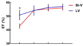
    Figure 1: LVEF was lower for those patients with right ventricular involvement

at admission day, although after appropriate treatment, their LVEF back

to almost normal when discharge from the hospital, usually 10 days at the

average, for the long-term prognosis, the deterioration of LVEF would appear

in the patients of right ventricular involvement TTS. Bi-V: Biventricular; LV:

Left Ventricle.
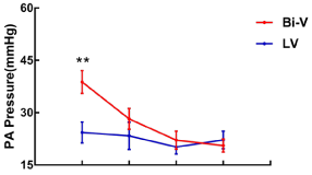
    Figure 2: Pulmonary Hypertension was higher for those patients with

right ventricular involvement at admission day, although after appropriate

treatment, their Pulmonary Hypertension back to almost normal when

discharge from the hospital, usually 10 days at the average, for the longterm

prognosis, the recovery of Pulmonary Hypertension would longer time

for the patients of right ventricular involvement TTS. Bi-V: Biventricular; LV:

Left Ventricle.
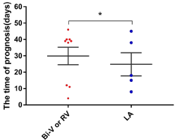
    Figure 3: Longer time of recovery needed and poor prognosis achieved for

patients with right ventricular involvement TTS. Bi-V: Biventricular; LV: Left

Ventricle.
Discussion
Major Findings
To our knowledge, this study is a small investigation of the comorbidities and prognostic impact of RV TTS. The study had 3 main findings: (1) patients with right ventricular involvement were more frequently older female, presented more commonly with typical TTS (apical ballooning), and with lower LVEF and higher BNP levels on admission; (2) Comorbidities such as Hydrothorax, Heart failure, Thrombus, Aneurysm, Mitral and tricuspid regurgitation (moderate or higher) could present in patients with RV TTS; (3) Extended recovery duration and higher mortality were associated with RV TTS followed up from discharge to 12-month. The results above are important in clinic as patients with those characteristics must be monitored closely after hospital admission, if identified earlier as RV TTS, which normally given the higher risk of potential clinical comorbidities, higher mortality and extended recovery, must be treated appropriately. In addition, patients with longer recovery duration were more likely to form ventricular thrombus which was found recently [29-34], because of the heavier myocardial injury and longer impairment of LV function [35-37]. Generally acknowledged, complicated and acute processes of the disease adversely impact the recovery rate [38-44], our patients went through prolonged recovery normally with higher level of Pulmonary artery pressure levels and lower level of LVEF supports this interpretation Schwarz et al recently reported [45]. It may just take more time to recover from a persistently high Pulmonary artery pressure and severely low LVEF than from a mild impairment of pump function.
Study Limitations
This study is based on a single center registry, the department of cardiology, Affiliated Drum Tower Hospital, Medical School of Nanjing University from 2012 to 2018, the number of patients was small. Moreover, the dichotomous classification with or without right ventricular involvement may influence our results because of the unclear priori diagnostic criterion to diagnose RV TTS [37-39]. And the vast majority follow-ups were performed by echocardiography because cardiac magnetic resonance imaging was not performed in all patients for some objective reasons. Recovery estimates were assessed using Kaplan–Meier curves, given the retrospective limitation of assessing the exact time to recovery, however, multivariable covariates associated with the right ventricular involvement assessment of survival was not performed because of the limited number of events at follow-up, which may prevent to establish an accurate and reliable model.
Conclusions
In summary, there are many differences between RV TTS and left ventricular involvement TTS alone. The current research and clinical data on RV TTS are basically derived from case reports and limited case studies, large-scale clinical data are lacking. Therefore, the epidemiology, pathogenesis, and prognosis of right ventricular involvement with or without RV TTS remain unclear, so there is no consistent diagnostic criterion. However, patients with the RV TTS, normally given the higher risk of potential clinical comorbidities, higher mortality and extended recovery, which should be treated appropriately in the early stage. What we need to do is strengthen both basic and clinical research on RV TTS meanwhile with the development and promotion of new technologies in regard to cardiac ultrasound, CMR and other imaging technology, in the future, to help developing RV TTS Patient management, reducing comorbidities events, and improving patient prognosis.
Funding
This work was funded by the Funds for Distinguished Young Scientists in Nanjing [JQX15002]; Natural Science Foundation of China [81870291]; Natural Science Foundation of China [81870204].
References
- Ramaraj R. Stress cardiomyopathy: aetiology and management. Postgraduate Medical Journal. 2007; 83: 543-546.
- K Tsuchihashi 1, K Ueshima, T Uchida, N Oh-mura, K Kimura, et al. Transient left ventricular apical ballooning without coronary artery stenosis: a novel heart syndrome mimicking acute myocardial infarction. Angina Pectori-Myocardial Infarction Investigations in Japan. J Am Coll Cardiol. 2001; 38: 11-18.
- Gianni M, Dentali F, Grandi AM, Sumner G, Hiralal R, Lonn E. Apical ballooning syndrome or takotsubo cardiomyopathy: a systematic review. European heart journal. 2006; 27: 1523-1529.
- Sharkey SW, Lesser JR, Maron MS, Maron BJ. Why not just call it tako-tsubo cardiomyopathy: a discussion of nomenclature. Journal of the American College of Cardiology. 2011; 57: 1496-1497.
- Ghadri J, Wittstein IS, Prasad A, Sharkey S, Dote K, Akashi YJ, et al. International Expert Consensus Document on Takotsubo Syndrome (Part I): Clinical Characteristics, Diagnostic Criteria, and Pathophysiology. European Heart Journal. 2018; 39: 2032-2046.
- Bonnemeier H, Schäfer U, Schunkert H. Apical ballooning without apical ballooning. European heart journal. 2006; 27: 2246-2246.
- Robles P, Alonso M, Huelmos AI, Jiménez JJ, Bescós LL. Images in cardiovascular medicine. Atypical transient left ventricular ballooning without involvement of apical segment. Circulation. 2006; 113: e686-8.
- Haghi D, Papavassiliu T, Flüchter S, Kaden JJ, Pörner T, Borggrefe M, et al. Variant form of the acute apical ballooning syndrome (takotsubo cardiomyopathy): observations on a novel entity. Heart. 2006; 92: 392-394.
- Nyui N, Yamanaka O, Nakayama R, Sawano M, Kawai S. ‘Tako-Tsubo’ transient ventricular dysfunction: a case report. Japanese circulation journal. 2000; 64: 715-719.
- Pastromas S, Koulouris S, Gavaliatsis IP, et al. Tako Tsubo cardiomyopathy with right ventricular involvement. Hospital Chronicles. 2009; 4: 181-183.
- Eitel I, Schuler G, Gutberlet M, Thiele H. Biventricular stress-induced (takotsubo) cardiomyopathy with left midventricular and right apical ballooning. International journal of cardiology. 2011; 151: e63-e64.
- Novak G, Kross K, Follmer K, Brofferio A, Shirani J. Transient Biventricular Apical Ballooning: A Unique Presentation of the “Broken Heart”. Clinical Cardiology. 2007; 30: 355-358.
- Mrdovic I, Kostic J, Perunicic J, Asanin M, Vasiljevic Z, Ostojic M. Right ventricular takotsubo cardiomyopathy. Journal of the American College of Cardiology. 2010; 55: 1751.
- Stähli BE, Ruschitzka F, Enseleit F. Isolated right ventricular ballooning syndrome: a new variant of transient cardiomyopathy. European heart journal. 2011; 32: 1821-1821.
- Burgdorf C, Hunold P, Radke PW, Schunkert H, Kurowski V. Isolated right ventricular stress-induced (Tako-Tsubo) cardiomyopathy. Clin Res Cardiol. 2011; 100: 617-619.
- Finocchiaro G, Kobayashi Y, Magavern E, Zhou JQ, Ashley E, Sinagra G, et al. Prevalence and prognostic role of right ventricular involvement in stressinduced cardiomyopathy. Journal of cardiac failure. 2015; 21: 419-425.
- Kagiyama N, Okura H, Tamada T, Imai K, Yamada R, Kume T, et al. Impact of right ventricular involvement on the prognosis of takotsubo cardiomyopathy. European heart journal cardiovascular Imaging. 2016; 17: 210-216.
- Elesber AA, Prasad A, Bybee KA, Valeti U, Motiei A, Lerman A, et al. Transient cardiac apical ballooning syndrome: prevalence and clinical implications of right ventricular involvement. Journal of the American College of Cardiology. 2006; 47: 1082-1083.
- Eitel I, von Knobelsdorff-Brenkenhoff F, Bernhardt P, Carbone I, Muellerleile K, et al. Clinical characteristics and cardiovascular magnetic resonance findings in stress (takotsubo) cardiomyopathy. JAMA. 2011; 306: 277-286.
- Sung J, Hong S, Chung I, Lee HY, Lee JH, Kim H, et al. Rupture of Right Ventricular Free Wall Following Ventricular Septal Rupture in Takotsubo Cardiomyopathy with Right Ventricular Involvement. Yonsei Medical Journal. 2017; 58: 248.
- Daoko J, Rajachandran M, Savarese R, Orme J. Biventricular takotsubo cardiomyopathy: case study and review of literature. Texas Heart Institute journal. 2013; 40: 305-11.
- Sung J, Hong S, Chung I, Lee HY, Lee JH, Kim H, et al. Rupture of Right Ventricular Free Wall Following Ventricular Septal Rupture in Takotsubo Cardiomyopathy with Right Ventricular Involvement. Yonsei Medical Journal. 2017; 58: 248.
- Scally C, Ahearn T, Rudd A, Neil CJ, Srivanasan J, Jagpal B, et al. Right Ventricular Involvement and Recovery After Acute Stress-Induced (Takotsubo) Cardiomyopathy. The American journal of cardiology. 2016; 117: 775- 780.
- Madias JE. Recurrent right ventricular takotsubo syndrome in a diabetic patient with dysautonomia. Chest. 2014; 146: e73.
- Chandorkar A, Codolosa JN, Lippmann ML, Pressman GS, Cruz JPS. Recurrent right ventricular takotsubo cardiomyopathy in a patient with recurrent aspiration. Echocardiography. 2014; 31: E240-242.
- Sumida H, Morihisa K, Katahira K, Sugiyama S, Kishi T, Oshima S. Isolated Right Ventricular Stress (Takotsubo) Cardiomyopathy. Internal Medicine. 2017; 56: 2159-2164.
- Elikowski W, Malek-Elikowska M, Rózanska P, Fertala N, Zawodna M. Isolated right ventricular takotsubo cardiomyopathy: a case report and literature review. Polski merkuriusz lekarski : organ Polskiego Towarzystwa Lekarskiego. 2016; 41: 283-286.
- Burgdorf C, Hunold P, Radke PW, Schunkert H, Kurowski V. Isolated right ventricular stress-induced (“Tako-Tsubo”) cardiomyopathy. Clin Res Cardiol. 2011; 100: 617-619.
- JOE B, HWANG H, PARK C, JIN E, SOHN I, CHO J, et al. Takotsubo cardiomyopathy recurrence with left ventricular apical ballooning following isolated right ventricular involvement: A case report. Experimental and Therapeutic Medicine. 2013; 6: 260-262.
- Santoro F, Ieva R, Di Martino LF, Musaico F, Scarcia M, et al. Incomplete leaflet coaptation and tricuspid regurgitation mechanismyopathy. Int J Cardiol. 2014; 177: e99-101.
- Ghadri J, Cammann VL, Templin C. The International Takotsubo Registry: Rationale, Design, Objectives, and First Results. Heart failure clinics. 2016; 12: 597-603.
- Nishikawa S, Ito K, Adachi Y, Katoh S, Azuma A, Matsubara H. Ampulla (‘takotsubo’) cardiomyopathy of both ventricles: evaluation of microcirculation disturbance using 99mTc-tetrofosmin myocardial single photon emission computed tomography and doppler guide wire. Circulation journal : official journal of the Japanese Circulation Society. 2004; 68: 1076-80.
- Kurisu S, Inoue I, Kawagoe T, Ishihara M, Shimatani Y, Mitsuba N, et al. Takotsubo-like transient biventricular dysfunction with pressure gradients. Internal medicine. 2005; 44: 727-732.
- Gill D, Liu K. Takotsubo cardiomyopathy associated with Miller-Fisher syndrome. The American journal of emergency medicine. 2017; 35: 1012.
- Haghi D, Athanasiadis A, Papavassiliu T, Suselbeck T, Fluechter S, et al. Right ventricular involvement in Takotsubo cardiomyopathy. Eur Heart J. 2006; 27: 2433-2439.
- Rodrigues AC, Guimaraes L, Lira E, Oliveira W, Monaco C, Cordovil A, et al. Right Ventricular Abnormalities in Takotsubo Cardiomyopathy. Echocardiography. 2013; 30: 1015-1021.
- Heggemann F, Hamm K, Brade J, Streitner F, Doesch C, Papavassiliu T, et al. Right Ventricular Function Quantification in Takotsubo Cardiomyopathy Using Two-Dimensional Strain Echocardiography. PLoS ONE. 2014; 9: e103717.
- Liu K, Carhart R. “Reverse McConnell’s sign?”: a unique right ventricular feature of Takotsubo cardiomyopathy. The American journal of cardiology. 2013; 111: 1232-1235.
- Vizzardi E, Bonadei I, Piovanelli B, Bugatti S, D’Aloia A. Biventricular Tako-Tsubo cardiomyopathy: Usefulness of 2D speckle tracking strain echocardiography. Journal of Clinical Ultrasound. 2014; 42: 121-124.
- Citro R, Caso I, Provenza G, Santoro M, Gregorio G, Bossone E. Right ventricular involvement and pulmonary hypertension in an elderly woman with tako-tsubo cardiomyopathy. Chest. 2010; 137: 973-975.
- Korlakunta H, Butkevich A, Muthupillai R, Cheong BYC. Biventricular takotsubo cardiomyopathy: cardiac magnetic resonance imaging as useful diagnostic tool. Texas Heart Institute journal. 2011; 38: 88-9.
- Lei J, Sun Z, Lyu L, Green RG, Scalzetti E, Feiglin D, et al. Mechanical interventricular dependency supports hemodynamics in tako-tsubo cardiomyopathy. Journal of thoracic disease. 2018; 10: 3027-3038.
- Liu K, Krone RJ. What truly causes the adverse outcome in Tako-Tsubo cardiomyopathy?. JACC. Cardiovascular imaging. 2014; 7: 742-743.
- Liu K. Letter by Liu regarding article, “systolic and diastolic mechanics in stress cardiomyopathy”. Circulation. 2015; 131.
- Becher T, El-Battrawy I, Baumann S, Fastner C, Behnes M, LoΒnitzer D, et al. Characteristics and long-term outcome of right ventricular involvement in Takotsubo cardiomyopathy. International journal of cardiology. 2016; 220: 371-375.