Case Report
Austin J Clin Cardiolog. 2022; 8(3): 1097.
Percutaneous Therapy of Functional Interrupted Aortic Arch with 3-1 Dimensional Printing Guidance
Mao Y, Li L, Zhai M, Ma Y, Jin P, Liu Y* and Yang J*
Department of Cardiovascular Surgery, Xijing Hospital, Air Force Medical University, Xi’an, China
*Corresponding author: Jian Yang, Department of Cardiovascular Surgery, Xijing Hospital, Air Force Medical University, 127 Changle West Road, Xi’an 710032, Shaanxi, China
Yang Liu, Department of Cardiovascular Surgery, Xijing Hospital, Air Force Medical University, 127 Changle West Road, Xi’an 710032, Shaanxi, China
Received: September 02, 2022; Accepted: October 06, 2022; Published: October 13, 2022
Abstract
Functional interrupted aortic arch is a congenital malformation characterized by a complete separation between the ascending aorta and the descending aorta. Untreated patients are in danger of having late complications including cerebral hemorrhage, aneurysm formation, and aortic regurgitation. Traditionally, classical surgical therapies included bypass grafting or orthotopic repair. Herein, we report a simplified percutaneous therapy for functional interrupted aortic arch with a retrograde crossing technique and without an incremental-sized expandable balloon. With the guidance of 3-dimensional printing, percutaneous treatment with a covered stent is a feasible, safe, and effective alternative to surgery with excellent short- and midterm results in selected patients with favorable anatomy.
Introduction
Functional Interrupted Aortic Arch (IAA) is a congenital malformation characterized by a complete separation between the ascending aorta and the descending aorta. The presence of functional IAA in adults is extremely rare [1,2]. Patients with functional IAA can present with refractory hypertension and upper limb/lower limb systolic gradients. Untreated patients are in danger of having late complications including cerebral hemorrhage, aneurysm formation, and aortic regurgitation. Traditionally, classical surgical therapies included bypass grafting or orthotopic
repair. Transcatheter treatments for coarctation of the aorta have become more common in recent decades. Stenting therapy has also been used for functional IAA in limited cases with complicated and challenging techniques [3-5]. Due to high surgical risk, we simulated the procedure using a 3-dimensional (3D) printed model. During the procedures, all of these therapies used antegrade crossing techniques and dilation with a incremental-sized balloon. We report a simplified percutaneous therapy for functional IAA with a retrograde crossing technique and without an expanding incremental-sized balloon.
Case Presentation
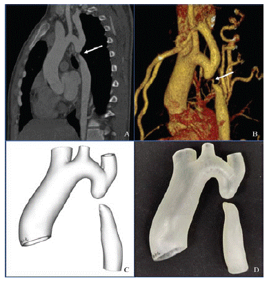
A 17-year-old male presented with refractory hypertension and increasing dyspnea on exertion. The cardiac function was New York Heart Association functional class II. During the clinical examination, a bounding pulse was noted in the upper limbs, and the blood pressure in the upper limbs was 210/110 mm Hg. Femoral and all peripheral pulses in the lower limbs were absent, and the blood pressure was unrecordable. No pulse was found in the lower limbs. Echocardiography revealed mild to moderate aortic regurgitation. The left ventricular ejection fraction was depressed at 45% with dilatation of the left ventricle. Computed tomography angiography showed an interrupted aortic arch just distal to the origin of the left subclavian artery with a gap of 12 mm between the proximal and distal segments (Figure 1A, B).

    Figure 1: Preoperative computed tomography angiographic scans. A. Median

sagittal section. B. Three-dimensional reconstruction. C. Preprocedural

3-dimensional reconstructed model. D. 3-Dimensional printed model.
3-Dimensional Printing and Simulation
A 3D-printed model was reconstructed by computed tomography angiography (Figure 1C). After the image data in DICOM format were collected, Materialise Mimics version 21.0 software (Materialise, Leuven, Belgium) was used to segment the images and export files into the Standard Tessellation Language (STL) format. Materialise 3-Matic software (Leuven, Belgium) was used for post processing of STL format files. Finally, STL files were imported into a Polyjet 850 multimaterial full-color 3D printer (Stratasys, Inc., Eden Prairie, MN, USA) (Figure 1D). The 3D-printed model was followed for platform simulation. Postsimulation analysis showed that the procedure would be feasible with a thorough preprocedural plan.
Procedures
Percutaneous therapy of the interrupted aortic arch was planned. The procedure was performed with the patient under local anesthesia. We gained access to the left radial artery and the right femoral artery during the procedure. The upper limb/lower limb systolic gradients were 110 mm Hg (185/110 mm Hg in the UL and 75/60 mm Hg in the LL). Simultaneous antegrade and retrograde aortograms were done at the upper and lower ends of the interrupted aorta in diagonally opposite projections (LAO 50) to analyze the relation between the proximal and distal segments (Figure 2A).

    Figure 2: Fluoroscopy during the operative procedure. A. Simultaneous antegrade and retrograde aortograms. B. The gap was perforated retrogradely. C. The

guidewire was snared from the radial entry. D. A 14 Fr straight sheath was advanced. E. The interrupted lesion was repaired with the covered Cheatham platinum

stent. F. Postoperative aortogram.
A Judkins 6 Fr guiding catheter was firmly advanced retrogradely to the floor of the distal segment via femoral access. The gap was perforated retrogradely with a 260-cm 0.032-inch Terumo soft wire and a 5 Fr multipurpose diagnostic catheter (Boston Scientific Corporation, Marlborough MA, USA). Then the Terumo soft wire was snared from the radial entry. Next, the lesion was dilated gently with a 6 Fr straight sheath (Cook Medical, Bloomington, IN, USA) via a tensile soft-wire loop, followed by advancement of a 14 Fr straight sheath (Cook Medical) (Figure 2B-D).
Then the lesion was repaired with a 39-mm covered Cheatham Platinum stent (NuMED Inc., Cross Roads, TX, USA) inserted with a 20-mm balloon-in-balloon catheter (NuMED Inc.) (Figure 2E, F;). There were no residual gradien- or procedure-related complications. At the 6-month follow-up examination, the patient was asymptomatic, and his blood pressure was controlled without any medications. Postoperative computed tomography angiography and the 3D printed model showed a properly positioned stent with no complications (Figure 3).

    Figure 3: Postoperative computed tomography angiography. A. Median

sagittal section. B. Three-dimensional reconstruction. C. Postprocedural

3-dimensional reconstructed model. D. 3-Dimensional printed model.
Discussion
Functional IAA is an extremely rare congenital malformation in adults. This anomaly is an extreme form of aortic coarctation, characterized by a total luminal and anatomical interruption between the ascending and descending sections of the thoracic aorta [6]. Transcatheter treatment of aortic coarctation with endovascular stents has become an effective alternative to surgery in recent decades. The prognosis was encouraging based on the results of a previous clinical trial. However, percutaneous stenting of functional IAA is technically challenging. The risks and potential complications related to the use of the percutaneous procedure for functional IAA include the creation of a false lumen, injury to the vascular wall, or disruption and acute vascular compromise [2,7,8]. Therefore, these procedures should be attempted only by experienced operators in carefully selected cases of functional IAA with favorable anatomy. In addition, all necessary equipment, including covered stents, must be readily available in the cardiac catheterization laboratory. The presence of a backup surgical team is also necessary to ensure immediate access to a cardiac surgeon in case of complications.
In the limited number of previously published cases, operators always used antegrade crossing techniques and dilation with an incrementally sized balloon [3-5]. In this case, a simplified procedure was performed with limited equipment costs. The retrograde approach was conducted by crossing the occluded segment with a regular guide wire instead of a thin one. In previous articles, a 0.014-inch or even a 0.008-inch guidewire was used. The malformation of functional IAA is different from a chronic total occlusion. The thin wire might not be necessary for crossing. Also, incremental expansion of the balloon was not performed during the procedure. Only a 6 Fr sheath was advanced to expand the lumen; it was followed by a 14 Fr sheath. Based on the anatomy of functional IAA, there should be a potentially expandable lumen located in the stent. Finally, a covered rather than an uncovered stent, which was used in previously published cases, was deployed at the occluded segment. Covered stents help in a bailout procedure when complications related to coarctoplasty, such as dissection, aortic rupture, or aneurysm formation, occur [8].
In addition, with the continuous development of material science and imaging technology, the application of medical 3D printing technology is becoming much more extensive. In recent years, 3D printing has played an important role in the treatment of orthopedics, stomatology, plastic surgery, and other fields [9,10]. Because the cardiovascular system is pulsatile and flexible, with elaborate, complex anatomical structures, development of 3D printing technology in this field has been relatively slow. As the technology has progressed, procedures have become much more minimally invasive, but an intervention could not look straight into the anatomical structure of the pathological changes occurring in patients. Cardiovascular 3D printing technology can display the anatomy in different parts of the heart individually, accurately, and intuitively. Tong et al. used 3D printing to help guide the treatment of aortic arch disease by using modified prefenestrated/branched stent grafts [11]. Futhermore, 3D printing, used as a tool to carry out preprocedural planning and training, is critical for assessing factors such as anatomy, function, and the delivery capacity of the device. Therefore, it may have the potential to diminish clinicians’ learning curves.
Conclusion
In conclusion, with the guidance of 3D printing, percutaneous treatment of functional IAA with a covered stent is a feasible, safe, and effective alternative to surgery with excellent short- and midterm results in selected patients with favorable anatomy.
Clinical Trial Registration
ClinicalTrials.gov Protocol Registration System (NCT02917980).
Funding
This work was supported by the National Key R&D Program of China (No. 2020YFC2008100), the Shaanxi Province Innovation Capability Support Plan – Innovative Talent Promotion Plan (No. 2020TD-034), and the Discipline Boosting Program of Xijing Hospital (No. XJZT18MJ69).
Acknowledgements
We would like to thank Make Medical Technology Co., LTD. (Xi’an, China) for supplying the 3D printed models.
Conflict of Interest
The authors have no conflicts of interest to declare.
Data Availability Statement
The original contributions presented in the study are included in the article/supplementary material, further inquiries can be directed to the corresponding author.
References
- Messner G, Reul GJ, Flamm SD, Gregoric ID, Opfermann UT. Interrupted aortic arch in an adult: Single-stage extra anatomic repair. Tex Heart Inst J. 2002; 29: 118–121.
- Kosucu P, Kosucu M, Dinç H, Korkmaz L. Interrupted aortic arch in an adult: Diagnosis with MSCT. Int J Cardiovasc Imaging. 2006; 22: 735–739.
- Suárez de Lezo J, Romero M, Pan M, Suárez de Lezo J, Segura J, Ojeda S, Pavlovic D, Mazuelos F, López Aguilera J, Perez SE. Stent repair for complex coarctation of aorta. JACC Cardiovasc Interv. 2015; 8: 1368–79.
- Moorthy N, Ananthakrishna R, Nanjappa MC. Percutaneous stenting of interrupted aortic arch to treat compressive myelopathy. Catheter Cardiovasc Interv. 2014;.84:.815–819.
- Goel PK, Moorthy N. Percutaneous reconstruction of interrupted aortic arch in an adult. JACC Cardiovasc Interv. 2013; 6: e21–2
- Mishra PK. Management strategies for interrupted aortic arch with associated anomalies. Eur J Cardiothorac Surg. 2009; 35: 569–576.
- Hijazi ZM. Catheter intervention for adult aortic coarctation: be very careful! Catheter Cardiovasc Interv. 2003; 59: 536–537.
- Butera G, Heles M, MacDonald ST, Carminati M. Aortic coarctation complicated by wall aneurysm: the role of covered stents. Catheter Cardiovasc Interv. 2011; 78: 926–32.
- Maglara E, Angelis S, Solia E, Apostolopoulos AP, Tsakotos G, et al. Threedimensional( 3D) printing in orthopedics education. J Long Term Eff Med Implants. 2020; 30: 255-258.
- Nesic D, Schaefer B M, Sun Y, Saulacic N, Sailer I. 3D printing approach in dentistry: The future for personalized oral soft tissue regeneration. J Clin Med. 2020; 15: 2238.
- Tong YH, Qin Y, Yu T, Zhou M, Liu C, et al. Three-dimensional printing to guide the application of modified prefenestrated stent grafts to treat aortic arch disease. Ann Vasc Surg. 2020; 66: 152-159.