Short Communication
Austin J Clin Cardiolog. 2022; 8(3): 1100.
Flow-Mediated Dilation Can be used as an Indicator for Assessing Severity and Deaths by Covid-19 at the Initial Hours of Hospitalization
Oliveira MR, Goulart CL, Back GD and Borghi-Silva A*
Cardiopulmonary Physiotherapy Laboratory, Physiotherapy Department, Federal University of Sao Carlos, Brazil
*Corresponding author: Borghi-Silva A Cardiopulmonary Physiotherapy Laboratory, Physiotherapy Department, Federal University of Sao Carlos, UFSCar, Rodovia Washington Luis, KM 235, Monjolinho, CEP: 13565-905, Sao Carlos, SP, Brazil
Received: November 17, 2022; Accepted: December 22, 2022; Published: December 28, 2022
Abstract
Assessment of Flow-Mediated Dilation (FMD) in patients hospitalized for COVID-19 will assist with regard to early identification of markers at the onset of SARS-CoV-2 infection. Therefore, the early evaluation of simple markers, obtained at the bedside before the most serious manifestations are already installed, can help health professionals to act preventively to save lives, direct care in the initial stage of infection, helping in the prognosis and diagnosis. Given the importance and relevance of this technique in this population at this time and the need to better understand the pathophysiology of COVID-19 as well as its damage to the endothelium, we performed an evaluation of FMD in different severity of COVID-19 in recently hospitalized patients. A total of 100 patients were enrolled in the study and were divided into two groups according to the severity of COVID- 19. The results provide new evidence that patients with COVID-19 classified as severe/critical have greater endothelial dysfunction and that FMD may be a simple marker, helping in the prognosis and diagnosis and, consequently, in the prevention of thrombotic events.
Keywords: COVID-19; Hospitalization; Endothelial Dysfunction.
Introduction
Severe acute respiratory syndrome coronavirus 2 (SARSCoV- 2) occurs from entry is the respiratory tract, in which the type 2 Angiotensin Converting Enzyme (ACE2) is a functional receptor sequestered [1]. The progression of systemic inflammation and over activation of immune cells can trigger a “cytokines storm” that culminates in increased levels of inflammatory markers [2]. The disease progresses to a severe form in 10–30% of patients infected, requiring hospitalization and potential Intensive Care Unit (ICU) treatment [3]. Another feature is activated coagulation with thrombus formation, indicating the fundamental roles played by endothelial damage [4].
The endothelium contributes in vascular homeostasis, regulating vascular tone, leukocyte adhesion, smooth muscle cell growth and platelet aggregation, playing a protective role for the blood vessel. The endothelial damage that COVID-19 generated, also leads to immune super reactivity (cytokine storm). These are related to the decrease in nitric oxide (NO) by decreasing the activity of endothelial Nitric Oxide Synthase (eNOS). This decrease may cause less endothelium-dependent vasodilation, which may increase the risks of endothelial dysfunction, leading to endothelitis and cardiovascular diseases in patients with COVID-19 [5].
The repercussions of this virus on the cardiac system and specifically on the endothelium, it is of such importance that hospitalized patients infected with SARS-CoV-2 are evaluated at the beginning of their hospitalization. These patients should undergo tests that can predict the risk of aggravating their case early, avoiding complications from the virus and hospitalization, providing indicators for the sequence of therapies to be installed [4]. In addition, such early measures at the bedside may identify markers of adverse outcomes and death by COVID- 19. Among these evaluations and exams, we have the Flow- Mediated Dilation (FMD), this is a validated, non-invasive, safe technique, independent predictor of cardiac outcomes [6] and high reliability, as indicated by International Brachial Artery Reactivity Task Force e American College of Cardiology [7]. FMD is an important physiological stimulus regulating vascular tone and homeostasis of the peripheral circulation, therefore knowing the importance of early evaluation of FMD, we recommend the application of the method in patients with COVID-19.
This technique is used widely in hospitals and aims to evaluate the endothelial function from the ultrasound investigation of a medium-sized artery, usually the brachial artery, before and after a condition induced by high flow and shear stress by inflation and deflation of a cuff in the proximal region of the arm [8]. FMD brachial artery has been widely used in clinical research since 1992, when it was developed by Celermajeret al. [9]. There is sufficient evidence to suggest that this method provides adequate information on the health of the endothelium [8,10].
In our previously published study, we sought to assess endothelial function in relation to length of hospitalization and mortality in patients diagnosed with COVID-19 and compare with patients without COVID-19 [11]. We found the clinically relevant cutoff points for FMD% and FMDmm in patients with COVID-19. Specifically, we demonstrated FMDmm was significant predictor of mortality in patients with COVID-19, indicating patients with endothelial dysfunction are more likely to die in 10 days of hospitalization. As such, FMD measurement may be valuable in patients hospitalized with COVID-19 to determine those at particularly high risk for death [11]. Based on the positive results found in this study, we can justify the importance of applying FMD in patients with COVID-19. FMD is already well used in other pathologies, such as in sepsis in which Bonjorno et al. [12] were observed the influence of several clinical FMD variables (% FMD, peak FMD and delta FMD) for prognostic efficacy and mortality in sepsis in Intensive Care Unit (ICU) patients.
Given the importance and relevance of this technique in this population at the moment and the need to better understand the pathophysiology of COVID-19 as well as its damage to the endothelium, we aimed, with an analysis of a prospectively collected clinical database, to compare endothelial dysfunction in different severity of COVID-19 in recently hospitalized patients. These data are from a cohort study carried out in hospitals in the state of São Paulo (Brazil), accepted by the ethics committee (protocol number: 33265220.9.0000.5504).
Patients included in this analysis were over 18 years old, of both sexes, hospitalized within the first 48 hours in the ward or ICU of hospitals in the state of São Paulo - Brazil with a positive diagnosis for COVID-19 by means of the reverse transcriptase polymerase chain reaction in time (RT-PCR) from nasopharyngeal swab [13]. The exclusion criteria were: 1) Patients or family members who did not accept to participate in the study and did not sign Consent Form; 2) Patients under palliative care; 3) Readmission cases; 4) Patients using non-invasive ventilation during the time of evaluation; 5) Patients in the prone position during the assessment; and 6) Patients with intravenous accesses in both upper limbs, making it impossible to perform the FMD exam.
Clinical assessment and patient comorbidities were obtained from medical records and endothelial function was assessed by flow-mediated vasodilation of the brachial artery using ultrasound imaging (M-Turbo, Sonosite, Seattle, WA, USA). The FMD measurement protocol followed our previously published study [11].
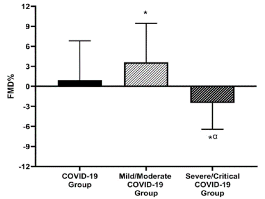
A total of 100 patients were included in study and were divided into two groups according to their severity of COVID-19, classified according to the World Health Organization (WHO) [14]. The results of this sample are shown in (Table 1 and Figure 1). It is possible to affirm that patients with Severe/critical COVID-19 (Severe/critical COVID-19 group) were predominantly male, and stayed more days hospitalized compared to the COVID- 19 group and the Mild/moderate COVID-19 group. Furthermore, the Severe/critical COVID-19 group had a higher number of deaths compared to the Mild/moderate COVID-19 group. In relation to vascular measurements, the Severe/Critical COVID- 19 group standing out with negative values of relative FMD (FMD%), having lower values in comparison with the COVID-19 group and Mild/moderate COVID-19 group. Such data emphasize the importance of evaluating these patients at the initial moment of their hospitalization, helping to predict the severity of the disease and the therapies to be used.
Variables
COVID-19 group
(n = 100)Mild/Moderate COVID-19 group
(n = 56)Severe/Critical COVID-19 group
(n = 44)Age (years)
54±16
54±15
53±16
Male
59 (59)
28 (50)
31 (70)*
Woman
41 (41)
28 (50)
13 (30)* a
BMI (Kg/m²)
30±7
29±6
31±8
Hospitalization days
13±11
9±9
19±12*a
Death, n (%)
21 (21)
7 (12)
14 (32)a
Vascular measures
Baseline artery diameter, mm
4.1±0.7
4.0±0.7
4.2±0.8
Baseline blood flow, cm/s
13±8
13±10
12±7
Hyperemic blood flow, cm/s
35±18
37±18
33±17
Shear stress
70±39
78±39
60±37
FMD, mm
0.02±0.23
0.13±0.22*
-0.10±0.17* a
BMI: Body mass Index; FMD: Flow-mediated dilation; mm: millimeters; *= different from the COVID-19 group;a=different from the Mild/Moderate COVID-19group.
Table 1: Clinical data and vascular measures in patients with COVID-19 and Control group.

    Figure 1: Comparison of relative FMD values (%) between

COVID-19 patients.

FMD: Flow-mediated dilation; mm: millimeters; *= different from

the COVID-19 group;a=different from the Mild/Moderate COVID-

19group.
Riou et al. also used FMD to verify whether there was a reduction in endothelium-dependent vasodilation in critically ill patients with COVID-19 after 3 months of infection and whether the severity of the infection could influence FMD indices. They concluded that these patients showed a reduction in FMD values, but no changes were found according to the severity of the infection. Furthermore, they highlighted the importance of studies with longer follow-up time and a larger sample to better understand the mechanisms involved in this virus. [15] Ergul et al. found that COVID-19 is an independent predictor of endothelial dysfunction as assessed by FMD. The authors state that COVID-19 can cause endothelial dysfunction independent of other risk factors and that FMD is a non-invasive, easy-to-apply and useful method to assess endothelial function [16]. Heubel et al. confirmed these findings, as when investigating non-critical patients hospitalized for COVID-19, they found endothelial dysfunction assessed by FMD [17].
In this context, we emphasize the relevance of this study and the difference from others already published, as the assessments took place at the initial moment of hospitalization, being an important outcome to guide the medical team in the choice of therapy. In this way, it is highly recommendable early endothelial function assessment in order that the staff initiates, when necessary, antithrombotic measures from the initial moment of hospitalization, in order to prevent thromboembolic events [11,18,19]. However, we must emphasize the care in carrying out this examination in hospitals, given the rapid contamination of SARS-CoV-2 from materials, professionals or patients. Therefore, it is recommended to clean the device and probe with a tissue soaked in 70% alcohol before and after each evaluation, as well as the use of individual protective equipment in the evaluators and, if possible, also in patients due to the proximity necessary for the examination [20].
In conclusion, this study provides novel evidence that patients with COVID-19 classified as severe/critical have greater endothelial dysfunction and that the FMD can be a simple marker, obtained at the bedside before the most serious manifestations performed to characterize the most severe patients in the initial stage of the infection, helping in the prognosis and diagnosis and consequently prevent thrombotic events.
Financial Support
This study is supported by Fundação de Amparo à Pesquisa do Estado de São Paulo – FAPESP, São Paulo, Brazil Process N◦2015/26/501-1, and by the Coordenação de Aperfeiçoamento de Pessoal de Nível Superior- Brasil (CAPES – 001), process N◦88887.507811/2020-00. Borghi-Silva A. is a recognized investigator of CNPq - 1B level.
References
- R Lu, X Zhao, J Li, P Niu, B Yang, et al. Genomic characterisation and epidemiology of 2019 novel coronavirus: implications for virus origins and receptor binding, Lancet. 2019; 395: 565–574.
- S Su, G Wong, W Shi, J Liu, ACK Lai, et al. Epidemiology, Genetic Recombination, and Pathogenesis of Coronaviruses, Trends Microbiol. 2016; 24: 490–502.
- C Huang, Y Wang, X Li, L Ren, J Zhao, et al. Wang, R. Jiang, Z. Gao, Q. Jin, J. Wang, B. Cao, Clinical features of patients infected with 2019 novel coronavirus in Wuhan, China, Lancet. 2020; 395: 497– 506.
- JJ Marini, L Gattinoni. Management of COVID-19 Respiratory Distress. JAMA. 2020; 7: 435–444.
- Z Varga, AJ Flammer, P Steiger, M Haberecker, R Andermatt, et al. Correspondence Endothelial cell infection and endotheliitis in, Lancet. 2019; 395: 1417–1418.
- TJ Anderson, SA Phillips. Science Direct Assessment and Prognosis of Peripheral Artery Measures of Vascular Function. Prog Cardiovasc Dis. 2014; 57: 497–509.
- MC Corretti, TJ Anderson, EJ Benjamin, CMs D Celermajer, F Charbonneau, et al. Guidelines for the Ultrasound Assessment of Endothelial- Dependent Flow-Mediated Vasodilation of the Brachial Artery A Report of the International Brachial Artery Reactivity Task Force. J Am Coll Cardiol. 2002; 39: 257–265.
- JB De Melo, J Albuquerque, DF Neto, R Cristina, A Campos, Estudo da Função Endotelial no Brasil : Prevenção de Doenças Cardiovasculares. Rev Bras Cardiol. 2014; 27: 120–127.
- D Celermajer, K Sorensen, V Gooch, D Spiegelhalter, O Miller, et al. Non-invasive detection of endothelial dysfunction in children and adults at risk of atherosclerosis. Lancet (London, England). 1992; 340: 1111–1115.
- Y Gunes, HA Gumrukcuoglu, S Akdag, H Simsek, M Sahin, et al. Vascular Endothelial Function in Patients with Coronary Slow Flow and the Effects of Nebivolol. Arq Bras Cardiol. 2011; 97: 275–280.
- MR Oliveira, GD Back, C da Luz Goulart, B Domingos, R Arena, et al. The endothelial function Provides early prognostic Information in patients with COVID-19: A cohort study. Respir Med. 2021; 106469.
- JC Bonjorno, FR Caruso, RG Mendes, TR Da Silva, TMP de Campos Biazon, et al. Noninvasive measurements of hemodynamic, autonomic and endothelial function as predictors of mortality in sepsis: A prospective cohort study. PLoS One. 2019; 14: 1–16.
- GM Bwire, MV Majigo, BJ Njiro, A Mawazo. Detection profile of SARS-CoV-2 using RT-PCR in different types of clinical specimens: A systematic review and meta-analysis. J Med Virol. 2021; 93: 719–725.
- WHO. Clinical management Clinical management Living guidance COVID-19. World Heal Organ. 2021.
- M Riou, W Oulehri, C Momas, O Rouyer, F Lebourg, et al. Reduced Flow-Mediated Dilatation Is Not Related to COVID-19 Severity Three Months after Hospitalization for SARS-CoV-2 Infection. J Clin Med. 2021; 10: 1318.
- E Ergül, AS Yilmaz, MM Ögütveren, N Emlek, U Kostakoglu, M çetin. COVID 19 disease independently predicted endothelial dysfunction measured by flow-mediated dilatation. Int J Cardiovasc Imaging. 2021.
- AD Heubel, AA Viana, SN Linares, VT do Amaral, NS Schafauser, et al. Determinants of endothelial dysfunction in non-critically ill hospitalized COVID-19 patients: a cross-sectional study. Obesity. 2021; 0–3.
- E Oikonomou, N Souvaliotis, S Lampsas, G Siasos, G Poulakou, et al. Endothelial dysfunction in acute and long standing COVID-19: A prospective cohort study. Vascul Pharmacol. 2022; 144: 106975.
- RL Flumignan, VT Civile, JD de S Tinôco, PI Pascoal, LL Areias, et al. Anticoagulants for people hospitalised with COVID-19. Cochrane Database Syst Rev. 2022.
- Colégio brasileiro de radiologia e diagnóstico por imagem, Cuidados específicos para serviços de ultrassonografia diagnóstica durante o surto de COVID-19, n.d.