
Case Series
Austin J Clin Cardiolog. 2023; 9(1): 1105.
Coronary Artery Anomalies, Myocardial Bridging Associated with Fistula to Pulmonary Artery Trunk. Case Reports
Marian Gaspar*
Department of Cardiac Surgery, University of Medicine and Pharmacy “Victor Babes”, Timisoara, Romania
*Corresponding author: Marian Gaspar Department of Cardiac Surgery, University of Medicine and Pharmacy “Victor Babes”, Timisoara, Romania. Tel: +40-723995444. Email: mariangaspar24@yahoo.ro
Received: February 13, 2023 Accepted: March 28, 2023 Published: April 04, 2023
Abstract
Between coronary artery anomalies, myocardial bridging means an epicardial coronary artery, mostly Left Anterior Descending artery (LAD), running through intramyocardial “tunnel” (usually in the middle segment), leading during systolic contraction, flow reduction, through the vessel. When this anomaly is associated with a coronary fistula, who “steal” more from the bloodstream, the symptoms are more pronounced and the management complex, surgical in particular. Despite the presence from birth remains a symptomatic and it becomes clinically manifest later in the third to fourth decade of the life, with a diverse palette of symptoms; angina, arrhythmias, acute myocardial infarction up to sudden death. Diagnosis and particular management, medical, interventional and surgical should avoid major cardiac complications and sudden death. We present two adult patients, with coronary artery bridging, one case associated with coronary artery fistula, LAD to pulmonary artery trunk, very symptomatic with surgical management and the second only myocardial bridging controlled with medication and supervision.
Keywords: Myocardial bridging; Coronary fistula
Introduction
This congenital anomaly in which a coronary artery, usually Left Anterior Descending (LAD), it follows a deep path in the myocardium bridge, “tunnel”, was described morphological several hundred years ago (Reyman, 1737), but first angiographic documented, later by Portmann and Iwig in 1960 [1]. The prevalence of myocardial “bridge” at angiography is lower (0,5–2,5%) than at autopsy (15–85%). The coronary filling flow in the most part occurs in diastole, systolic compression of the artery should have only a little impact on total effective myocardial perfusion, but more refined studies using frame-by-frame quantitative coronarography with IVUS study, reveal that compression of the vessel extending also into diastole and as the result affect the myocardial perfusion [2]. Coronary bridge syndrome are also common in hypertrophic cardiomyopathy, with a frequency of 25-80% and in the patient with orthotropic heart transplant 33%. Due to this association, sudden death has been described in young people, athletes. An intriguing clinical situation is myocardial infarction in young persons, without atherosclerotic lesions discernible by coronary angiography. In the absence of atherosclerosis, myocardial infarction may result from several aetiologies including; vascular spasm, transient dysrhythmia, drugs abuse, hypercoagulability and coronary thrombus formation, and dissection of acquired or congenital vascular anomalies [3]. The patients are admitted with; angina, arrhythmias, even acute myocardial infarction with left ventricular dysfunction or the worse, sudden death in young, active person [4,5]. Another congenital anomaly is coronary fistula, is an anomalous communication between one or two coronary arteries and a cardiac chamber or any of the great vessels (the coronary sinus, the superior vena cava, and the pulmonary artery). Prevalence, in general population is 0.002%, within all congenital heart disease 0.08–0.4% and 0.3–0.8% of all patients who undergo selective coronary angiography [6]. The most common site of drainage is the right ventricle, followed by the right atrium and the Pulmonary Artery (PA). The blood flow from the coronary, usually LAD to PA shunt, leads to ‘coronary steal’, drawing blood away from the normal coronary tree, the results are symptoms and signs of myocardial ischaemia.
Even more, if two anomalies are associated, coronary bridging and fistula, then the symptomatology is obvious and the management much more complex, medical, interventional and surgical [7].
We do not have any specific medical therapy for coronary fistula, this should be occluded by transcatheter embolization (coils, vascular plug, covered stent) or surgical intervention (dissection and ligature of fistula on the both sides).
We present two cases; first case a LAD bridging associated with fistulae between LAD and PA trunk, very symptomatic in spite of medical treatment and the second only with LAD bridge, managed with medication and surveillance.
Case Report 1
The first patient F. C, 46 years old male, presented with typical angina by exercise and at the rest, was admitted for diagnosis and management.
ECG: Sinus rhythm, intermediate QRS axis, HR=80bpm. Eco-cardiography. LV hypertrophy, FE:55%, mild mitral regurgitation, mild tricuspid regurgitation. Normal pericardial fluid.
Chest X-ray: Heart and lungs according to age. No pulmonary condensation or fluid collections.
In spite of complex medication; Aspirin 75mg/day, selective beta-blocker (Bisoprolol 5mg/day), Candesartan cilexetil –(Atacand) 16mg/day, diuretics 50/20mg/day, ivabradine (Corlentor) 5mgx2/day, atorvastatina (Zetovar) 40/10mg/day, to control angina, blood hypertension, mixt-dyslipidemia, the patient continueto have exercise and rest angina. The patient was admitted for more investigation.
Angiocoronarography: Right radial artery approach. Coronary system with right dominance. Left main, normal. Coronary arteries show marked spasm that improves with the administration of intracoronary NTG. Upon injection into the ACD, a marked negation of T waves is observed in the lower leads on the EKS. LAD presents in the middle segment, over a length of 4cm, an accentuated muscular bridge after administration of intracoronary NTG, with 80% stenotic systolic compression. A coronary fistula emerges from the proximal ADA that drains into the pulmonary circulation. Proximal and distal LAD, circumflex artery and RCA do not present lesions visible angiographically (Figure 1). Coronary artery fistulas between the LAD and the PA are rare congenital malformations. However, concomitant significant coronary artery stenosis and fistula can cause coronary steal phenomenon and this result in severe myocardial ischemia.

Figure 1: (A) Coronaro-angiography showing diastolic lumen dimensions are normal, (B) Typical systolic compression (red arrows) of the mid LAD, a long segment. (C) Associated a fistula from LAD to pulmonary artery trunk is reveals.
In such situation, bridging associated with coronary artery fistula, the treatment is interventional or surgical. Operation was performed through standard median sternotomy using heart- lung machine. After aortic was clamped, anterograde cardioplegia stopped the heart for clear inspection. First LAD was release, starting in distal superficial segment and continuing carefully proximal dissection using a beaver, for 8cm segment.
The fistula was clearly identified on the surface of the right ventricle reaching the proximal portion of the main pulmonary trunk above the pulmonary valve. After that, the fistula was tender dissectedin special origin from LAD, then closed using two stitches of 5/0 polypropylene (Figure 2).
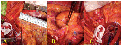
Figure 2: (A) Using heart-lung machine, the heart is stopped by anterograde cardioplegia, LAD inspected and dissected, myotomy for approximately 8cm starting distally and carefully proximal, (B) Then the LAD-to- PA fistula is identified, (C) Isolated and sutured on the both sides.
In case of untimely dissection some complications can be associated with myotomy; injury to the artery, incomplete myotomy, and right ventricul perforation. To avoid right ventriculotomy, the myotomy should be located anterior toward the left side of the artery.
Association of coronary by-pass, with Left Internal Mammary Artery (LIMA) to LAD is advised in case of proximal severe atherosclerotic stenosis, otherwise due to competitive flow, graft can be obstructed. The postoperative and subsequent evolution was good, without complications and with the disappearance of symptoms.
Case Report 2
The second patient I.G, 54 years old male, hospitalized for precordial pain with radiation in the left shoulder, occurring after meals, upon admission, patient in good clinical condition, without precordial pain, without signs of heart failure with AP 115/70mmHg, AV=80bpm. ECG, sinus rhythm, AV=96bpm, intermediate QRS axis, ST depression 1mm in V6.
Cardiac Echocardiography: Ascending aorta=26mm, aortic anulus 19mm, LV of normal dimensions, VD=30mm, VS=41mm, SIV=11mm, PPLV=10mm, with good systolic function EF=55%, without disturbances of parietal kinetics. LA and right cavities of normal size. Mitral insufficiency degree one, mitral flow E=0, 76. Pericardial fluid 7mm.
Biochemistry: LDL cholesterol-135mg/dl, HDL cholesterol=33mg/dl, cholesterol-204mg/dl, Triglyceride 176mg/dl.
Coronary angiography: does not show coronary stenoses on the RCA, LCA, but shows a "muscular bridge" with LAD constriction in the middle portion, after the administration of NTG (Figure 3).

Figure 3: (A) Coronary angiography highlights normal vessel on diastole and (B) The LAD “muscular bridge” in the middle segment, with obvious constriction when nitroglycerin is administered.
Management: In this case, the treatment was only medical, with B-blockers, aspirin, combination of two lipid-lowering drugs (Leridip and Sortis), hypotensive medication (Candesartan cilexetil - Atacand, 8mg/day) and periodic surveillance with readmission in case of angina attacks. Nitrates are contraindicated in patients with myocardial bridging. Nitroglycerin has been shown to accentuate systolic compression of bridged segments, and indeed is used as an agent for provocation of these lesions, like in our case.
Discussion
Myocardial bridging of the Left Anterior Descending (LAD) coronary artery occurs in 1%-4.5% of coronary angiographies [8]. But the association with coronary fistula is very rare and gives it a particular, clinical and therapeutic significance. In case of myocardial bridging, the majority of the patients are asymptomatic. When they become symptomatic with angina, myocardial ischemia, myocardial infarction, left ventricular dysfunction, myocardial stunning, paroxysmal AV blockade, exercise-induced ventricular tachycardia and sudden cardiac. Young patients, usually without high risk factors for atherosclerosis (diabetes mellitus, dyslipidemia, smoking), come to the emergency department complaining of chest pain and fatigue, usually after physical exertion or stress.
Then, even the constriction is very severe or in 90% of the cases, atherosclerotic lesions are associated, located in the proximal part of the vessel possibly due to mechanical shear stress (Haager and all) or is associated with other anomalous like in our case, coronary artery fistula, which accentuated myocardial “steal” and ischemia (first described 1865 by Krause).
Diagnosis: with the new imaging techniques; cardiac echocardiography, echo-stress, coronary cineangiography, Fractional Flow Reserve (FFR), Intravascular Ultrasound, (IVUS), 256 slices- Computer Tomography, it has led to improved identification and functional quantitation of myocardial bridging and associated disease [9]. Coronary cineangiography, remain the most used technique for diagnosing of myocardial bridging.
IVUS, is more sensitive than angiography (in one study detected bridging in 23% of patients, while angiographic systolic compression was only apparent in 3%). The muscle tunneled segment of artery clearly demonstrates systolic compression that persists into diastole. Also, when combined with provocation testing with nitroglycerin, dobutamineor rapid atrial pacing.
The treatment: of myocardial LAD bridging is complex, the patients are usually young and may present with severe, usually atypical, symptoms often attributed to other causes. Once the diagnosis is made, medication is considered first-line therapy, beta-blockers, calcium- channels blockers, Aspirin, lipid-lowering agents (if the case). Volume loading may also reduce compression of the tunneled segment, whereas administration of nitroglyceride may aggravate compression and ischemia [9]. For this reason, nitrates are contraindicated in patients with myocardial bridging and indeed is used as an agent for provocation of these lesions like we did in our second case.
The interventional cardiologist (Stables et al., 1995) first reported coronary stenting as an interventional approach to severe myocardial bridging refractory to medication but multiple cases of coronary complication, perforation and stent fracture in a stented myocardial bridge have been reported, perhaps related to stent oversizing. In patients with persistence of symptoms in spite of right medication or associated coronary disease (atherosclerotic stenosis, coronary fistula), surgical management is the choice.
Surgical technique for myocardial bridging include; myotomy (Binet 1975) associated or not with Coronary Artery Bypass Graft surgery (CABG). This is a relatively easy surgical procedure, LAD dissection and release from distal part to proximal area. However, risks complication must be taken in account, LAD damage, perforation into the right ventricle, graft occlusion in the presence of competitive flow on LIMA to LAD graft, arrhythmia and later vessel fibrosis.
Coronary artery fistula to the great vessel and heart chambers, single or in association with others anomalous should be addressed by interventional or surgical closure [10].
Conclusion
Surgical approach of LAD bridging associated with closure of the coronary artery fistula is indicated when the both anomalies are associated. Dissection and ligation of the fistula, with exploration of the pulmonary artery is a safe and effective method of treatment of coronary artery fistula to the pulmonary artery. The myotomy is enough for LAD bridging. In case of mild bridging symptoms, medication and surveillance can be the choice.
References
- JoseMaria Perez-Pomares, Jose Luis de la Pompa, Diego Franco, Deborah Henderson, Siew Yen Ho, et al. Congenital coronary artery anomalies: a bridge from embryology to anatomy and pathophysiology—a position statement of the development, anatomy, and pathology ESC Working Group. Cardiovascular Research. 2016; 109: 204–216.
- Michael S Lee, Cheng-Han Chen. Myocardial Bridging: An Up-to-Date Review. J Invasive Cardiol. 2015; 27: 521–528.
- Jeffrey Dwyer. Coronary artery bridging as an etiology for non- atherosclerotic myocardial infarction: A review of literature and case history. J Cardio Case Rep. 2019.
- Stefan Mohlenkamp, Waldemar Hort, Junbo Ge, Raimund Erbel. Update on Myocardial Bridging. Circulation. 2002; 106: 2616-2622.
- Chiara Ripa, Maria Cristina Melatini, Fabiola Olivieri, Roberto Antonicelli. Myocardial bridging: A ‘forgotten’ cause of acute coronary syndrome – a case report. Int J Angiol. 2007; 16: 115-118.
- Khurram Butt, Ali Agha, Ryan Parente, Joseph Limback, Jeremy R Burt. Anomalous Coronary Anatomy with Fistula Diagnosed on Coronary Computed Tomography Angiography. Cureus. 2019; 11: e4403.
- Alper Sami Kunt. Coronary Artery and Pulmonary Artery Fistula Originated from Significant Stenosis in the Left Anterior Descending Artery.Case Report. Case Reports in Emergency Medicine. 2013; 2013: 298156.
- Chigozirim N Ekeke, Stephen Noble, Ernest Mazzaferri, Juan A Crestanello. Myocardial bridging over the left anterior descending: Myotomy, bypass, or both?. The Journal of Thoracic and Cardiovascular Surgery. 2015; 149: e57-8.
- Arjun Kanwal, Ankit B Shah. Myocardial Bridging in Adult. American College of Cardiology. 2020.
- Li Wan, Qingyu Wu. Myocardial bridge, surgery or stenting?. Interactive Cardio Vascular and Thoracic Surgery. 2005; 4: 517–520.