Case Report
Austin J Clin Case Rep. 2014;1(4): 1020.
Nodular Fasciitis of the Breast Can Mimic Cancer
Khalid Alhajri1*, Eyad Alkharashi1, Hussam Binyousef1, Haitham Nasser2
1Department of surgery, Prince Sultan Military Medical City, Saudi Arabia
2Department of pathology, Prince Sultan Military Medical City, Saudi Arabia
*Corresponding author: Khalid Alhajri, Department of surgery, Breast and endocrine surgery unit, Prince Sultan Military Medical City, Riyadh, Saudi Arabia
Received: May 31, 2014; Accepted: June 25, 2014; Published: June 27, 2014
Abstract
Nodular fasciitis of the breast is a benign pathological entity that can mimic breast cancer. We present the case of a 42-year-old female who was referred to our institution with right breast pain for 3 years. Radiology demonstrated a highly suspicious lesion. Initially, the needle biopsy showed a spindle cell proliferation, but after wide local excision, the final pathology result was nodular fasciitis. Nodular fasciitis of the breast is a rare lesion where local excision is the preferred treatment approach, in the proper clinical context.
Keywords: Nodular fasciitis; Breast; Cancer
Case Presentation
Nodular fasciitis of the breast is a rare benign spindle cell proliferation that can present as a mass lesion and is sometimes related to breast trauma [1]. It can be mistaken for a breast carcinoma clinically, radiologically and histologically due to its vague clinical, radiological, and histological features [2].
A 42-year-old previously healthy female, presented to our breast clinic with a history of breast pain for 3 years. She gave a history of trauma to the right breast for more than 15 years prior to presentation. Family history was unremarkable. Physical examination revealed no palpable lesions. There were no palpable lesions, skin changes or nipple discharge. Mammography showed an area of focal asymmetry with associated architectural distortion at around 12 o’clock of the right breast. Ultrasound showed a 3 cm, tubular, hypoechoic and hypervascular mass. The anterior portion appears cystic with angular margins and spicules that extended to the subcutaneous tissue. The picture, being highly suspicious for malignancy, was coded as BIRADV.
Based on the above information, a core needle biopsy was performed and was reported as spindle cell proliferation. The differential diagnosis included phyllodes tumor and fibromatosis and excisional biopsy was recommended.
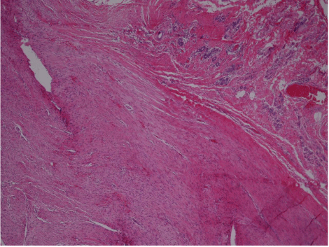
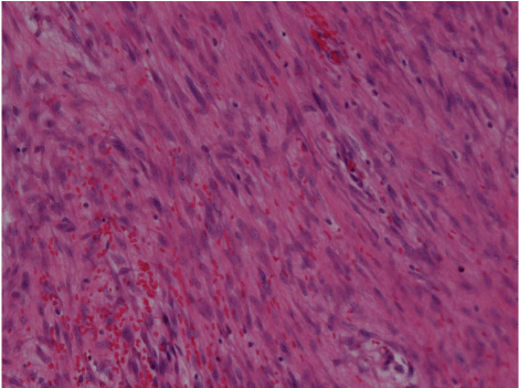
The case was discussed in the tumor board meeting and the recommendation was to go ahead with a wide local excision. It was carried out with k-wire localization. The final pathology was Nodular fasciitis of the breast. The pathology demonstrated a myofibroblastic proliferation arising in the breast tissue with occasional extravasated red blood cells and rare typical mitotic figures (Figure 1,2). The tumoral cells stained negative for pankeratin, smooth muscle actin, CD34, desmin and beta-catenin.
Figure 1: Low magnification: Well-circumscribed tumor (left) with pushing borders adjacent to normal breast tissue (right).
Figure 2: High magnification: Proliferating myofibroblasts arranged focally in a tissue culture-like pattern with extravasated red blood cells.
Discussion
Nodular fasciitis, which was first described by Konwaller et al. [3], is a benign proliferation of fibroblasts in the subcutaneous tissues and commonly associated with fascia.
Nodular fasciitis of the breast can mimic breast cancer. It appears as an irregular or spiculated lesion on mammography and ultrasound. The exact cause of nodular fasciitis of breast is still unknown. It is likely to be triggered by injury, though a history of trauma has been elicited in few cases such as this one [1]. For proper diagnosis, the lesion requires core needle biopsy. The key histological features are tissue culture-like spindle cell proliferation and extravasated red blood cells, and whenever present a myxoid stroma, chronic inflammatory cells, capillary proliferation, and vascular channels [1,4,5].
Management of nodular fasciitis can be either conservative or surgical; and in the latter excisional biopsy is preferred. A conservative approach is appropriate if the lesion has a typical clinical appearance, with the core biopsy consistent with the proper diagnosis [1]. Spontaneous regression has been observed [4]. However, if these criteria are not met, excisional biopsy is the most appropriate treatment, and no further management is necessary [5]. Recurrence following surgical excision is rare [6].
In summary, we present in this paper a new case of nodular fasciitis arising in the right breast of a middle-aged female, with a past long history of trauma to the same breast. This is a rare lesion that can mimic breast cancer. And correct diagnosis can only be made on tissue examination. We always recommend excisional biopsy for such lesions.
References
- Brown V, Carty NJ. A case of nodular fascitis of the breast and review of the literature. Breast. 2005; 14: 384-387.
- Dahlstrom J, Buckingham J, Bell S, Jain S. Nodular fasciitis of the breast simulating breast cancer on imaging. Australas Radiol. 2001; 45: 67-70.
- KONWALER BE, KEASBEY L, KAPLAN L. Subcutaneous pseudosarcomatous fibromatosis (fasciitis). Am J Clin Pathol. 1955; 25: 241-252.
- Tulbah A, Baslaim M, Sorbris R, Al-Malik O, Al-Dayel F. Nodular fasciitis of the breast: a case report. Breast J. 2003; 9: 223-225.
- Squillaci S, Tallarigo F, Patarino R, Bisceglia M. Nodular fasciitis of the male breast: a case report. Int J Surg Pathol. 2007; 15: 69-72.
- Batsakis JG, el-Naggar AK. Pseudosarcomatous proliferative lesions of soft tissues. Ann Otol Rhinol Laryngol. 1994; 103: 578-582.

