Case Report
Austin J Clin Case Rep. 2014;1(5): 1023.
Elevated CEA and Medullary Cell Carcinoma
Azhar Ali Malik1*, Ali Elhouni2, Bachar Afandi3 and Rya Almazroei4
Department of internal medicine, Tawam Hospital, UAE, (1-4)
*Corresponding author: Azhar Ali Malik, Department of internal medicine Division of Endocrinology, Tawam Hospital Al ain UAE
Received: May 23, 2014; Accepted: June 30, 2014; Published: July 01, 2014
Keywords
CEA; Medullary cell carcinoma; CT
25 years old female was investigated for a nonspecific abdominal pain history and was found to have elevated CEA (carcinogenic embryonic antigen). The work up for gastrointestinal malignancy included upper and lower gastrointestinal endoscopy and a contrast enhanced CT abdomen. The endoscopy findings were inconclusive while the CT abdomen revealed a 1cm nodule in left adrenal gland, which was of doubtful clinical significance, and not likely cause of elevated CEA Figure 1.
On clinical examination she is a lady of average build and height. Her systemic examination including neck and abdominal examination were unremarkable.
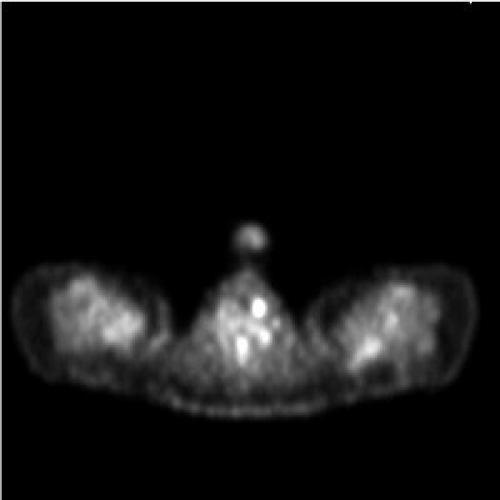
Prompted by concerns of rising CEA whole body PET scan was requested, which showed a small thyroid nodule in right side of neck Figure 2, which was confirmed on subsequent ultrasound of thyroid. Her thyroid nodule biopsy showed medullary cell carcinoma. This was not surprising in setting of elevated CEA, and positive uptake on PET scan, hence, explaining the elevated CEA .Because of positive thyroid biopsy, RETS gene testing and serum calcitonin, level was requested. The genetic testing was negative for MEN 2 (Medullary endocrine neoplasia), but calcitonin level was significantly elevated (>20ng/ ml). She has total thyroidectomy on 12/9/2012. Post operatively she had transient hypoparathyroidism and her serum calcitonin and CEA fall to zero level. She is on replacement therapy for post surgical hypothyroidism. Her follow up calcitonin CEA, level and urine metanephrine levels are within normal limits (Table1). CT abdomen, at 6 months follow up, showed a small increase in left adrenal nodule and it remained non functional.
Figure 1 :
    Dec 2011
    6 March 2012
    11June 2013


    CEA
    16.9ng/ml
    25ng/ml
    0.5g/ml


    Calcitonin
    >20ng.ml
    <1ng/ml
    1.2ng/ml


    Urine nor adrenaline

    157mmol/24hrs
      27mcg/24hrs
    100mmol/24hrs


    Urine adrenaline

    <27mmol/24hrs
      <5mcg//24hrs
    <30mmol/24hrs


    Urine Dopamine

    1263nmml/24hrs
      193mcg/24hrs
    1300nmmo/24hrs
Table 1: comparison of results of CEA and calcitonin before and after surgery.
Whole body PET scan Feb 2012
No abnormal uptake in abdomen to pelvis to indicate occult malignancy no abnormal activity in presumed small left adrenal adenoma. Focal uptake at inferior aspect of left thyroid lobe concerning for metabolically active thyroid lesion. Recommended us of thyroid, CT abdomen 6/March /2012
A left hypodense adrenal mass 10x10 mm most likely represent incidentiloma few Para aortic lymph nodes largest measure 6mm on short axis
September 30 / November / 2013 follow up CT Abdomen
Left adrenal mass lesion 1.5 x 1.2cm slightly increased in size compared to previous CT study.
Medullary thyroid carcinoma and elevated CEA
MTC are neuroendocrine tumours of parafollicular or C cells of thyroid. The tumours occur usually in sporadic form but a quarter of cases are hereditary. The tumours may be part of multiple neo-endocrine neoplasias (MEN2). The most common presentation is solitary thyroid nodule (70-95% of patients) [1]. The diagnosis is made by fine needle aspiration biopsy of thyroid nodule. The sensitivity of fine needle aspiration is 50 to 80 %, although higher sensitivity can be obtained by addition of immunohistochemical staining with calcitonin [2]. The mutation in RET proto-oncogene are known to be cause of MEN2 and familial Medullary thyroid carcinoma.
Calcitonin is stored in secretory granules of C cells. The sensitivity and specificity of calcitonin being close to 100% for early detection of medullary carcinoma [3]. However calcitonin negative cases of MTC have been reported [4]. Anyway in some cases it is possible to observe false positive for high serum calcitonin levels in adult individuals. Conditions related to high calcitonin levels are renal insufficiency, hypergastrenemia (omeprazole), hypercalcemia, neuroendocrine tumours, inflammation and possible Hashimoto”s thyroiditis [5]. Abnormal levels of calcitonin can be confirmed by pentagastrin stimulation test which only stimulates calcitonin secretion. In 2006 European guidelines recommended calcitonin screening [6]. 2010 summary of recommendations from European and American societies suggest that calcitonin screening should be considered before performing thyroidectomy for thyroid nodules [7].
CEA was first detected in 1965, when Gold and Freeman isolated it from colon cancer tissue [8]. C .It is another tumour marker used in follow up of MTC (Medullary thyroid carcinoma). CEA is a cytosilic enzyme tumour marker which is not specific biomarker for MTC, being generally expressed by many endocrine and non endocrine tumours. In MTC it is considered to have lower diagnostic accuracy than calcitonin. CEA is also a tumour marker of gastrointestinal malignancy. The patient with elevated CEA should be extensively investigated for gastrointestinal malignancy as was done in our case. In absence of gastrointestinal cause of elevated CEA, the possibility of tumour of neuroendocrine origin should be considered. A case series of 4 patients has been presented by Deepak Abraham et al., in his paper, “Medullary thyroid carcinoma presenting as initial elevation of CEA. Assessment of calcitonin and CEA doubling time post operatively provide sensitive measurement of prognosis and aggressiveness of metastatic MTC [9] Raised CEA level indicated the presence of large primary tumour, regional lymph node metastases and systemic metastases [10]. There is no available date in literature on percentage of patient of medullary thyroid carcinoma presenting as elevated CEA.
Medullary cell carcinoma is the third commonest thyroid tumour. Survival rates are worse than those of papillary or follicular thyroid cancer, but if the lesion is confined to thyroid, 10 years survival of 90% is not uncommon. The tumour arises from parafollicular cells (also called C cells) which produce calcitonin. Medullary cell carcinoma may arise sporadically or in association with other endocrinopathies (MEN-2). The initial work up of MTC includes ultrasound of neck and fine needle aspiration biopsy. Calcitonin is a very sensitive tumour marker. Though CEA is not a sensitive tumour marker for MTC (Medullary cell carcinoma) in the absence of gastrointestinal causes of its elevation, the possibilities of neuroendocrine origin especially MTC should be considered. All patients with histology proven MTC should undergo RET proto-oncogene testing and work up for other tumours of MEN2 should be considered.
References
- Saad MF, Ordonez NG, Rashid RK, Guido JJ, Hill CS Jr, Hickey RC, et al. Medullary carcinoma of the thyroid. A study of the clinical features and prognostic factors in 161 patients. Medicine (Baltimore). 1984; 63: 319-342.
- Bugalho MJ, Santos JR, Sobrinho L. Preoperative diagnosis of medullary thyroid carcinoma: fine needle aspiration cytology as compared with serum calcitonin measurement. J Surg Oncol. 2005; 91: 56-60.
- Tashijan AH Jr, Howland BG, Melvin KE, Hill CS Jr. Immunoassay of human calcitonin. N Engl J Med. 1970; 283: 890-895.
- Bockhorn M, Frilling A, Rewerk S, Liedke M, Dirsch O, Schmid KW, et al. Lack of elevated serum carcinoembryonic antigen and calcitonin in medullary thyroid carcinoma. Thyroid. 2004; 14: 468-470.
- Borget I, De Pouvourville G, Schlumberger M. Editorial: Calcitonin determination in patients with nodular thyroid disease. J Clin Endocrinol Metab. 2007; 92: 425-427.
- Pacini F, Schlumberger M, Dralle H, Elisei R, Smit JW, Wiersinga W. European Thyroid Cancer Taskforce. European consensus for the management of patients with differentiated thyroid carcinoma of the follicular epithelium. Eur J Endocrinol. 2006; 154: 787-803.
- American Thyroid Association (ATA) Guidelines Taskforce on Thyroid Nodules and Differentiated Thyroid Cancer, Cooper DS, Doherty GM, Haugen BR, Kloos RT, Lee SL, Mandel SJ. Revised American Thyroid Association management guidelines for patients with thyroid nodules and differentiated thyroid cancer. Thyroid. 2009; 19: 1167-1214.
- Gold P, Freedman So. Demonstration Of Tumor-Specific Antigens In Human Colonic Carcinomata By Immunological Tolerance And Absorption Techniques. J Exp Med. 1965; 121: 439-462.
- Barbet J, Campion L, Kraeber-Bodéré F, Chatal JF; GTE Study Group. Prognostic impact of serum calcitonin and carcinoembryonic antigen doubling-times in patients with medullary thyroid carcinoma. J Clin Endocrinol Metab. 2005; 90: 6077-6084.
- Rougier P, Calmettes C, Laplanche A, Travagli JP, Lefevre M, Parmentier C, et al. The values of calcitonin and carcinoembryonic antigen in the treatment and management of nonfamilial medullary thyroid carcinoma. Cancer. 1983; 51: 855-862.
