Case Report
Austin J Clin Case Rep. 2014;1(6): 1026.
Bizarre Laryngo-Pharyngeal Manifestation in Disseminated Tuberculosis
Manish Gupta1*, Monica Gupta2, Dinesh Kumar1, Shivani Jindal3
1Department of ENT, Gian Sagar Medical College & Hospital, India
2Department of Medicine, Gian Sagar Medical College & Hospital, India
3Department of Pathology, Gian Sagar Medical College & Hospital, India
*Corresponding author: Manish Gupta, Department of ENT, Gian Sagar Medical College & Hospital, 1156-C, Govt. Medical College & Hospital Campus, Sector 32-B, Chandigarh 160030, India
Received: June 24, 2014; Accepted: July 04, 2014; Published: July 07, 2014
Abstract
Tuberculosis is known by the adage “the great mimic” because of its multisystem polymorphic manifestations. In countries with high tuberculosis burden, the medical practitioner may encounter such bizarre presentations that it is almost like rediscovering tuberculosis. We present a case of young lady presenting with absolute dysphagia, multiple ulceroproliferative lesions involving both oropharynx and larynx in addition to concurrent miliary tuberculosis. The biopsy from oropharyngeal lesions clinched the diagnosis and the patient responded to antitubercular chemotherapy. The simultaneous occurrence of ulceroproliferative oropharyngeal lesions and miliary tuberculosis in an immunocompetent patient is intriguing and extremely rare.
Keywords: Laryngeal tuberculosis; Pharyngeal tuberculosis; Uvular destruction; Military dissemination
Introduction
Tuberculosis (TB) still remains a deadly scourge in the Indian subcontinent. It is an infectious granulomatous disease caused by Mycobacterium tuberculosis, an acid-fast bacillus that is transmitted primarily via the respiratory route. It may present as primary or post-primary TB. Usually the primary focus is lungs and then the disease spreads secondarily to other organs through self inoculation via infected sputum, blood and lymphatic system.
Extrapulmonary tuberculosis (EPTB) can occur as an isolated entity or along with a pulmonary focus as in disseminated tuberculosis. Oropharyngeal TB lesions are infrequent and seen only in 0.05-5% of total TB cases [1]. It is thought to be secondary to direct inoculation of bacteria presented in the coughed out sputum, and is usually observed in patients with sputum positive cavitatory pulmonary TB. Our case was unique that oropharyngeal TB was observed without pulmonary cavitatory disease, but concurrently with disseminated (miliary) TB.
Case Presentation
A 23-year-old female visited our outpatient department with complaints of painful ulcerations and difficulty in swallowing since the past 6 months. The dysphagia was initially to solids and had progressed to liquids and even saliva. This was associated with cough and mild expectoration. She did not divulge any history of fever, loss of appetite or weight loss, chest pain or blood in sputum. There was no history to suggest change in voice or difficulty in breathing. There was no history of nasal blockage, discharge or earache. There was no significant illness in the past and family history was not contributory.
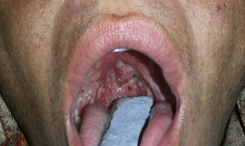
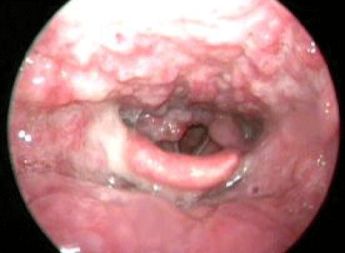
On oropharyngeal examination, multiple exophytic red granular tissues over uvula and posterior pharyngeal wall were noticed, with slough covered ulcers over soft palate in addition to thickened tonsillar pillars (Figure 1). The lesions were tender and bled on touch. The examination of larynx by angled rigid endoscope revealed similar lesions involving whole of posterior pharyngeal wall, right arytenoids and aryepiglottic fold, while the cords and epiglottis were spared (Figure 2). On neck examination, bilateral firm, mobile upper deep cervical lymph nodes sized 2x1.5 cm2 and left middle deep cervical 1x1cm2 were palpable. Patient was malnourished and weighed 40 kg at the time of admission.
Figure 1 : Clinical picture of oropharynx showing multiple exophytic red granular tissues over uvula and posterior pharyngeal wall, with slough covered ulcers over soft palate and thickened tonsillar pillars.
Figure 2 : Endoscopic picture showing granular lesion involving whole of posterior pharyngeal wall, right arytenoids and aryepiglottic fold.
Erythrocyte sedimentation rate (ESR) was elevated; 50mm/ hour (Westergren method) and the Tuberculin test yielded an induration of 18x 18 mm. Rest of the biochemical, hematological investigations were normal. Serology for HIV was negative. Throat swab demonstrated gram positive cocci in clusters, with no evidence of acid fast bacilli. Suspecting the lesion to be malignant, biopsy was taken from the friable part of the uvula.
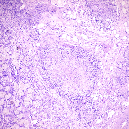
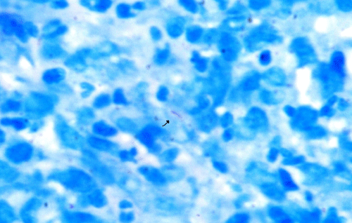
The histopathology showed tissue lined with stratified squamous epithelium with ulcerations and dense acute exudates. Subepithelial tissue showed ill defined epithelioid cell granulomas with Langhans and foreign body type giant cells along with area of caseation necrosis and fibrosis suggestive of TB. Surrounding tissue showed dense lymphoplasmacytic infiltrate and lobules of seromucinous glands (Figure 3). Zeihl- Neelsen (ZN) stain was positive for typical acid fast Mycobacterium tuberculosis (Figure 4). The sputum for AFB was negative.
Figure 3 : Microscopic picture of histopathology slide showing epithelioid cell granulomas with Langhans type giant cells. Surrounding tissue shows dense lymphoplasmacytic inflammatory infiltrate and seromucinous glands. (H&E 40X).
Figure 4 : High magnification microscopic picture showing presence of acid fast bacilli (Mycobacterium tuberculosis) on Zeihl -Neelsen stain. (100X).
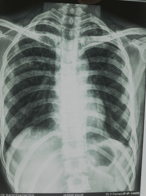
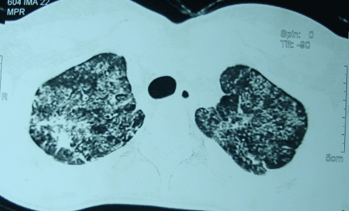
Chest X-ray showed bilateral discrete fine nodular densities (reticulonodular shadows) signifying miliary TB (Figure 5). Non- Contrast computed tomography of the chest showed miliary nodules involving both lungs diffusely, showing confluence at places with tree in bud pattern again indicative of miliary TB (Figure 6). Fine needle aspiration cytology from cervical node showed dense lymphoplasmacytic infiltrate with caseation necrosis.
Figure 5 : Chest X-ray showing bilateral discrete fine retico-nodular densities.
Figure 6 : The Plain Computed Tomogram Chest showed miliary nodules involving both lungs diffusely, showing confluence at places with tree in bud pattern.
The patient received two months of intensive antitubercular regimen consisting of isoniazid (200mg), rifampicin (400mg), pyrazinamide (1200mg) and ethambutol (600mg), all once daily, followed by four months of maintenance therapy with isoniazid and rifampicin. Within one month of starting antitubercular therapy patient showed improvement, with the dysphagia subsiding within two weeks of treatment and the oropharyngeal lesions showing resolution after one month of therapy.
Discussion
Tuberculous lesions of pharynx have become so infrequent that it is virtually a forgotten entity. The primary involvement of pharynx is very rare due to multiple factors. First, the bacterium is unable to invade the intact mucosa and the repeated swallowing action clears the throat. The presence of salivary enzymes, tissue antibodies and oral saprophytes also provide protection against such invasion. Any trauma or inflammatory condition or poor oral hygiene may provide a route for bacteria to invade [2].
When the oropharyngeal tissue is involved secondary to pulmonary disease, it can be by exogenous (infected sputum) or endogenous (blood stream) extension. The organ involved following endogenous spread includes lymph nodes, meninges, kidneys, adrenals, bones, fallopian tubes, and epididymis. The larynx and pharynx are typically infected by the bacilli in the sputum coughed up subsequent to lung infection. The oropharyngeal TB lesion consists of satellite ulcers with undermined edges and a granulating floor [3].
The similar case of laryngo-pharyngeal tuberculosis mentioned in literature had pregnant female presenting with odynophagia with neither clinical, radiological pulmonary findings nor immunocompromised [4].
The differential diagnoses of such atypical ulcerative lesions are reactive and traumatic ulcers, malignancy, deep fungal infections, sarcoidosis, and Wegener’s granulomatosis [1]. Usually on presentation the ulceroproliferative lesion appears to be malignant, especially when not responding to antibiotics. In such a situation tissue biopsy is indicated to trounce the diagnostic dilemma.
Conclusion
This case is an ideal illustration of the bizarre and atypical presentations of EPTB, re-establishing the fact that Mycobacteria can infect virtually any human organ. It is often challenging for the dealing physicians to discern the myriad presentations of TB. With the resurgence of tuberculosis worldwide, this sinister disease can no longer be overlooked.
References
- Dadgarnia MH, Baradaranfar MH, Yazdani N and Kouhi A. Oropharyngeal tuberculosis: an unusual presentation. Acta Medica Iranica. 2008; 46: 521-524.
- Iype EM, Ramdas K, Pandey M, Jayasree K, Thomas G, Sebastian P, et al. Primary tuberculosis of the tongue: report of three cases. Br J Oral Maxillofac Surg. 2001; 39: 402-403.
- Von Arx DP, Husain A. Oral tuberculosis. Br Dent J. 2001; 190: 420-422.
- Sá LC, Meirelles RC, Atherino CC, Fernandes JR, Ferraz FR. Laryngo-pharyngeal Tuberculosis. Braz J Otorhinolaryngol. 2007; 73: 862-866.
