Case Report
Austin J Clin Case Rep. 2014;1(6): 1028.
Renal Infarction Secondary to Renal Artery Ectasia
Yedla S and Ansari N*
Department of Medicine, Jacobi Medical Center, USA
*Corresponding author: Ansari N, Department of Medicine, Division of Nephrology, Jacobi Medical Center, 1400 Pelham Parkway South, Bronx, NY. 10461, USA
Received: June 14, 2014; Accepted: July 14, 2014; Published: July 17, 2014
Abstract
We describe a case of renal infarction secondary to renal artery ectasia. A 45 year old male presented to emergency department with sudden onset of left flank pain for one day. Physical examination was significant for tenderness in left upper quadrant and left costovertebral angle with no guarding. Laboratory tests revealed leukocytosis, creatinine 1.4mg/dl, high LDH, and microscopic hematuria. CT abdomen revealed wedge shaped hypodensities in left kidney compatible with renal infarction and focal nonenhancing regions within left renal artery suspicious for thrombus. Subsequent work-up was negative for cardiac and hypercoagulable causes of renal infarction. Urine toxic screen was negative for cocaine. Renal angiogram demonstrated bilateral renal artery ectasias, with left upper pole infarction. He was referred to vascular surgery for endovascular/ surgical intervention. Renal infarction typically occurs due to thromboembolic disease but rare causes like renal artery ectasia as in our patient should also be considered in the differential diagnosis.
Keywords: Renal Infarction; Renal artery aneurysm; Renal artery ectasia
Case Presentation
A 46 year old male, with past medical history of peptic ulcer disease (PUD) presented to the emergency department with severe left sided abdominal pain. The pain was colicky in nature, which progressively worsened over next two days. Patient had taken famotidine with no relief. He recalled having had subjective fevers, chills and intermittent dysuria during the week prior to presentation. His social and personal history was not significant.
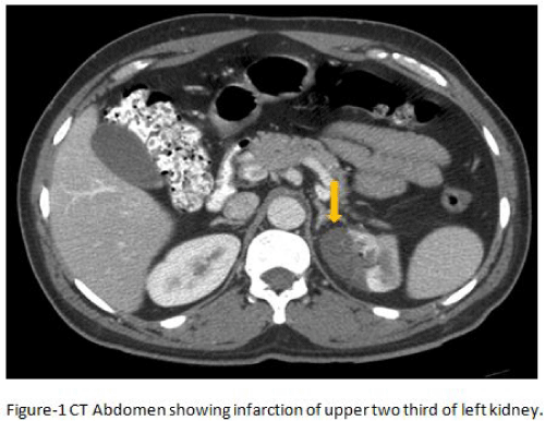
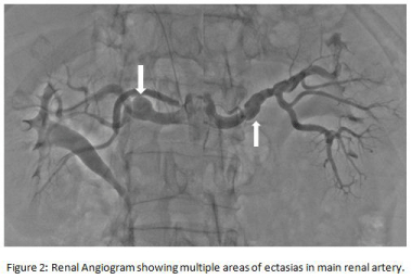
In emergency room his vital signs showed temperature 101.4F, heart rate 106 per minute, respiratory rate 18per minute, blood pressure 152/107mmHg, O2 saturation 97% (room air). On physical examination, he appeared extremely uncomfortable and diaphoretic lying flat in bed. Heart and lung examinations were normal. Abdomen was distended and hyperresonant with tenderness in left upper and lower quadrants. Left costovertebral tenderness was elicited on examination. His laboratory data revealed WBC 12.6 × 10⁹/l, creatinine 1.4 mg/dl, mild transaminitis, high lactate dehydrogenase (LDH-574U/L), and cholesterol 298 mg/dl, LDL 195 mg/dl, triglycerides 205 mg/dl, and HDL 53 mg/dl. Urine cocaine was absent on toxicology screen. EKG showed normal sinus rhythm. Urinalysis was positive for blood with no proteinuria. Sepsis work up was negative. CT abdomen showed wedge shaped hypodense areas along the medial aspect of the left kidney with perinephric stranding and non contrast enhancing regions within the left renal artery suspicious for thrombotic plaque (Figure 1). Further workup for renal infarction did not reveal any hypercoagulable state. Vasculitis work up was inconclusive. Holter monitoring showed normal sinus rhythm. Transthoracic and transesophageal echocardiogram did not show any evidence of vegetations or clots. Carotid duplex revealed no atherosclerotic plaque in both carotid arteries. Diagnostic renal angiogram demonstrated bilateral renal artery ectasia of 8mm in mid left renal artery and 6mm in left upper branch with left upper pole infarct. Right renal artery showed an aneurysm in mid portion measuring 11mm (Figure 2). The patient on admission was started on enoxaparin and transitioned to coumadin. He was discharged on aspirin and plavix due to the thrombotic event and referred to vascular surgery for further surgical intervention. Unfortunately the patient did not follow up with medical appointments after discharge from the hospital.
Figure 1 :
Figure 2 :
Discussion
Renal infarction is very rare (0.007%-1.4% in various studies) [6,7]. Because of its nonspecific presentation, it is often mistaken for other more common disorders such as ureterolithiasis, pyelonephritis, appendicitis and diverticulitis. Acute renal infarction should be suspected in all patients with persistent flank pain; especially in those at risk for thromboembolic events accompanied by an increase in LDH level [1]. Most of renal infarctions are diagnosed incidentally during radiological procedures performed to elucidate cause of acute abdominal pain.
Common causes of renal infarction include underlying cardiac diseases like atrial fibrillation (AF), ventricular aneurysm and dilated cardiomyopathy, cocaine use (both intravenous and nasal) [3] and hypercoagulable disorders [2,3,14]. Renal infarction can be associated with trauma and rare vascular anomalies like renal artery aneurysm/ renal artery ectasia. Atrial fibrillation and cocaine use are the major risk factors for renal infarction. Acute embolic renal infarction can be seen in 2% of patients with AF [12].
Flank pain and upper abdominal pain are the common symptoms in renal infarction often accompanied with fever, nausea or vomiting. Occasionally, patients may have gross hematuria [8]. Blood pressure may be acutely elevated in some patients due to activation of renin-angiotensin mechanism. Physical examination can reveal costovertebral angle tenderness. Serum LDH is the most sensitive marker for renal infarction [15,16]. Proteinuria and hematuria can be seen in some patients [13]. Several other serum markers can be elevated in renal infarction like alkaline phosphatase, fibrinogen, C-reactive protein and aspartate, but their sensitivity is low.
Imaging studies remain the main diagnostic tool for renal infarction. Contrast enhanced CT scan, isotope scan, and angiography is helpful in diagnosing renal infarction. The sensitivity of these imaging studies vary and were found to be 80%, 97% and 100% for computed tomography(CT), renal isotope and angiography respectively [13]. Though renal angiography remains the gold-standard for diagnosis, abdominal contrast CT is the diagnostic modality of choice at this time. The characteristic findings in CT scan are usually a wedge-shaped, peripheral, non enhancing area seen on the affected kidney.
Renal artery aneurysms, previously thought to be uncommon, have been encountered more frequently in recent years. The Incidence of renal artery aneurysm is 0.01% and 1% in both autopsy and angiographic studies [10]. Common causes of renal artery aneurysm include congenital malformation of the kidneys or associated vessels, atherosclerosis, fibromuscular dysplasia, polyarteritis nodosa, pregnancy, and trauma [11]. These aneurysms can be unilateral or bilateral. Aneurysms can occur in main renal artery or its main branches and can be fusiform or saccular in shape. Most of the patients with renal artery aneurysm are asymptomatic. Complications of renal artery aneurysms appear to be rare and can result in renal infarction due to thrombosis, renovascular hypertension, dissection of aneurysm and arteriovenous fistula formation [11]. Renal artery aneurysms can be diagnosed with 3D Contrast enhanced magnetic resonance angiography (CE MRA), spiral CT, and duplex sonography [22,5]. Contrast enhanced magnetic resonance angiography remains preferable to other non-invasive modalities in initial diagnosis, therapeutic planning and follow-up [22,4].
Treatment of renal artery aneurysm/ectasia is indicated to prevent potential complications such as renal artery thrombosis, renal infarction and rupture of aneurysm. Treatment is indicated for aneurysms greater than 2.0 cm in diameter and for symptomatic patients with flank pain or hematuria, pregnancy, refractory hypertension caused by the aneurysm and expanding aneurysms [19- 21]. Surgical management consists of aneurysmectomy with or without revascularization. With current improvements in endovascular technology, multiple techniques like transcatheter embolization and stent grafts have become popular treatment modality. However, the treatment modality needs to be individualized to the pertinent renal vascular anatomy with available technology [9].
Early diagnosis and timely intervention of renal infarction can minimize the loss of renal function [17]. In majority of patients, satisfactory outcomes can be achieved through anticoagulation with heparin followed by warfarin [13]. Heparin use alone often results in some degree of renal recovery with minimal morbidity [18]. Thrombolysis and thrombectomy had been shown to be beneficial for improving renal function in few patients with unilateral and bilateral renal artery occlusion [10].
Conclusion
Renal infarction should be suspected in a patient with persistent flank or abdominal pain with elevated serum LDH and/or hematuria who are at high risk for a thromboembolic event. Contrast enhanced CT scan should be performed to make the diagnosis. Renal angiography may be indicated in patients with renal infarction suspected to be due to underlying renal vascular abnormality. Prompt recognition and timely intervention of acute renal infarction is important since anticoagulation, thrombolysis or embolectomy may reduce the loss of renal function and future risk of chronic kidney disease.
References
- Huang CC, Lo HC, Huang HH, Kao WF, Yen DH, Wang LM, et al. ED presentations of acute renal infarction. Am J Emerg Med. 2007; 25: 164-169.
- Lessman RK, Johnson SF, Coburn JW, Kaufman JJ. Renal artery embolism: clinical features and long-term follow-up of 17 cases. Ann Intern Med. 1978; 89: 477-482.
- Mochizuki Y, Zhang M, Golestaneh L, Thananart S, Coco M. Acute aortic thrombosis and renal infarction in acute cocaine intoxication: a case report and review of literature. Clin Nephrol. 2003; 60: 130-133.
- Vosshenrich R, Fischer U. Contrast-enhanced MR angiography of abdominal vessels: is there still a role for angiography? Eur Radiol. 2002; 12: 218-230.
- Shonai T, Koito K, Ichimura T, Hirokawa N, Sakata K, Hareyama M. Renal artery aneurysm: evaluation with color Doppler ultrasonography before and after percutaneous transarterial embolization. J Ultrasound Med. 2000; 19: 277–280.
- Hoxie HJ, Coggin CB. Renal Infarction: Statistical study of two hundred and five cases and detailed report of an unusual case. Arch Intern Med. 1940; 65: 587.
- Paris B, Bobrie G, Rossignol P, Le Coz S, Chedid A, Plouin PF. Blood pressure and renal outcomes in patients with kidney infarction and hypertension. J Hypertens. 2006; 24: 1649-1654.
- Bande D, Abbara S, Kalva SP. Acute renal infarction secondary to calcific embolus from mitral annular calcification. Cardiovasc Intervent Radiol. 2011; 34: 647-649.
- Etezadi V, Gandhi RT, Benenati JF, Rochon P, Gordon M, Benenati MJ, et al. Endovascular treatment of visceral and renal artery aneurysms. J Vasc Interv Radiol. 2011; 22: 1246-1253.
- Walker JS, Dire DJ. Vascular abdominal emergencies. Emerg Med Clin North Am. 1996; 14: 571-592.
- Lumsden AB, Salam TA, Walton KG. Renal artery aneurysm: a report of 28 cases. Cardiovasc Surg. 1996; 4: 185-189.
- Meyrier A, Hill GS, Simon P. Ischemic renal diseases: new insights into old entities. Kidney Int. 1998; 54: 2-13.
- Hazanov N, Somin M, Attali M, Beilinson N, Thaler M, Mouallem M, et al. Acute renal embolism. Forty-four cases of renal infarction in patients with atrial fibrillation. Medicine (Baltimore). 2004; 83: 292-299.
- Domanovits H, Paulis M, Nikfardjam M, Meron G, Kürkciyan I, Bankier AA, et al. Acute renal infarction. Clinical characteristics of 17 patients. Medicine (Baltimore). 1999; 78: 386-394.
- Antopolsky M, Simanovsky N, Stalnikowicz R, Salameh S, Hiller N. Renal infarction in the ED: 10-year experience and review of the literature. Am J Emerg Med. 2012; 30: 1055-1060.
- Kansal S, Feldman M, Cooksey S, Patel S. Renal artery embolism: a case report and review. J Gen Intern Med. 2008; 23: 644-647.
- Safian RD, Textor SC. Renal-artery stenosis. N Engl J Med. 2001; 344: 431-442.
- Gasparini M, Hofmann R, Stoller M. Renal artery embolism: clinical features and therapeutic options. J Urol. 1992; 147: 567-572.
- Calligaro K, Dougherty M. Renal artery aneurysms and Arteriovenous fistulae. Vascular Surgery 6th ed. Rutherford R, editor. Elsevier; Pennsylvania. 2005; 1861-70.
- Henke PK, Cardneau JD, Welling TH 3rd, Upchurch GR Jr, Wakefield TW, Jacobs LA, et al. Renal artery aneurysms: a 35-year clinical experience with 252 aneurysms in 168 patients. Ann Surg. 2001; 234: 454-462.
- English WP, Pearce JD, Craven TE, Wilson DB, Edwards MS, Ayerdi J, et al. Surgical management of renal artery aneurysms. J Vasc Surg. 2004; 40: 53-60.
- Fink C, Hallscheidt PJ, Hosch WP, Ott RC, Wiesel M, Kauffmann GW, et al. Preoperative evaluation of living renal donors: value of contrast-enhanced 3D magnetic resonance angiography and comparison of three rendering algorithms. Eur Radiol. 2003; 13: 794-801.

