Case Report
Austin J Clin Case Rep. 2014;1(7): 1034.
Minimally Invasive Approach of Bladder Paraganglioma: Case Report and Review of Literature
Matthias Beysens1, Bruno Lapauw2, Pieter-Jan Gykiere3, Sven Dekeyzer4, Nicolaas Lumen1 and Karel Decaestecker1*
1Department of Urology, University Hospital Ghent, Belgium
2Department of Endocrinology, University Hospital Ghent, Belgium
3Department of Nuclear Medicine, University Hospital Ghent, Belgium
4Department of Radiology, University Hospital Ghent, Belgium
*Corresponding author: Karel Decaestecker, Department of Urology, University Hospital Ghent, De Pintelaan 185, 9000 Ghent, Belgium.
Received: July 10, 2014; Accepted: Aug 05, 2014; Published: Aug 07, 2014
Abstract
Purpose: Paraganglioma of the bladder is a rare neoplasm that usually presents with hypertension, headache and dizziness after micturition.
Case Presentation: We present the case of a 22-year-old man with typical symptoms of bladder paraganglioma.
We performed the first robot-assisted laparoscopic tumorectomy without opening of the bladder mucosa. Additionally, we give an overview of the literature on minimally invasive approaches for bladder paraganglioma.
Conclusions: This article gives an overview of the symptoms, diagnostic tools and surgical treatment of bladder paraganglioma. It is a rare neoplasm that, however it has a typical presentation, can be easily missed based on patient history alone.
Keywords: Bladder paraganglioma; Pheochromocytoma; Minimally invasive surgical procedure
Introduction
Pheochromocytoma is a neuro-endocrine tumor that usually develops in the adrenal medulla. Extra-adrenal pheochromocytomas, also called paragangliomas (which account for 5% to 10% of pheochromocytomas), are most often found in the retroperitoneum, thorax and urinary bladder [1].
In the bladder wall it originates from the sympathetic plexus of the detrusor muscle. Bladder paragangliomas are extremely rare and constitute less than 0,05% of bladder tumors [2].
Typical symptoms are headache, palpitations, sweating, blurred vision and hypertension, all related to the act of micturition [1].
Diagnosis of a paraganglioma can be made in almost all patients with the demonstration of elevated measurements of plasma fractionated metanephrines and/or urinary plasma fractionated metanephrines and catecholamine’s (measured on a 24-hour urine collection).
Nuclear imaging in combination with computer tomography (CT) or magnetic resonance imaging (MRI) may be required to localize and fully delineate the extent of the disease.
Standard treatment is open partial or radical cystectomy. Minimal approaches are reported, but only in case reports.
We give a literature overview of all minimal-invasive cases and report the first case of a robot-assisted laparoscopic tumorectomy without opening the bladder.
Case Presentation
A 22-year-old man was referred to our urological department with complaints of severe headache, palpitations and dizziness after micturition occurring for 2 months. His complaints were initially this severe, that he even refused to micturate and a transurethral catheter was placed in another hospital. His medical history and physical examination were completely normal. He had no urinary tract symptoms and no hematuria.
A 24-hour urine collection was performed and revealed an elevated amount of norepinephrine, together with elevated normetanephrine levels on a plasma sample, suggesting a paraganglioma.
Imaging
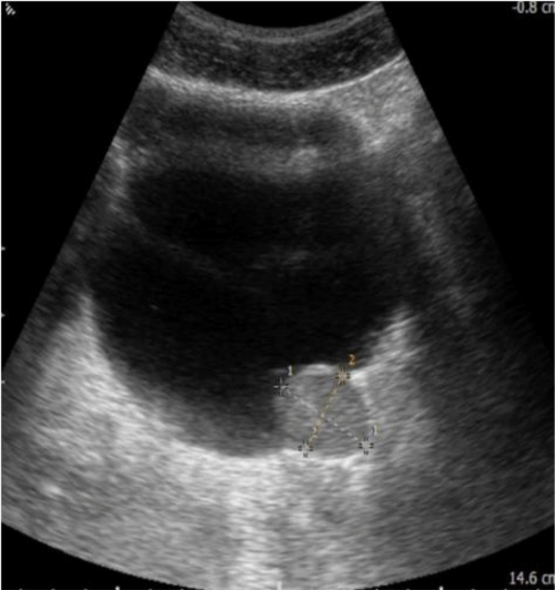
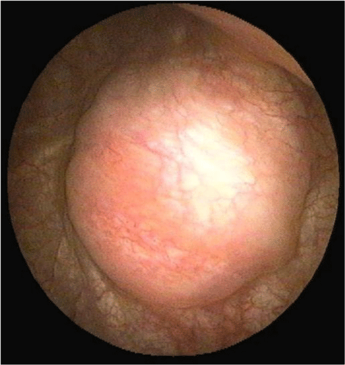
On ultrasonography, a 2x3-cm, slightly hypoechogenic mass was seen embedded in the left detrusor wall (Figure 1). On cystoscopy, the same mass was seen lying under the mucosa in the left side of the bladder (Figure 2). The lesion was estimated to be 5 cms away from the ureteral orifices.
Figure 1 : Ultrasonography.
Figure 2 : Cystoscopic view.
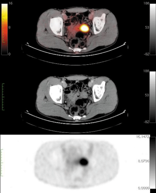
A preoperatively performed 18F-fluorodeoxyglucose (FDG) positron emission tomography-computed tomography (PET-CT) scan showed no other PET-positive locations than the tumor in the bladder wall (Figure 3).
Figure 3 : FDG PET-CT.
Pre-operative preparation
The patient was admitted three days before surgery. Preoperative preparation with alpha-adrenergic blocking agents (phenoxybenzamine 2x10mg/day) and pre-operative fluid loading (NaCl 0.9% 2L/24h) was given to restore the arterial blood pressure and intravascular volume to normal levels in order to prevent intraoperative and post-operative blood pressure fluctuations.
Operative technique
Under general anesthesia, a robot-assisted laparoscopy was performed using a 4-armed Da Vinci Si platform (Intuitive Surgical). We used our common “robot-assisted laparoscopic radical prostatectomy” patient positioning and port placement. The patient was installed in the lithotomy position. The procedure started with the placement of a 12-mm umbilical port for the camera. Pneumoperitoneum was achieved to a pressure of 12mmHg. Three additional 8-mm ports were placed under direct vision for the 3 robot arms (one in the right iliac fossa and 2 in the left iliac fossa) and a 12-mm and 5-mm port for the assistant in the right iliac fossa and near to the camera port respectively. The patient was placed in 30O Trendelenburg position.
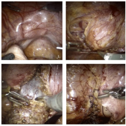
Inspection of the pelvis showed a well-marginated extraperitoneal mass in the left side of the bladder wall. The peritoneum was incised along the left medial umbilical ligament and lifted from the bladder and the mass. The left vas deferens, ureter and neurovascular bundle were identified. Then the dissection of the tumor was started, without direct manipulation to avoid hypertensive crisis. Blood supply to the tumor was sealed with an ultrasonic/bipolar device (Thunderbeat, Olympus). The mass was encircled with a combination of this sealing device and unipolar electrocoagulation. It showed to be well embedded in detrusor muscle; a safety margin of normal detrusor tissue was left on the tumor. However, there was a clear margin between the tumor and the bladder mucosa (Figure 4). As a result, the tumor could be macroscopically completely removed without opening the bladder. The specimen was put in an endocatch bag. The detrusor layer of the bladder was closed in two layers with an absorbable polyfilament suture (Vicryl 2/0). Filling the bladder with 200cc of water showed no leakage or diverticula. The specimen was removed by extending the incision of the 12-mm assistant port in the left fossa and sent for anatomopathological examination.
Robot-assisted laparoscopic tumorectomy.
- Intraperitoneal view of the bladder with tumoral mass (asterix) on the left side.
- Starting dissection with identifying and controlling the vascularisation of the paraganglioma, with minimal manipulation of the tumour.
- Detrusurotomy and dissecting the plane between tumour and bladder mucosa.
- After removal of the tumour, the detrusor was closed with a running suture and a bladder filling test showed no leakage or diverticula.
Figure 4 :Robot-assisted laparoscopic tumorectomy.
- Intraperitoneal view of the bladder with tumoral mass (asterix) on the left side.
- Starting dissection with identifying and controlling the vascularisation of the paraganglioma, with minimal manipulation of the tumour.
- Detrusurotomy and dissecting the plane between tumour and bladder mucosa.
- After removal of the tumour, the detrusor was closed with a running suture and a bladder filling test showed no leakage or diverticula.
During the operation, there was only a short and slight rise in arterial blood pressure (160mmHg systolic) following minimal tumor manipulation that could be easily controlled by the administration of antihypertensive drugs (sodium nitroprusside maximum of 5mcg/ kg/min).
Post-operative period
The patient made a quick post-operative recovery. Immediately post-operative, he was slightly hypotensive (90mmHg systolic), but within six hours the patient became and remained normotensive without any further medication. The urinary catheter was removed and he could leave the hospital on the second postoperative day without the need for further antihypertensive treatment.
Follow-up
A clinical follow-up visit 3 weeks postoperatively showed normal blood pressure and absence of symptoms relating to a hypertensive crisis or related to micturition. The patient showed no lower urinary tract symptoms related to the robot-assisted laparoscopic bladder surgery. A new 24-hour urine collection showed normal amounts of catecholamines.
Histopathological report
The final histopathological examination showed a paraganglioma embedded in detrusor muscle. The tumor measured 2.5x2.2x1.8 cm and there were negative surgical margins.
The tumoral mass consisted of groups of epithelioid cells lying in an organoid pattern (so called ‘Zellballen’ or cell clusters). These groups of cells were divided by thin fibrovascular septae. Immunohistochemical staining was used to confirm the definitive diagnosis. NSE (neuron-specific enolase), chromogranin and synaptophysine were strongly positive in the neoplastic cells, all neuroendocrine markers. SDHB (succinate dehydrogenase B-subunit)-staining was negative. Further exploration with genetic testing of the SDHB-gene is ongoing. There was no family history of pheochromocytoma/paraganglioma syndrome.
Discussion
Pheochromocytoma is a neuro-endocrine tumor that usually develops in the adrenal medulla. It is believed to arise from embryonic rests of chromaffin cells in the sympathetic plexus. Extra-adrenal pheochromocytomas, also called paragangliomas (which account for 5% to 10% of pheochromocytomas), are most often found in the retroperitoneum, thorax and urinary bladder [1]. Bladder paragangliomas are extremely rare and constitute less than 0,05% of bladder tumors [2]. They may occur more frequently with familial syndromes, including the von Hippel-Lindau (VHL) syndrome and succinate dehydrogenase B (SDHB) mutations [3].
Typical symptoms (present in 65% to 75% of patients) are headache, palpitations, sweating, blurred vision and hypertension, all related to the act of micturition. These symptoms are caused by an increased catecholamine release due to compression of the tumor in the bladder wall. Painless hematuria is also a common symptom, present in 55% to 58% of cases [1].
Diagnosis of a paraganglioma can be made in almost all patients with the demonstration of elevated measurements of plasma fractionated metanephrines and/or urinary plasma fractionated metanephrines and catecholamine’s (measured on a 24-hour urine collection).
Nuclear imaging in combination with computer tomography (CT) or magnetic resonance imaging (MRI) may be required to localize and fully delineate the extent of the disease. 123/131I-metaiodobenzylguanidine (MIBG) single photon emission tomography (SPECT) is the conventional nuclear imaging technique for catecholamine-secreting tumors with a sensitivity of 80 – 95% and a specificity of 90 – 95% for non-metastatic paraganglioma. MIBG is a guanidine analogue, which is chemically similar to norepinephrine, and accumulates in adrenergic cells. Positron emission tomography (PET) with a variety of different tracers is superior to SPECT due to a higher sensitivity. Most paragangliomas, as the one in our case, show avidity for 18F-fluorodeoxyglucose (FDG), a glucose analogue - even in cases with a low proliferation rate and 6-(18F)fluoro-L-dihydroxyphenylalanine (DOPA), the catecholamine precursor [4,5].
A biopsy is not recommended, as this could lead to a hypertensive crisis. A transurethral resection of the tumor entails the same risk.
Careful preoperative preparation is very important. Intravascular volume is contracted from the chronic sustained effect of the elevated catecholamine levels and should be re-expanded prior to removal of the neoplasm. If not, profound postoperative hypotension may result. Blood pressure is first controlled with alpha-blockade using phenoxybenzamine. Beta-blockade may be added to counteract the rebound tachycardia of the former [6]. Blood pressure control and a skilled anesthesia team are essential during operation. Surges in arterial blood pressure can be managed with titrated doses of sodium nitroprusside and nitroglycerine and boluses of esmolol and labetalol [7].
Standard treatment for localized or locally advanced bladder paraganglioma is surgery.
Approximately 10% of paragangliomas/pheochromocytomas are malignant. Physicians should be aware of poor prognostic indicators, such as large tumor size, advanced stage, SDHB-mutation and multifocal tumors [2]. If the tumor is limited to the bladder and is benign-looking, partial resection of the bladder can be performed, keeping the tumor intact [2]. Whether a pelvic lymphadenectomy should be done in every case is not clear. In case of proven malignancy, a pelvic lymphadenectomy can be performed in a secondary procedure. For more advanced disease (extravesical extension or clear pelvic lymphadenopathy), radical cystectomy with pelvic lymph nodal dissection has been advocated. In case of metastatic disease, surgical treatment is rarely curative, but it may prolong survival by reducing comorbid conditions, like severe hypertension [2]. Adjunct therapeutic options are limited, including 131I-MIBG radiation therapy for MIBG-avid metastases and combination chemotherapy (cyclophosphamide, vincristine and dacarbazine) [6].
Histologically, it is almost impossible to distinguish malignant and benign tumors. In the literature, cases are reported of patients who showed metastases of a malignant paraganglioma several decades after partial cystectomy [8]. For this reason, life-long follow-up is always necessary, even when the initial lesion is solitary and the surgical resection is complete. A review in 2013 by Beilan et al. showed that 15 of 75 patients (20%) had local or distant disease recurrence after a mean follow-up of 35 months. Patients who presented with localized paraganglioma of the bladder had a significantly improved survival compared to patients who presented with locally advanced or metastatic disease [2].
No clear guidelines exist on how to do follow-up. Examinations can include monitoring of urinary vanillylmandelic acid (VMA), metanephrine and catecholamine levels or imaging of abdomen and pelvis. Others recommend an annual MIBG-scan [6].
Because of the small number of cases and the smaller number of cases followed up, the prognosis of urinary bladder paraganglioma is not well established. Overall 5-year survival for patients with metastatic pheochromocytoma or paraganglioma is approximately 50% [9].
More than 180 cases of bladder paraganglioma have been reported in the literature, but reports of minimally invasive surgical approach are sparse. In total, only 11 cases of (robot-assisted) laparoscopic partial cystectomy for bladder paraganglioma have been published [1,3,10-18] (Table 1). Kozlowski et al. performed the first laparoscopic partial cystectomy for a bladder paraganglioma in 2001 [11]. They combined a laparoscopic with a transurethral approach. Bozbora et al. were the first to only use laparoscopy for partial cystectomy for a bladder paraganglioma [1]. In 2009, Huang et al described a pre-peritoneal laparoscopic approach for a bladder paraganglioma [14].
    Authors
    Year
    Approach
    Tumor    localization
    Operation


    Kozlowski et    al [11]
    2001
    Laparoscopy+transurethral    cystotomy
    Anterolateral    left
    Partial    cystectomy


    Gaur et al [18]
    2002

    Laparoscopy+transurethral    cystotomy
    Posterior    wall
    Partial    cystectomy


    Bozbora et    al [1]
    2006
    Laparoscopy
    Anterolateral    right
    Partial    cystectomy


    Dilbaz et al [17]


    2006
    Laparoscopy
    Left bladder    wall
    Partial    cystectomy


    Dhawan et al [16]
    2008
    Laparoscopy    with laparoscopic ultrasonography
    Posterolateral    right
    Partial    cystectomy


    Huang et al [14]
    2009
    Pre-peritoneal    laparoscopy

    Partial cystectomy


    Xu et al [15]
    2010
    Laparoscopy
    Bladder dome
    Partial    cystectomy


    Nayyar et al [12]
    2010
    Robot-assisted    laparoscopy
      +transurethral    cystotomy
    Left bladder    wall incl VUJ
    Partial    cystectomy, distal ureterectomy and ureteric reimplantation


    Kang et al [13]
    2011
    Robot-assisted    laparoscopy
    Anterior    bladder wall
    Partial    cystectomy + bilateral pelvic lymphadenectomy


    Luchey et al [10]
    2012
    Robot-assisted    laparoscopy

    Partial    cystectomy + lymphadenectomy


    Weintraub [3]
    2013
    Robot-assisted    laparoscopy +cystoscopy
    Anterior    bladder wall
    Partial    cystectomy
Table 1: Overview of literature on minimally invasive approach for bladder paraganglioma.
In 2010, Nayyar et al. reported the first robot-assisted partial cystectomy, distal ureterectomy and ureteric reimplantation for a paraganglioma in the vesicoureteric junction [12]. In 2011, Kang et al. also reported on a robot-assisted partial cystectomy for bladder paraganglioma [13]. In 2012, Luchey et al. performed a robot-assisted partial cystectomy and pelvic lymphadenectomy for metastatic bladder paraganglioma [10]. More recently, a video of partial cystectomy for bladder paraganglioma was published by Weintraub et al. [3]. To our knowledge, this case is only the fifth robot-assisted laparoscopic partial cystectomy performed for bladder paraganglioma. To be precise, we performed a robot-assisted laparoscopic tumorectomy for paraganglioma in the bladder wall.
Originally, we planned to do a classic partial cystectomy, but as the tumor presented with a clear margin between detrusor and bladder mucosa, we decided to leave the bladder mucosa unopened. This was made possible by the fine manipulation and perfect view that can be only achieved laparoscopically with a robot-assisted approach. This entails a shorter need for post-operative transurethral urinary catheterisation and significant reduction in morbidity for the patient. Clear resection margins were achieved and the tumor was removed intact, with results probably comparable to classic open surgical approach. Because of the low malignant potential of this type of tumors (<10%) and negative additional imaging techniques, we decided not to perform a pelvic lymphadenectomy. However, a life-long follow-up is necessary.
Conclusion
Paraganglioma of the bladder wall is a very rare tumor with a typical presentation. However, it could be easily missed by clinicians who are not aware of this symptomatology. We present this case to demonstrate the feasibility of a minimally invasive approach for this kind of tumors. We performed a tumorectomy without the need for a partial cystectomy, which was made possible by a robot-assisted laparoscopic intervention and resulted in minimal operative morbidity for the patient and complete relief of his symptoms.
References
- Bozbora A, Barbaros U, Erbil Y, Kiliçarslan I, Yildizhan E, Ozarmagan S, et al. Laparoscopic treatment of hypertension after micturition: Bladder pheochromocytoma. JSLS. 2006; 10: 263-266.
- Beilan JA, Lawton A, Hajdenberg J, Rosser CJ. Pheochromocytoma of the urinary bladder: a systematic review of the contemporary literature. BMC Urol. 2013; 13: 22.
- Weintraub SM, Ricketts CJ, Vourganti S, Shuch B, Nix JW, Stamatakis L, Rosen P, Linehan WM, Agarwal PK. Robot-Assisted Partial Cystectomy for Treatment of Bladder Paraganglioma. Videourology, 2013.
- Timmers HJ, Chen CC, Carrasquillo JA, Whatley M, Ling A, Havekes B, et al. Comparison of 18F-fluoro-L-DOPA, 18F-fluoro-deoxyglucose, and 18F-fluorodopamine PET and 123I-MIBG scintigraphy in the localization of pheochromocytoma and paraganglioma. J Clin Endocrinol Metab. 2009; 94: 4757-4767.
- TaÏeb D, Neumann H, Rubello D, Al-Nahhas A, Guillet B, HindiÉ E, et al. Modern nuclear imaging for paragangliomas: beyond SPECT. J Nucl Med. 2012; 53: 264-274.
- Pahwa HS, Kumar A, Srivastava R, Rai A. Unsuspected pheochromocytoma of the urinary bladder: reminder of an important clinical lesson. BMJ Case Rep. 2012; 2012.
- Pandey R, Garg R, Roy K, Darlong V, Punj J, Kumar A, et al. Perianesthetic management of the first robotic partial cystectomy in bladder pheochromocytoma. A case report. Minerva Anestesiol. 2010; 76: 294-297.
- Kato H, Suzuki M, Mukai M, Aizawa S. Clinicopathological study of pheochromocytoma of the urinary bladder: immunohistochemical, flow cytometric and ultrastructural findings with review of the literature. Pathol Int. 1999; 49: 1093-1099.
- Huang H, Abraham J, Hung E, Averbuch S, Merino M, Steinberg SM, et al. Treatment of malignant pheochromocytoma/paraganglioma with cyclophosphamide, vincristine, and dacarbazine: recommendation from a 22-year follow-up of 18 patients. Cancer. 2008; 113: 2020-2028.
- Luchey A, Zaslau S, Talug C. Robotic assisted partial cystectomy with pelvic lymph node dissection for metastatic paraganglioma of the urinary bladder Can J Urol. 2012; 19: 6389-6391.
- Kozlowski PM, Mihm F, Winfield HN. Laparoscopic management of bladder pheochromocytoma. Urology. 2001; 57: 365.
- Nayyar R, Singh P, Gupta NP. Robotic management of pheochromocytoma of the vesicoureteric junction. JSLS. 2010; 14: 309-312.
- Kang SG, Kang SH, Choi H, Ko YH, Park HS, Cheon J, et al. Robot-assisted partial cystectomy of a bladder pheochromocytoma. Urol Int. 2011; 87: 241-244.
- Huang Y, Tian XJ, Ma LL. Pre-peritoneal laparoscopic partial cystectomy of the bladder pheochromocytoma. Chin Med J (Engl). 2009; 122: 1234-1237.
- Xu DF, Chen M, Liu YS, Gao Y, Cui XG. Non-functional paraganglioma of the urinary bladder: a case report. J Med Case Rep. 2010; 4: 216.
- Dhawan DR, Ganpule A, Muthu V, Desai MR. Laparoscopic management of calcified paraganglioma of bladder. Urol J. 2008; 5: 126-128.
- Dilbaz B, Bayoglu Y, Oral S, Cavusoglu D, Uluoglu O, Dilbaz S, et al. Laparoscopic resection of urinary bladder paraganglioma: a case report. Surg Laparosc Endosc Percutan Tech. 2006; 16: 58-61.
- Gaur DD, Sinha RY, Trivedi S, Prabudesai MR. Combined laparoscopic and transurethral partial cystectomy for juxta-ureteral pheochromocytoma of the bladder. Minim Invasive Ther Allied Technol, 2002; 11: 19-22.


