Case Report
Austin J Clin Case Rep. 2014;1(8): 1036.
Multiple Recurrent Congenital Mesoblastic Nephroma (CMN) in an Infant
Govani DR1, Patel RV2 and Philip I2*
1Medical Student, University of Birmingham Medical School, UK
2Department of Pediatric Surgery, Royal Belfast Hospital for Sick Children, UK
*Corresponding author: Isaac Philip, Department of Pediatric Surgery, Royal Belfast Hospital for Sick Children, 180-184 Falls Road, Belfast, County AntrimBT12 6BE, Northern Ireland, UK
Received: July 10, 2014; Accepted: Aug 10, 2014; Published: Aug 11, 2014
Letter to the Editor
We wish to present a case of multiple recurrent congenital mesoblastic nephroma (CMN) in an infant and indicate that adjuvant chemotherapy consisting of doxorubicin and carboplatin is of benefit for long-term disease-free survival in recurrent CMN cases.
A one year-old girl born at 36/40 weeks gestation was noted to have increasing abdominal distension, pallor and weight loss of 4 weeks duration. She looked pale and was hypertensive (157/97). A firm non-tender mass with visible engorged veins and smooth surface was palpable in whole of the right side of abdomen extending from rib cage to the iliac crest. It was crossing the midline and measured 13 cm in diameter.
Full blood count showed hemoglobin of 110 g/L biochemistry including renal and liver functions, bone profile and tumour markers (alpha fetoproteins, beta human chorionic gonadotropins) were normal except elevated calcium at 3mmol/L. A radiograph of the chest and abdomen suggested a large mass in the right side of the abdomen (Figure 1). An ultrasound scan of abdomen showed a large mass occupying almost the entire right side of the abdomen with solid and cystic change within it surrounded by a pseudo capsule arising from the site of the right kidney. Left kidney was normal and measured 73mm in length.
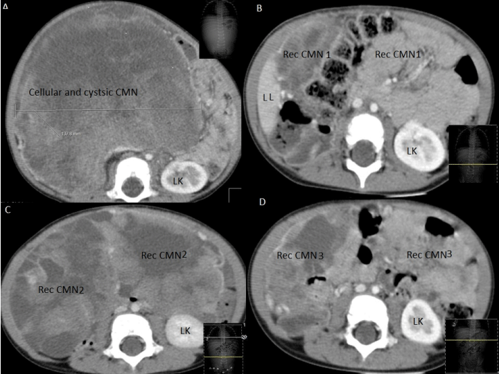
CAT scan of chest and abdomen (Figure 1A) showed features of congenital mesoblastic nephroma (CMN) with solid and cystic components. There was a large abdominal mass extending from the right hypochondrium into the pelvis, that filled the entire right side of the abdomen and central part of the abdomen. It originated from the right kidney; enhancing renal cortex was visible stretched around the periphery of the kidney posterolaterally. The mass was of mixed attenuation, it has areas of enhancement and large areas of low attenuation in keeping with necrosis. The abdominal aorta was displaced antero laterally to the left by the mass. The inferior vena cava was displaced and compressed by the mass.
Figure 1 : Babygram- Note massive right abdominal mass displacing bowel gas and crossing the midline.
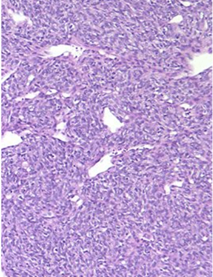
CMN with predominant cellularity was confirmed by ultrasound (USS) guided needle biopsy. She was started on atenolol for hypertension and IV fluids for hypercalcaemia. She underwent right radical nephrectomy and discharged home on day five. She was monitored with USS and the first recurrence was noted after 6 months (Figure 1B). This was unresponsive to first-line chemotherapy and underwent excision of recurrent CMN.
A second recurrence followed in 2 months in the paracolic region (Figure 1C) and she underwent re-excision. She had full metastatic work up including bone scans which were negative.
She developed a 3rd recurrence in another 3 months abutting left lobe of liver, posterior to the stomach encircling splenic vessels (Figure 1D) and underwent excision of recurrent CMN with splenectomy. The original radical nephrectomy and recurrent resections were macroscopically complete but microscopically, the tumour marginswere involved. She has been commenced on adjuvant chemotherapy consisting of doxorubicin and carboplatin for 3 cycles and she is recurrence free at 31 months of age.
Figure 2 : CT scan showing A. original CMN B 1st recurrence C. 2nd recurrence, D. 3rd recurrence, LK=Left Kidney, LL=Left lobe of liver, Rec=Recurrent, CMN=Congenital mesoblastic nephroma.
Figure 3 : Microphotograph showing cellularity in the background of congenital mesoblastic nephroma.
CMN is a benign tumour and nephrectomy is usually curative. However, local recurrence and/or distal metastasis have been reported and adjuvant chemotherapy and /or radiotherapy may be useful [1-3]. There were only two mixed CMN in the United Kingdom Children’s Cancer and Leukaemia Group (UKCCLG) series [4]. Sarcoma based chemotherapy has good effect in such cases [5].
CMN is usually a benign renal tumour and multiple local recurrences are extremely uncommon. Although CMN is usually cured by surgery with nephrectomy those with mixed cellularity are the rarest with tendency to have local recurrence. Routine adjuvant treatment is not routinely necessary. However, our case indicates that chemotherapy is of benefit and can produce long-term disease-free survival in recurrent disease.
References
- Loeb DM, Hill DA, Dome JS. Complete response of recurrent cellular congenital mesoblastic nephroma to chemotherapy. J Pediatr Hematol Oncol. 2002; 24: 478-481.
- Levin NP, Damjanov I, Depillis VJ. Mesoblastic nephroma in an adult patient: recurrence 21 years after removal of the primary lesion. Cancer. 1982; 49: 573-577.
- Steinfeld AD, Crowley CA, O'Shea PA, Tefft M. Recurrent and metastatic mesoblastic nephroma in infancy. J Clin Oncol. 1984; 2: 956-960.
- England RJ, Haider N, Vujanic GM, Kelsey A, Stiller CA, Pritchard-Jones K, et al. Mesoblastic nephroma: a report of the United Kingdom Children's Cancer and Leukaemia Group (CCLG). Pediatr Blood Cancer. 2011; 56: 744-748.
- Patel Y, Mitchell CD, Hitchcock RJ. Use of sarcoma-based chemotherapy in a case of congenital mesoblastic nephroma with liver metastases. Urology. 2003; 61: 1260.


