Case Report
Austin J Clin Case Rep. 2014;1(8): 1037.
Bilateral Taurodontism in Primary Molar: a Case Report
Nagpal R1* and Manuja N2
1Department of Conservative Dentistry, Kothiwal Dental College and Research Centre, India
2Department of Pediatric Dentistry, Kothiwal Dental College and Research Centre, India
*Corresponding author: Nagpal R, Department of Conservative Dentistry, Kothiwal Dental College and Research Centre, Moradabad-244001, Uttar pradesh, India
Received: July 10, 2014; Accepted: Aug 10, 2014; Published: Aug 11, 2014
Abstract
Taurodontism is a rare dental anomaly in which the involved tooth has an enlarged and elongated pulp chamber with apical displacement of the pulpal floor. Endodontic treatment of taurodont teeth is challenging and requires special handling because of the proximity of canal orifices and apical displacement of roots. In this article, a case of bilateral taurodontism of primary mandibular first molar is presented.
Keywords: Taurodontism; Primary mandibular first molar; Elongated pulp chamber
Introduction
Taurodontism (Bull-like tooth) is a rare morphologic variation which causes the occluso-apical elongation of the pulp chamber and reduction of the root size. This term was first introduced by Sir Arthur Keith in 1913 [1]. This variation is thought to be caused by the failure of Hertwig’s epithelial sheath diaphragm to invaginate at the proper horizontal level. The resulting tooth has an elongated and enlarged pulp chamber which lacks the constriction at the cementoenamel junction giving it a rectangular shape and apical displacement of the bifurcation or trifurcation with short roots [2]. Taurodontism can occur alone, limited to one or more teeth or it can be associated with various syndromes like Down’s syndrome and Klinefelter’s syndrome [3]. Taurodontism may be unilateral or bilateral and affects permanent teeth more frequently than primary teeth. The prevalence of taurodontism is reported to range from 2.5% to 11.3% of the human population. Taurodontism may be classified as mild, moderate and severe (hypo, meso and hyper respectively) based on the degree of apical displacement of the pulpal floor [4].
Presenting here is a case report of a six year old boy with bilateral taurodontism in relation to the primary mandibular first molar.
Case Presentation
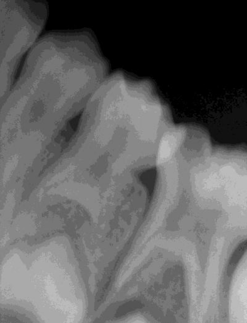
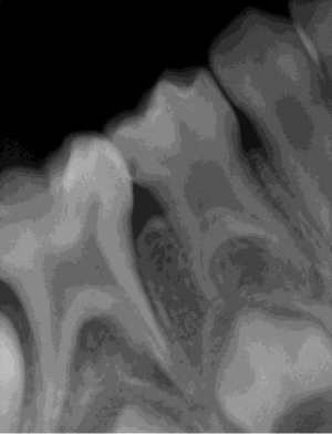
A six year old boy reported to the pediatric dental clinic with complaint of pain in the lower right posterior region of the jaw. His medical and dental history was noncontributory. Clinical examination revealed large carious lesion in relation to right mandibular primary second molar. Intraoral periapical radiographs revealed endodontically involved right primary second molar. Besides this, an unusual morphology of both primary mandibular first molars was also evident on the radiographs. These teeth showed elongated pulp chambers and apical displacement of bifurcation with short roots suggesting of taurodontism (Figures 1 and 2). The right primary second molar was treated by pulpectomy procedure. For this procedure, first local anesthesia was administered and access opening was done. Then coronal and radicular pulp was extirpated. Working length was determined and biomechanical preparation was done with copious irrigation. Now the root canals were obturated with resorbable material (zinc oxide eugenol) and the tooth was restored with stainless steel crown.
Figure 1 : Periapical radiograph showing taurodontism in right primary first molar with elongated pulp chamber and short roots.
Figure 2 : Periapical radiograph showing taurodontism in left primary first molar with elongated pulp chamber and short roots.
Discussion
Taurodontism is an anomaly of multi-rooted teeth characterized by enlargement of the apical portion of the pulp chamber. Keith described the taurodontic tooth as having tendency towards enlargement of the body of the tooth at the expense of the roots [1]. A taurodont tooth shows wide variation in the size of the pulp chamber, apically positioned canal orifices, varying degrees of obliteration and canal configuration. Therefore, taurodontism presents a challenge during negotiation, instrumentation and obturation in root canal treatment [5].
References
- Keith A. Problems relating to the Teeth of the Earlier Forms of Prehistoric Man. Proc R Soc Med. 1913; 6: 103-124.
- Mangron JJ. Two cases of taurodontism in modern human jaws. Br Dent J. 1962; 113: 309–312.
- Ahmet G, Filiz N, Kamil G, Metin A. Taurodontism in association with super numerary teeth. J Clin Pediatr Dent. 1999; 23: 151–153.
- Kosinski RW, Chaiyawat Y, Rosenberg L. Localized deficient root development associated with taurodontism: case report. Pediatr Dent. 1999; 21: 213-215.
- Tsesis I, Shifman A, Kaufman AY. Taurodontism: an endodontic challenge. Report of a case. J Endod. 2003; 29: 353-355.

