Case Report
Austin J Clin Case Rep. 2014;1(8): 1038.
Pneumo-omentocele - a Sign of Silent Lethal Neonatal Posterior Gastric Perforation
Govani DR1, Patel RR2, Patel RV3*, More B4, Bhimani SD5 and Vaghela SV6
1Medical Student, University of Birmingham Medical School, UK
2Department of Pediatrics, Saurashtra University, India
3Department of Ped Surgery, Saurashtra University, In
4Department of Paediatric Surgery, Leicester Royal Infirmary, UK
5Department of Ped Cardiology, Saurashtra University, India
6Department of Ped CT Surgery, Saurashtra University, India
*Corresponding author: Patel RV, Department of Surgery, Postgraduate Institute of Child Health and Research and K T Children Government Hospital, Saurashtra University, Rajkot 360001 Gujarat, India
Received: July 10, 2014; Accepted: Aug 10, 2014; Published: Aug 11, 2014
Abstract
A case of silent lethal posterior neonatal gastric perforation in a term neonate who had severe respiratory distress syndrome following meconium aspiration and difficult intubation at birth has been reported. He underwent very successful and costly treatment of meconium aspiration with extracorporeal membrane oxygenation (ECMO) but succumbed to unrecognized silent posterior gastric perforation although clinically he deteriorated progressively following oral feeding, inflammatory markers started climbing and the radiological findings of lobulated collection of air in the two layers of greater omentum of the lesser sac boundary well outside the bowel outline were not timely co-ordinated leading to septicaemia, multiple organ failure and death.
Keywords: New born; Stomach; Perforation; Lobulated; Pneumoperitoneum; Intubation; Meconium aspiration; ECMO; Pneumo-omentocele; Lesser sac
Introduction
Neonatal gastrointestinal perforations in general are relatively uncommon mainly seen in preterm babies as isolated perforation or as a consequence of necrotizing enterocolitis and gastric perforation in particular are rare and presents with catastrophic sequel and has very high mortality rates [1-15].
Case Presentation
A term male neonate had meconium aspiration syndrome at birth. His condition was poor, with APGAR scores of 2, 5 and 5 at 1, 5 and 10 minutes respectively. The infant had a difficult intubation and required aggressive resuscitation with intubation, ventilation, 100% oxygen and nil by mouth with intravenous fluids and antibiotics.
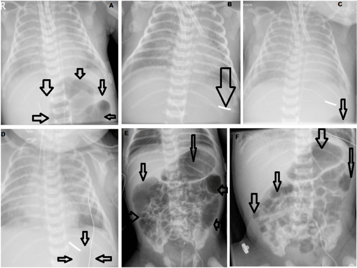
He was transferred to the regional extracorporeal membrane oxygenation (ECMO) service. He had a veno-arterial ECMO cannula inserted, radiograph confirmed position of the cannula but an abnormal loculated gas shadow was appreciated with the tip of the nasogastric tube beyond the shadow itself (Figure 1A).
Figure 1 : A. Loculated gas collection without ant gastric or bowel outline suggesting free gas B-C. Tip of the nasogastric tube too lateral and beyond the abnormal gas shadow. D. Post ECMO reduced air shadow and NG tube withdrawn. E. Sudden increase in the loculated air behind the stomach shadow, both colonic flexure areas and the borders of greater omentum in the flanks on starting feeds. F. Reduced air shadow following nil by mouth.
After ECMO therapy the gas shadow diminished, but the nasogastric tube was seen far laterally so was subsequently withdrawn (Figure 1B & C). The infant recovered on day 5 of ECMO therapy and the cannula was removed, but the abnormal shadow in the left upper abdomen continued (Figure 1D).
He was transferred to the paediatric intensive care unit and feeds via nasogastric tube (NGT) were initiated. He had tachypnoea, and had PCO2 retention soon after initiating the NGT feeds. On the following day, he developed vomiting; abdominal distension and fresh blood in the nasogastric tube despite proton pump inhibitors being given intravenously. Laboratory investigations confirmed a drop in haemoglobin, as well as an increased C-reactive protein despite receiving antibiotics. Infant was kept nil by mouth.
An abdominal radiograph showed marked abnormal gas behind the stomach shadow, as well as both flexures and loin areas (Figure 1E). He was referred to a senior surgical registrar for the evaluation and management of necrotising enterocolitis (NEC). The repeat abdominal radiograph showed improvement in gas pattern without any features of NEC but abnormal gas shadow continued (Figure 1F).
Feeds were resumed. He was transferred back to his original regional hospital where he continued to deteriorate despite full resuscitation measures and inotropic support with multi organ failure and died on the following day. Post-mortem examination revealed posterior gastric perforation with pneumo-omentocele and gross lesser sac milk peritonitis.
Discussion
It could be spontaneous, iatrogenic, ischaemic and congenital muscle weakness or anomaly of the interstitial cells of Kajal. Neonatal gastric perforation presents with severe symptoms and it is difficult to miss it in the normal circumstances but our case was exception as the perforation remained silent and the radiological features of free or tension pneumoperitoneum were missing due to not being fed since birth and covered with antibiotics for respiratory problem.
Neonatal posterior gastric perforation could be silent, lethal and can cause limited pneumo-omentocele rather than pneumoperitoneum [1-3].
Neonates with acute respiratory distress, difficult intubation and requiring aggressive resuscitation are high risk for gastric perforation, it may remain silent before starting feeds with rapid deterioration on introduction of feeds and high index of suspicion and proactive approach may help detect and treat it.
A small perforation in a term neonate may wall off in the lesser sac only and gas may be able to escape in between two layers of greater omentum as tissue planes are all loose in neonates and this new sign is useful to look out for. Recently first case of loculated lesser sac pneumoperitoneum secondary to walled off posterior gastric perforation in the new born has been reported [4].
Our case had several odds in the presentation and management. Initial difficult intubation and accidental oesophageal intubation of the endotracheal tube or traumatic nasogastric tube injury would have caused the small posterior perforation. The infant was very seriously ill and was nil by mouth since birth for over 5 days till his respiratory meconium aspiration syndrome was successfully treated with ECMO. During ECMO therapy most of the abdominal bowel gas gets absorbed and they have gasless abdomen but in our case subtle radiological signs of the loculated extra luminal gas shadows in awkward locations continued but were missed as the baby did not have any symptoms.
The gastric perforation no matter how small or how well walled off in the neonatal period, milk feeding sets the trend of rapid septicaemia and they deteriorate very quickly and prove lethal invariably if the window of opportunity has been missed.
An intensive care patient referral should not be taken lightly, review of whole course of events-clinical, laboratory and serial imaging with timing and effect of various interventions and senior input from multidisciplinary panel with lateral thinking about other possibilities other than one referred may allow detection and intervention.
Conclusion
Avoidance of intubation related iatrogenic injuries-tracheal or gastric, early detection of such incidents in the immediate perinatal period, early treatment of underlying pathophysiology, and protection of the neonatal gastric distension for at risk patients are essential in the management of neonatal gastric perforation.s
Acknowledgement
We are grateful to Dr Nayan Kalavadia MD, Consultant Neonatal Intensivist, Dr Anil Patel MD, Consultant neonatologist, Dr G M Faldu MD, Consultant Neonatal Anesthetist, Dr J T Faldu, Consultant Neonatal Pathologist and Dr Kailas Dalsania MD, Consultant Neonatal Radiologist for their help and expertise in treating this complicated patient successfully.
References
- Lawther S, Patel R, Lall A. Neonatal gastric perforation with tension pneumo-peritoneum. J Ped Surg Case Reports. 2013; 1: 14-16.
- Patel RV, Kumar H, More B, Rajimwale A. Giant neonatal gastric perforation in a preterm baby. BMJ Case reports [in press].
- Govani DR, Kumar H, Scott V, Patel RR, Patel RV and Doshi S. Spontaneous Neonatal Posterior Gastric Perforation with Tension Pneumoperitoneum of Lesser Sac. Austin J Clin Case Rep. 2014; 1: 2.
- Ghribi A, Krichene I, Fekih Hassen A, Mekki M, Belghith M, Nouri A, et al. Gastric perforation in the newborn. Tunis Med. 2013; 91: 464-467.
- Kshirsagar AY, Vasisth GO, Ahire MD, Kanojiya RK, Sulhyan SR. Acute spontaneous gastric perforation in neonates: a report of three cases. Afr J Paediatr Surg. 2011; 8: 79-81.
- Lin CM, Lee HC, Kao HA, Hung HY, Hsu CH, Yeung CY, et al. Neonatal gastric perforation: report of 15 cases and review of the literature. Pediatr Neonatol. 2008; 49: 65-70.
- Kara CS, Ilçe Z, Celayir S, Sarimurat N, Erdogan E, Yeker D, et al. Neonatal gastric perforation: review of 23 years' experience. Surg Today. 2004; 34: 243-245.
- Kuremu RT, Hadley GP, Wiersma R. Neonatal gastric perforation. East Afr Med J. 2004; 81: 56-58.
- Oztürk H, Onen A, Otçu S, Dokucu AI, Gedik S. Gastric perforation in neonates: analysis of five cases. Acta Gastroenterol Belg. 2003; 66: 271-273.
- Pigna A, Iannella E, Gentili A, Libri M, Lima M, Baroncini S, et al. Gastric perforation in a newborn. Pediatr Med Chir. 2003; 25: 66-68.
- Jawad AJ, Al-Rabie A, Hadi A, Al-Sowailem A, Al-Rawaf A, Abu-Touk B, et al. Spontaneous neonatal gastric perforation. Pediatr Surg Int. 2002; 18: 396-399.
- Leone RJ Jr, Krasna IH. 'Spontaneous' neonatal gastric perforation: is it really spontaneous? J Pediatr Surg. 2000; 35: 1066-1069.
- Khan TR, Rawat JD, Ahmed I, Rashid KA, Maletha M, Wakhlu A, et al. Neonatal pneumoperitoneum: a critical appraisal of its causes and subsequent management from a developing country. Pediatr Surg Int. 2009; 25: 1093-1097.
- Asabe K, Oka Y, Kai H, Shirakusa T. Neonatal gastrointestinal perforation. Turk J Pediatr. 2009; 51: 264-270.
- Kuremu RT, Hadley GP, Wiersma R. Gastro-intestinal tract perforation in neonates. East Afr Med J. 2003; 80: 452-455.
