Case Report
Austin J Clin Case Rep. 2014;1(8): 1039.
Oral Myiasis-A Case Report
Navneet Sharma1, Divye Malhotra2, Manjunatha BS3 and Jasjit Kaur4*
1Department of Oral Medicine and Radiology, Himachal Dental College, India
2Department of Oral and Maxillofacial Surgery, Himachal Dental College, India
3Department of Oral and Maxillofacial Pathology, KM Shah Dental Collage & Hospital, India
4Department of Prosthodontics and Crown & Bridge, Himachal Dental College, India
*Corresponding author: Jasjit Kaur, Department of Prosthodontics and Crown & Bridge, Himachal Dental College, Sunder Nagar, Himachal Pradesh, India
Received: July 25, 2014; Accepted: August 20, 2014; Published: August 22, 2014
Abstract
Myiasis is a rare condition caused due to larvae infestation of body tissues. This can be caused by several species of Dipteran fly larvae and may be secondary to serious medical conditions. Common predisposing factors for oral myiasis are incompetent lips, poor oral hygiene, severe halitosis, anterior open bite, mouth breathing, facial trauma, extraction wounds, ulcerative lesions and carcinoma. Here, we describe a case of oral myiasis in the anterior palatal region in a mentally challenged patient caused by the larvae of Musca Nebulo whose clinical features, management and treatment outcome are discussed and case was followed up periodically.
Keywords: Oral myiasis; House fly; Necrosis
Introduction
The term myiasis (Greek: myi= fly, asis= disease) is applied to infestation of living tissues of humans and animals, by dipterous larvae [1]. Zumpt descriptively defined myiasis as “the infestation of live human and vertebrate animals with dipterous larvae, which, at least for a certain period, feed on the host’s dead or living tissue, liquid body substances, or ingested food” [2]. Myiasis is well recognized in the animals, but rare in humans [2,3]. Human myiasis is extremely rare in developed countries, but it is not an uncommon parasitic infestation in the tropics and subtropics [4]. Myiasis can target any body tissue or cavity that is accessible to egg-laying and development of larvae, but most common anatomic sites involved are the nose, eye, lung, ear, anus, vagina, and more rarely, the mouth [3,5].
Oral myiasis was first described by Laurence in 1909 [5]. Predisposing factors for development of oral myiasis are anterior open bite, mouth breathing, neglected mandibular fractures, Cancrum Oris, extraction sockets and patient with neurologic deficit, diabetes, and peripheral vascular disease [3].
In this article a case of oral myiasis in the anterior palatal region in a patient with neurological deficit is presented.
Case Presentation
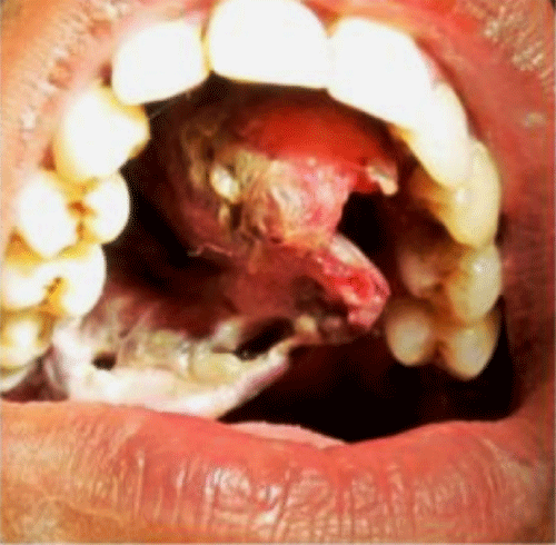
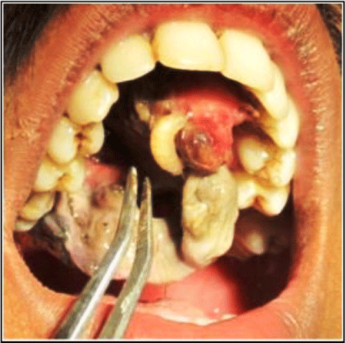
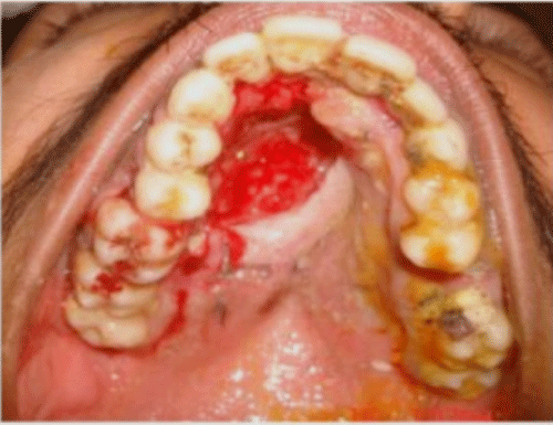
A 22 years old, mentally challenged male patient was referred to the Department of Oral Medicine, from a regional community health centre for assessment of the intraoral necrotic lesion. The patient’s mother stated that this condition started just a few days back and they showed to some local dentist, but his condition continued to worsen. There was no history of intraoral trauma or any evident previous lesion. On general physical examination patient was malnourished, anaemic, disoriented, and unable to communicate. Intra oral examination revealed presence of necrotic and ulcerative lesion on the palate, extending from incisors to first molar region (Figure 1). Lesion was painful and was accompanied by putrid and penetrating odor. The patient had an anterior open bite, inability to contact upper and lower lip, and poor oral hygiene. Necrotic lesion was cleaned with 10% hydrogen peroxide. On manipulation of tissue, several larvae were seen moving inside the lesion (Figure 2). These larvae were segmented, cylindrical, headless, and grayish-white in color. Whole of anterior hard palate was infected with larvae, causing separation of mucoperiosteum from underlynig bone, and the whole mass was hanging down from the roof of the oral cavity. There was evidence of tunneling produced by these larvae inside the necrotic mass. With all these clinical findings, this was an evident case of oral myiasis and the patient was hospitalized.
Figure 1 : Initial appearance of the lesion.
Figure 2 : Larvae seen moving inside the lesion on manipulation
Discussion
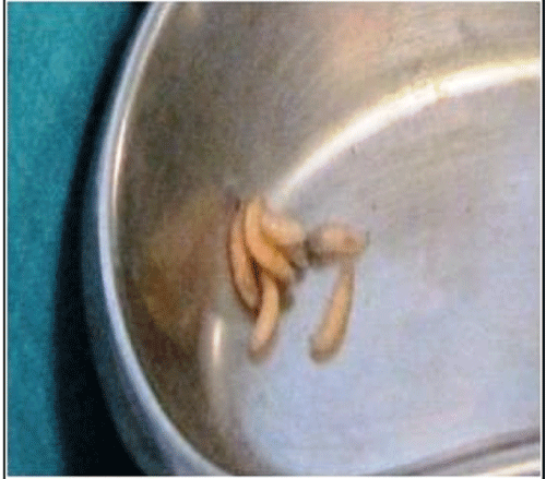
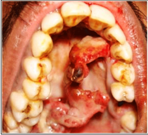
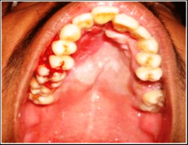
The wound was cleaned by 10% hydrogen peroxide, and larvae were manually removed with the help of serrated tweezers. Total 10 larvae were removed, and all were 6-10 mm in length (Figure 3). After removal of larvae, the wound was irrigated and cleaned with 1% iodine solution, and necrotic tissue and slough was removed under local anesthesia. The patient was prescribed analgesics and anti-inflammatory drugs, metronidazole 500mg eight hourly, and anti-tetanus serum under medical supervision. After 48 hours of removal of larvae and sloughed tissue, edema and inflammation were subsiding and there was detached periosteum hanging from the palate (Figure 4). The lesion was again cleaned with hydrogen peroxide and turpentine oil was applied to expel remaining larvae, if present. An incisional biopsy was planned, and acrylic retainer was fabricated to keep mucoperosteum in position for uneventful healing. The maggots were submitted to the Entomology Department and were later identified as larvae of Musca Nebulo fly. The case was followed up intermittently to observe healing (Figure 5,6).
Figure 3 : Photograph depicting retrieved larvae.
Figure 4 : Appearance of lesion after 48 hours.
Figure 5 : Intraoral photoraph after 4 weeks
Figure 6 : Phothgraph showing healing after 8 weeks
Myiasis is a rare clinical condition seen among the rural population living in close proximity to livestock and an environment conducive for growth of the flies. Oral myiasis is seen in people with predisposing factors such as mental disability, cerebral palsy, hemiparesis, and trauma to the oral cavity, loss of teeth, periodontitis, malocclusion, halitosis and mouth breathing [1-3].
Myiasis is caused by larvae of flies belonging to three families of Diptera, the Oestridae, Calliphordae and Sarcophagidae, which feed on living and dead tissues [6]. These flies can lay- over 500 eggs at a time. These eggs hatch to become larvae in less than one week and the life cycle is completed when the larvae turn into flies in about two to six weeks [2,7].
Depending upon organ or tissue involved myiasis can be cutaneous, intestinal, atrial, open wounds and external blood-suckers [8]. Whereas based on affinity of flies towards affected tissues, myiasis is classified as (a) obligatory, where larvae develop only in living tissue (b) facultative, the larvae develops in decaying tissue and may invade open wounds (c) accidental, the eggs or larvae are ingested and developed in the gastrointestinal tract [2,8].
Necrotic tissue and decaying food present in advanced periodontal disease will form a good environment for lying of eggs. After hatching, the larvae seek a warm and moist environment to further develop. The periodontal pocket fulfils these requirements and also gives mechanical protection to the larvae [8]. The larvae obtain their nutrition from the surrounding tissues and reside deeper into the soft issues by making tunnels, or separating the gingiva and mucoperiosteum from the bone [2], similar to our case.
During early developmental stage, the larva has segmental hooks which are directed backward. These hooks help the larva to anchor itself to the surrounding tissue [7]. In present case, larvae were residing deep into tunnels making their removal difficult. This can be explained because of the fact that, these larvae are photophobic in nature and burrow deeper into soft tissues by releasing toxins [2,5,7]. Necrotic ulceration, sloughing and excessive halitosis observed in this patient is due to the combined effect of bacterial proteolytic enzymes and toxins released by larvae. Itching and discomfort felt by patient is the result of crawling movement and increasing size of larvae [8].
The most important objective in management of myiasis is removal of all larvae. When there are multiple larvae in advanced stages of development and tissue destruction, local application of several substances have been used to ensure complete removal of all larvae. Use of normal saline, 0.2% chlorhexidine, iodoform, ethyl chloride, mercuric chloride, turpentine oil and ether has been suggested to compel the maggots wiggle out from host tissue [2,5,9]. Surgical debridement and necrotic tissue removal has also been advocated in literature [10].
Patient in our case report belongs to low socio-economic status and had poor and unhygienic living conditions. Anterior open bite, incompetent lips and dependency on parents for routine activities are main contributory factors for such infestation by flies.
Conclusion
Myiasis frequently affects people with low socioeconomic status, individuals with poor hygiene, unhealthy patients with psychiatric disorders and diabetic and immunocompromised individuals. Undoubtedly, preventive approach measures, including basic health care, access to primary health service, safe water and drainage are fundamental to prevent cases such as this one.
References
- Abdo EN, Sette-Dias AC, Comunian CR, Dutra CE, Aguiar EG. Oral myiasis: a case report. Med Oral Patol Oral Cir Bucal. 2006; 11: E130-131.
- Yeung C, Leung AC, Tsang AC. Oral myiasis. Hong Kong Dental Journal 2004; 1:35-36.
- Rossi-Schneider T, Cherubini K, Yurgel LS, Salum F, Figueiredo MA. Oral myiasis: a case report. J Oral Sci. 2007; 49: 85-88.
- Gabriel JG, Marinho SA, Verli FD, Krause RG, Yurgel LS, Cherubini K, et al. Extensive myiasis infestation over a squamous cell carcinoma in the face. Case report. Med Oral Patol Oral Cir Bucal. 2008; 13: E9-11.
- Sharma J, Mamatha GP, Acharya R. Primary oral myiasis: a case report. Med Oral Patol Oral Cir Bucal. 2008; 13: E714–716.
- Chadee DD, Rawlins SC. Dermatobia hominis myiasis in humans in Trinidad. Trans R Soc Trop Med Hyg. 1997; 91: 57.
- Sood VP, Kakar PK, Wattal BL. Myiasis in otorhinolaryngology with entomological aspects. J Laryngol Otol. 1976; 90: 393-399.
- Zeltser R, Lustmann J. Oral myiasis [corrected and issued with original paging in Int J Oral Maxillofac Surg 1988 Oct; 17(5):288-9]. Int J Oral Maxillofac Surg. 1989; 18: 288-289.
- Bhatt AP, Jayakrishnan A. Oral myiasis: a case report. Int J Paediatr Dent. 2000; 10: 67-70.
- Antunes AA, Santos Tde S, Avelar RL, Martins Neto EC, Macedo Neres B, Laureano Filho JR, et al. Oral and maxillofacial myiasis: a case series and literature review. Oral Surg Oral Med Oral Pathol Oral Radiol Endod. 2011; 112: e81-85.
