Case Report
Austin J Clin Case Rep. 2014;1(9): 1041.
“Isolated Innominate Artery Aneurysm: A Very Rare Finding”
Elia De Maria1*, Alina Olaru2 and Stefano Cappelli1
1Cardiology Unit, Ramazzini Hospital, Italy
2Department of Cardiology, University of Modena and Reggio Emilia, Italy
*Corresponding author: Elia De Maria, Cardiology Unit, Ramazzini Hospital, Via Molinari 1, 41012, Carpi (Modena), Italy
Received: July 31, 2014; Accepted: September 01, 2014; Published: September 03, 2014
Abstract
We describe the clinical case of an elderly woman with an aneurysm of right innominate artery, treated conservatively. The aneurysms of supraortic trunks and internal carotid artery are very rare (0.4-4% of all types of aneurysms); even rarer is the presence of aneurysms of subclavian and innominate artery (0.003%). Etiology is multifactorial and atherosclerosis accounts for 60% of cases. The clinical presentation is variable and the most common sign is the presence of a pulsatile mass on one side of the neck. In critically symptomatic patients, when rupture or embolization occurs, emergency treatment is mandatory. The most appropriate treatment is still controversial when aneurysm is asymptomatic and diameter is >30 mm.
Keywords: Aneurysm; Innominate artery; Supraortic trunks
Introduction
The aneurysms of supraortic trunks and internal carotid artery are very rare (0.4-4% of all types of aneurysms); isolated aneurysms of subclavian and innominate artery are even rarer, with an incidence reported in literature of 0.003% [1,2]. In this report we describe the case of an elderly female patient with an aneurysm of the right innominate artery and mild dilatation of the proximal part of homolateral subclavian artery, diagnosed after the appearance of a pulsatile mass on the right side of the neck.
Case Presentation
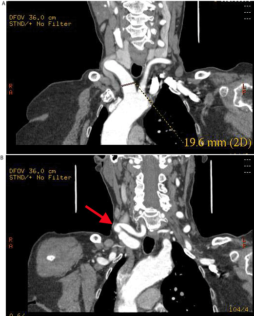
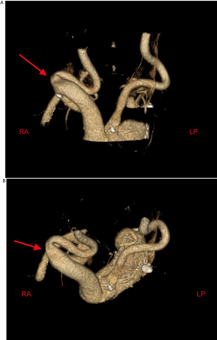
An 80-year-old woman was evaluated in our Hospital because of the appearance (few months before) of a pulsatile mass on the right side of the neck, associated with a vascular systolic murmur. She denied past or recent trauma to her neck or upper thorax; no central venous access had ever been placed or attempted. Her past medical history was only characterized by arterial hypertension, well controlled with Ramipril 5 mg daily. A recent echocardiogram showed a mild left ventricular hypertrophy with preserved systolic function, normal dimensions of aortic root and ascending segment, no evidence of valve disease. Blood exams were all within normal ranges (red and white blood cells, platelets count, coagulation tests, creatinine, electrolyte levels, thyroid and liver function tests, cholesterol level). For further evaluation she first underwent an Echo Doppler ultrasound evaluation of supraortic trunks that showed an enlargement of the innominate and right subclavian artery. Abdominal aortic aneurysm was excluded. Therefore a multiplanar Computed Tomographic Angiography (CTA) was performed. This exam showed an aneurysm of the right innominate artery (maximum diameter of 19.6 mm, with a tortuous course); also the proximal part of homolateral subclavian artery was dilated (13 mm diameter) (Figure 1A-1B). CTA was completed with a 3D reconstruction of arterial anatomy (Figure 2A-2B): right innominate artery dilatation and kinking were clearly evident. Infectious, autoimmune and connective tissue diseases were excluded as possible causes of aneurysm; in particular there was no clinical or hematological sign of bacterial, viral, mycotic infections (negative tests, negative blood cultures and negative antibodies for Staphylococcus species, syphilis, mycotic infection, tuberculosis, hepatitis A, B, and C). Rheumatoid Factor and autoimmune antibodies were all negative (anti-nuclear, anti-DNA, anti-mitochondria, anti-smooth muscle, anti-tireoglobulin, anti-thyroid peroxidase, anti-TSH receptor). Moreover there was no sign of connective tissue disorders like Marfan or Ehlers-Danlos syndromes. At this point an expert team discussion was carried out, together with a vascular surgeon. We all agreed for an initial conservative approach and follow-up with CTA after 6 months and 1 year. Antiplatelet therapy with aspirin 100 mg daily was started, in association with a beta-blocker (Nebivolol 5 mg daily). At 1 year follow up the patient was substantially asymptomatic (neck pulsation being less evident); a new CTA showed an unchanged situation and surgery has been postponed.
Figure 1A and 1B: Multiplanar Computed Tomographic Angiography of the neck showing aneurysm of 19.6 mm diameter of right innominate artery (1A), with an evident kinking (1B). RA: right-anterior projection. LP: left-posterior projection.
Figure 2A and 2B: 3D reconstruction of Computed Tomographic Angiography that clearly showed aneurysm and kinking of right innominate artery. RA: right-anterior projection. LP: left-posterior projection.
Discussion and Conclusion
The aneurysms of supraortic trunks and of internal carotid artery are very rare; their incidence ranges between 0.4 and 4% of all types of aneurysms. The most common site is carotid artery bifurcation followed by internal and common carotid artery [1,2]. Isolated aneurysms of subclavian and innominate artery are the most rare: only 3 cases have been described out of 1147 aneurysms -0.003%- in a case series [3]. Innominate artery (IA) aneurysms have been divided into 3 categories: type A, with no involvement of the origin of the IA (rare); type B, with involvement of the origin of the IA but not of the aorta (the most common); type C, with involvement of both the IA and the aorta [4]. Our patient displayed a type B phenotype, being the thoracic aorta within normal dimensions.
Etiology is multifactorial and has changed over the past decades: in the 1950s most IA aneurysms were syphilitic, massive lesions, difficult to treat and with a poor prognosis. With the modern imaging technologies, the diagnosis of small asymptomatic aneurysms has become increasingly common, especially during the workup for thoracic and abdominal aneurysm [3,4]. Nowadays atherosclerosis accounts for more than 60% of cases. Infective etiology (i.e. syphilis, mycotic aneurysm, tuberculosis) is decreasing, while autoimmune diseases (i.e. Beçhet Syndrome, Takayasu arteritis), connective tissue disorders (i.e Marfan and Ehlers-Danlos syndromes) and post-traumatic forms are increasing [3-5].
Clinical presentation is variable with a wide spectrum of symptoms ranging from headache and embolism (TIA or stroke) to symptoms caused by compression on surrounding structures (i.e. hoarseness, dyspnoea or dysphonia due to compression of the pharynx) and upper limb and facial edema due to superior vena cava syndrome. The most common objective sign is the presence of a pulsatile mass on one side of the neck with or without a cervical vascular systolic murmur. Complications are frequent: the risk of rupture is >10%; even higher is the risk of embolization (>50%). Embolism usually involves right upper extremities and brain, resulting in right hemispheric symptoms, right ocular deficits or vertebro-basilar syndrome [1,3,4].
Operative indications include ruptured or embolizing aneurysms and severely symptomatic patients; in critically ill patients (rupture, embolization) mortality is higher than 70%: in these cases emergency treatment is mandatory to reduce the risk of death or stroke. The most appropriate treatment is still controversial when the aneurysm is asymptomatic and its diameter is <30 mm [1,4]. In asymptomatic cases surgery has mainly the aim to prevent embolic complications: it can be considered when surgical risk is low, aneurysm is saccular, and its maximum transverse diameter is >30 mm [4]. The most common surgical technique is an open correction via median sternotomy with extension into the right neck; minimally invasive and endovascular approaches are emerging alternatives to open repair, but further data are required before a more widespread use [4,6,7].
In our case atherosclerosis was the presumed cause of the aneurysm, since we excluded infectious, autoimmune and connective tissue diseases. Considering the age of the patient, the “benign” presentation, the diameter of the lesion and the symptomatic improvement after medical therapy, we decided for a conservative approach. A possible criticism to our report can concern the definition of aneurysm itself: a dilatation of an artery of at least 50% of the normal diameter of the vessel. The “normal” diameter of brachio-cephalic trunk can reach 15 mm, so the values found in our case could not strictly fulfill the criteria to define an aneurysm. However Body Surface Area (BSA) of our patient was small (1.3 square meters, 45 kg weight x 150 cm height), so a 20 mm dilatation can be considered higher than 50% of normal diameter values corrected for BSA. Moreover the definition of aneurysm can overlap that of “dolico-mega” artery, a condition in which the dilatation of the vessel is associated with a preserved “parallelism” of the walls. In our case artery walls in the dilated segments were not exactly parallel (even if it was not so evident): for these reasons the definition of aneurysm seemed the most appropriate [8-10].
References
- Regina G, Greco L, Fullone M, Testini M, Caruso G, Rizzi R, et al. [The emergency treatment of aneurysms of the supra-aortic trunks and of the internal carotid]. Ann Ital Chir. 2000; 71: 469-473.
- GAY BB Jr, WALKER JF. Aneurysm of the innominate artery; review of clinical and radiologic findings in 18 cases. Radiology. 1953; 60: 804-813.
- Brewster DC, Moncure AC, Darling RC, Ambrosino JJ, Abbott WM. Innominate artery lesions: problems encountered and lessons learned. J Vasc Surg. 1985; 2: 99-112.
- Kieffer E, Chiche L, Koskas F, Bahnini A. Aneurysms of the innominate artery: surgical treatment of 27 patients. J Vasc Surg. 2001; 34: 222-228.
- Witz M, Yahel J, Lehmann JM. Subclavian artery aneurysms. A report of 2 cases and a review of the literature. J Cardiovasc Surg (Torino). 1998; 39: 429-432.
- Soylu E, Harling L, Ashrafian H, Anagnostakou V, Tassopoulos D, Charitos C, et al. Surgical treatment of innominate artery and aortic aneurysm: a case report and review of the literature. J Cardiothorac Surg. 2013; 8: 141.
- Stolf NA, Bittencourt D, Verginelli G, Zerbini EJ. Surgical treatment of ruptured aneurysms of the innominate artery. Ann Thorac Surg. 1983; 35: 394-399.
- MacLean AA, Vricella LA, Freischlag JA. Innominate artery aneurysm. Eur J Cardiothorac Surg. 2007; 32: 803.
- Thomas TV. Intrathoracic aneurysms of the innominate and subclavian arteries. J Thorac Cardiovasc Surg. 1972; 63: 461-471.
- Cury M, Greenberg RK, Morales JP, Mohabbat W, Hernandez AV. Supra-aortic vessels aneurysms: diagnosis and prompt intervention. J Vasc Surg. 2009; 49: 4-10.

