
Case Report
Austin J Clin Case Rep. 2014;1(9): 1042.
A Survivor of Chronic Disseminated Intravascular Coagulation (DIC) after a Gastric Bypass Surgery
Li W*, Salcedo V, Buendia J and Le C
Department of Family and Community Medicine, Texas Tech University Health Science Center, USA
*Corresponding author: Li W, Department of Family and Community Medicine, Texas Tech University Health Science Center, Odessa, TX 79763, USA
Received: July 31, 2014; Accepted: September 01, 2014; Published: September 03, 2014
Abstract
Though, the overall risk of death and adverse outcomes after bariatric surgery is low, the severity of complications and related mortality should never be underestimated. Here we report a 59-year-old Caucasian female who suffered but survived from chronic disseminated intravascular coagulation (DIC) three months after her laparoscopic Roux-en-Y gastric bypass (RYGBP) surgery. The patient developed intra-abdominal abscess and multiple venous thromboses. Most of workup for coagulopathy was negative except elevated D-dimer and fibrinogen to 2841 ng/ml and 860 mg/dL respectively. A chronic DIC was diagnosed. The patient was put on a heparin drip after exploratory laparotomy. Then she was successfully bridged to Coumadin and discharged. Surgical patients for gastric bypass with higher risks of venous thrombosis should be considered for aggressive peri-operative prophylactic anti-coagulation, and lower threshold for early work up.
Keywords: Chronic Disseminated Intravascular Coagulation (DIC); Gastric bypass surgery; Venous thrombosis; Venous Thromboembolism prophylaxis
Abbreviations
DIC: Chronic Disseminated Intravascular Coagulation; RYGBP: Roux-en-Y Gastric Bypass; VTE: Venous Thromboembolism
Case Presentation
This is a 59-year-old Caucasian female who presented to our emergency department because of abdominal pain that was worsening for 3 days. The patient had a BMI of 49.6 kg/m2 and received RYGBP gastric bypass surgery on August 15, 2013 at a hospital in New Mexico. After 5 days’ hospital course, the patient was discharged home uneventfully. She then followed up with his primary care physician in a different city in New Mexico on September 20 with a complaint of left upper quadrant and lower chest pain. CT of the abdomen and chest on the same day showed left pleural effusion, however no mass or other abnormality. A thoracentesis was performed and the cytology revealed reactive mesothelial cells in transudate. Cultures were negative for bacteria, AFB or fungi. Two weeks later, the patient experienced generalized abdominal pain, worsening over the following three days, accompanied by nausea, vomiting and diarrhea. She also complained of anorexia and weakness. Then patient came to our emergency department (ED), a university hospital in Texas on October 12, 2013. At initial assessment, patient’s vital signs were stable except for mild tachycardia. She appeared drowsy, lethargic and dehydrated. A generalized tenderness of abdomen was present. Laboratory studies showed leukocytosis and hypoalbuminemia. An CAT scan with intravenous contrast in ED was suggestive of multiple thrombosis of the superior mesenteric vein, portosplenic confluence, main portal vein, right and left portal veins, and splenic vein as well as multiple infarcts of the spleen and liver (Figure 1), multiple abscesses along the residual stomach and spleen (Figure 2), and mild left pleural effusion. A full workup for coagulopathy was performed. During admission, the patient developed acute abdomen and deteriorated quickly. General surgery was consulted and he suspected “peritonitis”, “small intestinal ischemia” and “portal vein thrombosis, splenic vein thrombosis, superior mesenteric vein thrombosis”. The general surgeon decided to perform an exploratory laparotomy emergently. The postoperative diagnoses were “left upper quadrant/left subdiaphragmatic abscess” and “Jejunal perforation at gastrojejunal anastomosis”. The general surgeon performed a laparotomy with “drainage of intra-abdominal abscess”, “Enterorrhaphy gastrojejunal anastomosis” and “insertion of 16-French jejunostomy feeding tube”. Unfractionated heparin instead of low molecular heparin was initiated as anti-coagulant postoperatively due to acute kidney injury. The coagulopathy studies came back later, included antinuclear antibody, rheumatoid factor, homocysteine level, lupus anticoagulant panel, protein C and protein S assay, cardiolipin antigen and antibodies, factor V Leiden mutation assay, complement 3 and 4 levels, prothrombin F2 gene mutation, MTHFR mutation C677T and MTHFR mutation A1298C assays. Though the dilute Russell’s viper venom time (dRVVT) screen test was abnormal in the lupus anticoagulant panel, the dRVVT MIX CONFIRM test was normal. All other tests were within normal limits, however, elevated D-dimer and fibrinogen of 2841 ng/ml and 860 mg/dL were observed. A diagnosis of chronic DIC secondary to VTE and intra-abdominal abscess was made.
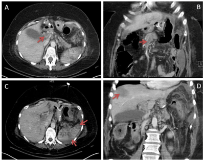
Figure 1: A. hepatic portal venous thrombosis; B. superior mesenteric vein thrombosis; C. splenic infarct; D. hepatic infarct.
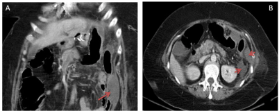
Figure 2: A. perifascial abscess; B. perirenal and perifascial abscess.
The patient required 7 days’ ventilator support in the intensive care unit (ICU) after exploratory laparotomy due to respiratory failure and severe sepsis. She was initially treated with meropenem and levaquin. When the peritoneal fluid culture came back positive for E Faecalis, Klebsiella and Candida, the antibiotics were replaced with daptomycin, avelox and micafungin. There was only one re-alimentation of jejunostomy. After Jejunostomy, the patient was on Glucerna 1.2 nutrition feeding at 10cc/hr through J-tube for 10 days. Three days after extubation, she started a PO full liquid diet, and then advanced as tolerated. After 23 days’ hospital course with 20 days’ ICU stay, the patient’s condition had been significantly improved with normalization of her renal and hepatic function. Then her INR became therapeutic and heparin was successfully bridged to Coumadin, She was subsequently discharged to a long term acute care facility for extended intravenous antibiotic therapy.
Discussion
Though the overall risk of death and adverse outcomes after bariatric surgery is low [1], the severity of complications and related mortality should never be underestimated. Since 1990, there has been increased performance of laparoscopic Roux-en-Y gastric bypass (RYGBP). It is currently considered the gold standard for the surgical treatment of morbid obesity in the United States. Romero- Lbargüengoitia et al. reported that the 3% mortality in 128 patients who went through Roux-en-Y gastric bypass surgery was mainly due to a leak of the anastomosis and/or intra-abdominal abscess. The cumulative incidence of VTE at 7, 30, 90, and 180 days was 0.3, 1.9, 2.1, and 2.1 % respectively in patients undergoing Roux-en-Y gastric bypass. Our patient probably developed VTE within one month after discharge.
To help identify the etiology of the VTE in this patient, an extensive work-up for coagulopathy was performed. Though cardiolipin antigen and antibodies tests were negative, dRVVT screen test was positive. dRVVT is one of the battery tests for lupus anticoagulant, with a specificity of 93% [2] and not affected by warfarin therapy [3]. However, the advanced mix confirmative dRVVT test was negative. In another words, no significant genetic thrombotic diathesis was identified in this patient. However, we noticed that both D-dimer and fibrinogen were elevated. So under what circumstances will those factors are elevated? As we know, chronic disseminated intravascular coagulation (DIC) can develop when blood is continuously exposed to tissue factor and compensation develops. The laboratory findings in chronic DIC are quite different from that of acute DIC. For example, acute DIC normally normally presents with reduction in platelet, plasma fibrinogen, plasma factor V, plasma factor VIII; prolongation in prothrombin time, activated partial thromboplastin time, and thrombin time as well as elevation of fibrin degradation products and D-Dimer. On the other hand, chronic DIC could have all laboratory findings above normal with the exception of elevated fibrin degradation products and D-Dimer. In this patient, all of her coagulopathy workup was normal except elevated D-Dimer and fibrinogen. In presence of intra-abdominal abscess, she gradually developed deep vein thrombosis, with elevated D-dimer and fibrinogen. Therefore, it is highly likely that she developed chronic compensated DIC. So far there are very limited case reports on intra-abdominal abscess induced DIC [4,5] except for a study by Gupta et al in 2011 showed that a bleeding disorder (OR = 2.23) is one of the most important risk factors of postoperative morbidity [6]. Generally, supportive care to maintain hemodynamic stability is essential. Heparin therapy should be applied to patients with chronic compensated DIC and predominantly thrombotic manifestations, as we did in this case. However, we do recognize that it could be quite costly and hazardous to perform a specific coagulation work-up before bariatric surgery for all patients.
Is there any way to prevent or avoid the severe complication as above? By using a standard preoperative screening method to identify the higher risk bariatric patients, a tailored perioperative prophylaxis might be able to improve the incidence and outcome in this subset population. Shepherd et al. reported that a prophylactic unfractionated heparin infusion protocol was effective in preventing postoperative thromboembolic events, especially in the highest risk gastric bypass patients [7]. For the patients with genetic hypercoagulable disorders such as Factor V Leiden mutation, prophylactic inferior vena cava filters proved to be preventative of recurrent PE [8]. In patients with a combination of risk factors (VSD, BMI ≥ 60 kg/m2, truncal obesity, OHS/SAS), prophylactic IVC filter placement is highly recommended [9]. Stroh c et al indicated that patients with higher BMI of average 51.5 kg/m2 who received more frequent VTE prophylaxis (low molecular weight heparin (LMWH) twice per day instead of once daily) had lower incidence of VTE postoperatively [10]. A recent study by Gupta et al. discussed numerous risk factors which could used to predict postoperative morbidity for patients undergoing gastric bypass and they developed a validated calculator for 30-day postoperative morbidity. In their module, factors associated with increased risk included recent MI/angina, dependent functional status, bleeding disorder, hypertension, BMI, and type of bariatric surgery [6]. Though many strategies or recommendations regarding perioperative prophylaxis in higher risk patients have been discussed, evidence-based data are lacking. There are no consented guidelines on the general approach currently. Otherwise this patient might have benefited from a preoperative IVC filter and / or an adjusted dose of heparin in consideration of her higher BMI (49.6 kg/m2) and advanced age. Randomized control studies are definitely in demand to optimize and standardize the interventional prophylaxis for thromboembolism in the near future.
Acknowledgement
We would like to thank Dr. Enrique Tobias for his generous help with the CAT image labeling.
References
- Longitudinal Assessment of Bariatric Surgery (LABS) Consortium, Flum DR, Belle SH, King WC, Wahed AS, Berk P. Perioperative safety in the longitudinal assessment of bariatric surgery. N Engl J Med. 2009; 361: 445-454.
- Ferro D, Saliola M, Quintarelli C, Valesini G, Basili S, Grandilli AM, et al. Methods for detecting lupus anticoagulants and their relation to thrombosis and miscarriage in patients with systemic lupus erythematosus. J Clin Pathol. 1992; 45: 332-338.
- Olteanu H, Downes KA, Patel J, Praprotnik D, Sarode R. Warfarin does not interfere with lupus anticoagulant detection by dilute Russell's viper venom time. Clin Lab. 2009; 55: 138-142.
- Chao A, Lin CT, Chang TC, Hsueh S, Lai CH. Choriocarcinoma with diffuse intraabdominal abscess and disseminated intravascular coagulation. A case report. J Reprod Med. 2002; 47: 689-692.
- Igawa T, Hakariya H. [A case of retroperitoneal abscess and disseminated intravascular coagulation as a complication of upper ureteral rupture caused by ureteral calculus]. Hinyokika Kiyo. 1996; 42: 525-528.
- Gupta PK, Franck C, Miller WJ, Gupta H, Forse RA. Development and validation of a bariatric surgery morbidity risk calculator using the prospective, multicenter NSQIP dataset. J Am Coll Surg. 2011; 212: 301-309.
- F Shepherd M, Rosborough TK, Schwartz ML. Unfractionated heparin infusion for thromboprophylaxis in highest risk gastric bypass surgery. Obes Surg. 2004; 14: 601-605.
- Atluri P, Raper SE. Factor V Leiden and postoperative deep vein thrombosis in patients undergoing open Roux-en-Y gastric bypass surgery. Obes Surg. 2005; 15: 561-564.
- Sapala JA, Wood MH, Schuhknecht MP, Sapala MA. Fatal pulmonary embolism after bariatric operations for morbid obesity: a 24-year retrospective analysis. Obes Surg. 2003; 13: 819-825.
- Stroh C, Luderer D, Weiner R, Horbach T, Ludwig K, Benedix F, et al. Actual situation of thromboembolic prophylaxis in obesity surgery: data of quality assurance in bariatric surgery in Germany. Thrombosis. 2012; 2012: 209052.