Case Report
Austin J Clin Case Rep. 2014;1(10): 1046.
Improvement of Diabetic Macular Oedema with Intravitreal Dobesilate: A Case Report
Cuevas P1,2*, Outeiriño LA2, Azanza C2, Angulo J3 and Giménez-Gallego G4
1Faculty of Medicine, Universidad Alfonso X, Spain
2Department of Ophthalmology, Hospital Day Pius XII, Spain
3Department of Histology Facility Research, Hospital Universitario Ramón y Cajal, Spain
4Department of Structure and Function of Proteins, Center for Biological Research, Spain
*Corresponding author: Cuevas P, Faculty of Medicine, Universidad Alfonso X, Spain
Received: August 10, 2014; Accepted: September 10, 2014; Published: September 11, 2014
Abstract
A single Intravitreal injection of Dobesilate seems to provide improvement in visual acuity and macular anatomy in a patient with diabetic macular oedema. To our knowledge, this is the first clinical case report using Intravitreal Dobesilate for diabetic macular oedema treatment. The therapeutic effect of Dobesilate may be due to the local inhibition of fibroblast growth factor.
Keywords: Diabetic retinopathy; Fibroblast growth factor; Dobesilate
Abbreviations
BCVA: Best-Corrected Visual Acuity; DME: Diabetic Macular Oedema; DR: Diabetic Retinopathy; ELM: External Limiting Membrane; FGF: Fibroblast Growth Factor; IS: Inner Photoreceptor Segment; OS: Outer Photoreceptor Segment; RPE: Retinal Pigment Epithelium; SD-OCT: Spectral-Domain Optical Coherence Tomography
Case Presentation
A 51-years old Caucasian diabetic type 2 man presented with great visual impairment in both eyes, previously treated with Intravitreal Avastin injections with unsatisfactory results. Complete ophthalmologic examination (including slit-lamp bio microscopy, tonometry, Best Corrected Visual Acuity (BCVA) measurement, and Fundus examination) was performed at baseline and at subsequent visits every 2 months until month 8th after treatment. Anterior segment examination was normal in both eyes. Fundus examination revealed diabetic macular oedema in both eyes and atherosclerotic retinal vessels. BCVA was 0.20 (OD) and 0.30 (OS) respectively, and spectral-domain optical coherence tomography (SD-OCT) showed typical signs of cystoids diabetic macular oedema. Central foveal thickness, as revealed by SD-OCT, was 269 and 465μm in OD and OS, respectively. Treatment with intravitreal Dobesilate was recommended first in the OS eye. After approval of our Institution Scientific Board, the patient signed an informed consent form, which included a comprehensive description of Dobesilate and the treatment procedure. Patient received an intravitreal solution of Dobesilate (150μl) under sterile conditions, following the International Guidelines for intravitreal injections [1]. Dobesilate was administered as a 12.5% solution of diethylammonium 2,5-dihydroxibenzenesulfonate (Dycinone®, Sanofi-Aventis, Paris, France, EU). Antibiotic eye drops were then applied. Patient returned to the outpatient clinic for routine post injection follow-up. No ocular side effects upon administration of Dobesilate or during the following days were observed.
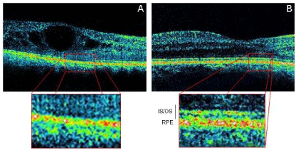
At baseline, SD-OCT showed significant macular oedema, with disintegration of the layered retinal structure and flattening of the central foveal depression. Several cystoids images were depicted (Figure 1A). Furthermore, the external limiting membrane (ELM), the inner segment and outer segment (IS/OS) photoreceptor junction appeared with disrupted integrity in some areas of the macula (Figure 1A). Morphological BCVA and SD-OCT data improved progressively along the follow-up. After 8 months of treatment, macular structure has improved, showing normal appearance and normal foveal depression; also the central macular thickness decreased to 203μm (Figure 1B) that was considered as normal, as in most referenced studies. Since integrity of IS/OS photoreceptor junction has a positive correlation with visual acuity [2,3] the appearance of the IS/OS photoreceptor junction depicted in Figure 1B suggested that intravitreal Dobesilate injection should have elicited a significant clinical improvement of the OS visual acuity. Effectively, 8 months after Dobesilate administration, visual acuity improved to 0.80.
Figure 1: Normalization of macular anatomy after a single intravitreal injection of Dobesilate. SD-OCT horizontal scan findings prior to treatment reveal macular oedema intraretinal cysts (A). SD-OCT scans before and after treatment was taken at the same orientation. Central foveal thickness was 465μm and BCVA 0.30. Eight months after treatment (B) central foveal thickness was 203μm and BCVA 0.80. Note the normalization of macular anatomy. IS: inner photoreceptor segment; OS: outer photoreceptor segment. RPE: retinal pigment epithelium. Rectangle areas in A and B show identical anatomical macular site.
Discussion
Diabetic retinopathy (DR) is the most common micro vascular complication of diabetes. In the USA, DR is the leading cause of blindness in working-aged adults, and currently affects approximately 150 million people worldwide. The World Health Organization projects that the number of people affected by DR will double by the year 2025 [4]. Diabetic macular oedema (DME) is the major vision-threatening complication of DR. Retinal micro vascular disturbance can lead to ischemia that in turn promotes inflammatory and angiogenesis interrelated processes, which remodel retinal tissue causing serious visual impairments. Vision loss in DR principally occurs due to neoformation of hyper permeable vessels, leading to macular oedema, retinal detachment and inner retinal and vitreous haemorrhage.
Basic research provides indications about the involvement of fibroblast growth factor (FGF) in DR [5-7]. It has also been described that the levels of FGF are higher in the vitreous from patients with DR in which neovascularization is evident [8]. Involvement of FGF, a well characterized inflammatory, angiogenic and vascular leakage-inducing protein [9,10] supports that intravitreal FGF inhibitors may be ideal candidates to treat DR by counteracting FGF over expression. It has been revealed that Dobesilate is the most effective member of a new family of efficient FGF inhibitors [11] that is effective when administered locally in inflammatory/angiogenesis-related conditions [12,13].
here are controversies over the use of oral Dobesilate (Doxium®), an old drug with a good safety profile in the treatment of DR [14-16]. More recently it has been reported in a double-blind multicenter trial that oral Dobesilate does not prevent or reduce the development of macular oedema during a 5-years follow-up period in patients with non proliferative DR [17]. The positive results described in the present case report seem in contradiction with these last results. Nevertheless, it should be taken into account that Dobesilate is a quite unstable compound at pH values above 5, and thus not too appropriate for both oral administration and systemic transportation to its biological targets. So, the different administration procedures of the mentioned Haritoglou et al. oral study [17] could be the reason of the outcome differences, as we have extensively discussed in previous studies. The therapeutic effects observed in this study are consistent with the anti-inflammatory and anti-angiogenic activities of Dobesilate, which have been observed in different pathologies and other retinopathies [12,13,18,19].
Conclusion
We demonstrate herein that the local inhibition of retinal over expressed FGF with a single-intravitreal administration of Dobesilate (a compound rediscovered as inhibitor of the FGF/FGFR system) restores retinal anatomy and improves vision in a DR patient. These results suggest that intervention/suppressors of inflammatory/ angiogenesis related to FGF over expression should be considered as a potential therapy in the treatment of degenerative retinopathies.
References
- Aiello LP, Brucker AJ, Chang S, Cunningham ET Jr, D'Amico DJ, Flynn HW Jr, et al. Evolving guidelines for intravitreous injections. Retina. 2004; 24: S3-19.
- Otani T, Yamaguchi Y, Kishi S. Correlation between visual acuity and foveal microstructural changes in diabetic macular edema. Retina. 2010; 30: 774-780.
- Maheshwary AS, Oster SF, Yuson RM, Cheng L, Mojana F, Freeman WR. The association between percent disruption of the photoreceptor inner segment-outer segment junction and visual acuity in diabetic macular edema. Am J Ophthalmol. 2010; 150: 63-67.
- King H, Aubert RE, Herman WH. Global burden of diabetes, 1995-2025: prevalence, numerical estimates, and projections. Diabetes Care. 1998; 21: 1414-1431.
- Hueber A, Wiedemann P, Esser P, Heimann K. Basic fibroblast growth factor mRNA, bFGF peptide and FGF receptor in epiretinal membranes of intraocular proliferative disorders (PVR and PDR). Int Ophthalmol. 1996; 20: 345-350.
- Paques M, Massin P, Gaudric A. Growth factors and diabetic retinopathy. Diabetes Metab. 1997; 23: 125-130.
- Simó R, Carrasco E, García-Ramírez M, Hernández C. Angiogenic and antiangiogenic factors in proliferative diabetic retinopathy. Curr Diabetes Rev. 2006; 2: 71-98.
- Sivalingam A, Kenney J, Brown GC, Benson WE, Donoso L. Basic fibroblast growth factor levels in the vitreous of patients with proliferative diabetic retinopathy. Arch Ophthalmol. 1990; 108: 869-872.
- Presta M, Andrés G, Leali D, Dell'Era P, Ronca R. Inflammatory cells and chemokines sustain FGF2-induced angiogenesis. Eur Cytokine Netw. 2009; 20: 39-50.
- Andrés G, Leali D, Mitola S, Coltrini D, Camozzi M, Corsini M, et al. A pro-inflammatory signature mediates FGF2-induced angiogenesis. J Cell Mol Med. 2009; 13: 2083-2108.
- Fernández IS, Cuevas P, Angulo J, López-Navajas P, Canales-Mayordomo A, González-Corrochano R, et al. Gentisic acid, a compound associated with plant defense and a metabolite of aspirin, heads a new class of in vivo fibroblast growth factor inhibitors. J Biol Chem. 2010; 285: 11714-11729.
- Cuevas P, Angulo J, Giménez-Gallego G. Topical treatment of contact dermatitis by pine processionary caterpillar. BMJ Case Rep. 2011; 2011.
- Cuevas P, Angulo J, Giménez-Gallego G. Long-term effectiveness of dobesilate in the treatment of papulopustular rosacea. BMJ Case Rep. 2011; 2011.
- Freyler H. Microvascular protection with calcium dobesilate (Doxium) in diabetic retinopathy. Ophthalmologica. 1974; 168: 400-416.
- Binkhorst PG, Van Bijsterveld OP. Calcium dobesilate versus placebo in the treatment of diabetic retinopathy: a double-blind cross-over study. Curr Ther Res Clin Exp. 1976; 20: 283-288.
- Ribeiro ML, Seres AI, Carneiro AM, Stur M, Zourdani A, Caillon P, et al. DX-Retinopathy Study Group. Effect of calcium dobesilate on progression of early diabetic retinopathy: a randomised double-blind study. Graefes Arch Clin Exp Ophthalmol. 2006; 244: 1591-1600.
- Haritoglou C, Gerss J, Sauerland C, Kampik A, Ulbig MW, CALDIRET study group. Effect of calcium dobesilate on occurrence of diabetic macular oedema (CALDIRET study): randomised, double-blind, placebo-controlled, multicentre trial. Lancet. 2009; 373: 1364-71.
- Cuevas P, Outeiriño LA, Angulo J, Giménez-Gallego G. Treatment of dry age-related macular degeneration with dobesilate. BMJ Case Rep. 2012; 2012.
- Cuevas P, Outeiriño LA, Azanza C, Angulo J, Giménez-Gallego G. Short-term efficacy of intravitreal dobesilate in central serous chorioretinopathy. Eur J Med Res. 2012; 17: 22.
