1University of Birmingham Medical School, UK
2Department of Ped Surgery, University Hospital of Wales, UK
3Department of Ped Surgery, Children Hospital of Pittsburgh of UPMC, USA
4Department of Ped Surgery, Texas Children’s Hospital Houston, USA
5Department of Ped Surgery, Saurashtra University, India
*Corresponding author: Patel RV, Department of Surgery, Postgraduate Institute of Child Health and Research and K T Children Government Hospital, Saurashtra University, Rajkot 360001 Gujarat, India
Received: November 07, 2014; Accepted: December 03, 2014; Published: December 04, 2014
Citation: Govani DR, Shrestha R, Sau I, Pimpalwar A, Milanovic D and Patel RV. Familial Infantile Hypertrophic Pyloric Stenosis: Old Wine (Ramstedt’spyloromyotomy) in the New Bottle (Changing Approaches). Austin J Clin Case Rep. 2014;1(12): 1059. ISSN : 2381-912X
Infantile hypertrophic pyloric stenosis is very common condition and its original treatment of Ramstedt’s pyloromyotomy has remained as gold standard and unchanged but the various approaches to perform the same procedure have evolved over last few decades. An unusual case of familial infantile hypertrophic pyloric stenosis has been reported which was performed by minimal invasion trans-umbilical laparoscopic approach and the remaining members of family had varying approaches being used have been presented with brief review of anatomical differences and scar migration phenomenon has been highlighted.
Keywords: Hypertrophic pyloric stenosis; Pyloromyotomy; Hypochloremic
Familial infantile hypertrophic pyloric stenosis in four generations in vertical family pedigree is rare [1]. We wish to report a case of familial infantile hypertrophic pyloric stenosis in four vertical generations all of which had the same operative procedure of Ramstedt’s pyloromyotomy but had different approached being used by different specialists’ generations of surgeons.
24-days-old male infant was referred to us from a district general hospital with the persistent projectile non-bilious vomiting of 3 days duration. There was family history of infantile hypertrophic pyloric stenosis to great grandfather, grandfather and father.
Test feed was positive with visible left to right upper abdominal peristalsis and a palpable pyloric tumor. Blood tests showed hypokalemic (K+ 3.2 mmol//L), hypochloremic (Cl-89 mmol/L), metabolic alkalosis (pH 7.56, HCO3 35 mmol/L, Base excess -13). Ultrasound scan confirmed infantile pyloric stenosis. Infant underwent laparoscopic pyloromyotomy after initial resuscitation and stabilisation uneventfully.
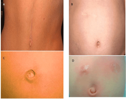
Interestingly, all four family members had the same surgical procedure of Ramstedt’s pyloromyotomy using different approaches-great grandfather had been operated by a general surgeon using vertical midline supraumbilical incision (Figure 1A), grandfather was operated by a general surgeon with paediatric interest by a supraumbilical transverse incision (Figure 1B), father was operated by a paediatric surgeon with a periumbilical skin crease incision (Figure 1C) and the infant had laparoscopic pyloromyotomy using umbilical port and two lateral direct instruments (Figure 1D). We have referred the family for the genetic counselling and further tests but family did not attend despite several reminders.
Interestingly they all got the same operative procedure performed using different approaches in vogue at the time of their ailment being treated. Familial aggregation and heritability has been described in Danish patients [2]. Ultrasound scan in diagnosis and minimal invasive approach in treatment has improved overall management [3].
Familial infantile hypertrophic pyloric stenosis in first degree family pedigree is exceptional rarity and although all of them had the same operative procedures and functional results, the cosmetic and overall satisfaction was seen in the last generation with early diagnosis and prompt correction due to awareness in the family.
Our cases are a strong reminder of the fact that growth affects relative size, positions and relations of the abdominal wall structures change from infant to adult in proportions expressed by not only increase in weight greater than length and expansion of surface area in between. Shape of the abdomen changes from being wider than it is in the neonate to being longer than it is wide in the adult.
Once the patent ductus arteriosus is closed after birth the growth of the lower half of the body gets accelerated. The ratio of the length from the xiphi sternum to the umbilical scar to that of from the umbilicus to symphysis pubis changes with growth of the lower half of the body having practical implications explaining preference for transverse abdominal incisions by paediatric surgeons and vertical ones by adult surgeons traditionally.
Our cases demonstrates practically the fact that abdominal supraumbilical scars of the surgical incisions not only lengthen but also migrate especially the supraumbilical transverse scar just above the umbilicus in neonatal period ended up very high over the costal margin and the vertical midline incision scar lengthened into the abdominal supraumbilical scars.
In summary, although rare, familial cases of infantile hypertrophic pyloric stenosis in a vertical pedigree in one family had the same gold standard operative surgical treatment. However, rapid advances in specialisation, early ultrasound diagnosis and minimal invasion surgery has changed the overall picture for better quality of care.
Clinical photographs. A. Great grandfather- vertical upper midline scar merging withumbilicus B. Grandfather- transverse supraumbilical scarnote lengthening and migration of both of these scars. C. Father-periumbilical skin crease scar with migration to right and D. laparoscopic pyloromyotomy in infant with umbilical port and bilateral direct instrument scars.
