Department of Obstetrics and Gynaecology, Sree Uthradam Thirunal Academy of Medical Sciences, India
*Corresponding author: Krishna PriyaLeela, Department of Obstetrics and Gynaecology, Shree Uthradam Thirunal Academy of Medical Sciences, Vattapara, Thiruvananthapuram, Kerala, India
Received: November 11, 2014; Accepted: December 03, 2014; Published: December 04, 2014
Citation: Leela KP. Cervical Circlage Suture Presenting As Foreign Body Uterus- Case Report. Austin J Clin Case Rep. 2014;1(12): 1060. ISSN : 2381-912X
Foreign body inside the uterus is a very rare condition. An interesting case of foreign body uterus was found in a 52 year old lady presented with abnormal uterine bleeding. On investigation an intrauterine foreign body was found which was removed under anesthesia. It was found to be a cervical cerclage suture put 23 years back.
Keywords: Cervical cerclage suture; Foreign body; Uterus
Foreign body uterus is a very rare condition. The usual foreign bodies that are found are IUCDs. Other foreign bodies found are broken laminaria tents, tips of curettes, nonabsorbable suture materials. Very rarely fetal bones after an incomplete abortion have been described.
Usually foreign bodies cause many symptoms like discharge per vaginum due to infection, menorrhagia, systemic infection and septicemia. Whenever a foreign body is suspected it is ideal to do a hysteroscopy to confirm the diagnosis. Here is an interesting case of foreign body uterus with no symptoms other than secondary infertility.
A 52 year old lady of Indian origin, P2L1 presented with excessive bleeding lasting for 15 days. She had regular periods before with normal bleeding. She had two normal deliveries. The first child died at 9 months .The second child is now 22 yrs old. After that she tried to conceive but never got pregnant again. She never went to any hospitals after her second delivery.
The patient underwent routine gynaec examination and everything was found to be normal. Pelvic ultrasound sound showed a normal sized uterus and normal ovaries. A longitudinal echogenic area was seen in the cavity suggestive of a foreign body in the uterus.
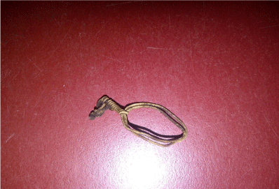
She was taken up for exploration of the uterine cavity under anesthesia and the foreign body was removed. The foreign body removed was examined carefully and it was found to be a cervical encirclage suture. On further interrogation she revealed history of encirclage done 23 yrs back during the second pregnancy. She doesn’t remember removing it before delivery.
Incompetent cervix is a well-known cause of second trimester spontaneous abortion [1]. Surgery is the principal form of therapy for premature cervical dilatation without labour. Several types of cerclage procedures are used; MacDonald and Shirodhkar are the most common. The McDonald procedure places a reinforcing purse-string suture around the proximal cervix. The Shirodkar operation places a reinforcing band around cervix at the level of internal os. Previously silk was used as suture material.
Now Mersilene tape or Prolene is used. Abdominal circlage may be appropriate in rare instances like traumatic cervical laceration, congenital shortening of cervix, previous failed vaginal procedure and advanced cervical effacement. This procedure places a band around the cervix at the level of internal os in an avascular space between branches of uterine artery. Cervical encirclage is usually done at around 14 weeks of pregnancy in recurrent 2nd trimester pregnancy loss [2].
The cerclage suture can be removed at 38 weeks or when fetal pulmonary maturity has been confirmed. It should be removed immediately in case labour starts earlier, membrane ruptures or there is fetal demise. Although some physicians leave a well-placed Shirodkar suture in place and delivered infants by Cesarean, evidence supporting this course of action is limited [2].
The complications of the procedure range from annoying to fatal. Complications include haemorrhage, rupture of membranes, infection including chorioamnionitis, abscess, cervical dystocia, uterine rupture, vesicovaginal fistula and fetal death [2].
Cervical laceration can occur if labour starts before removing the suture. Rare complications involving bladder has been reported due to migration of the cerclage when left in situ for long like bladder stone formation. Vesicovaginal fistula has also been reported as a complication. Suture migration can occur caudally also leading to loosening of the suture [3].
In this patient the cerclage suture migrated into the uterine cavity. This must have happened during her pregnancy itself, taking into consideration the fact that she delivered vaginally. Another possibility is that it must have got loosened and during delivery it must have got into the uterine cavity. Inside the uterus, it acted as a contraceptive device preventing further pregnancy. As it was an inert material it didn’t create any symptoms in the patient. It was an incidental finding when the patient was evaluated for abnormal vaginal bleeding after more than two decades.
A similar case has been discussed in the Hysteroscopy text book by Linda .D. Bradley where the patient presented with discharge per vaginum. Even after several blind procedures complaints persisted and only when they did a Hysteroscopy the diagnosis was made. The Mersilene tape that was used was then removed under guidance of Hysteroscope [4].
Cervical cerclage suture presenting as foreign body uterus is a very rare and interesting case. Obstetricians should give special care for patients for whom cerclage has been placed and make it a point to remove the cerclage in time. If the suture is displaced and not to be found in position at term extra efforts are to be taken to find it and remove it since if left in situ it can lead to complications like vesicovaginal fistula.
Picture of cervival cerclage suture removed from inside uterus.
