
Clinical Image
Austin J Clin Case Rep. 2023; 10(1): 1270.
Tenosynovitis of the Anterior Tibialis Tendon: Typical Clinical Radiological Correlation: A Case Report
*Corresponding author: Andour Hajar Radiology Department, Military Hospital Mohammed V Rabat, 10100, Morocco.
Received: November 23, 2022; Accepted: January 02, 2023; Published: January 09, 2023
Clinical Image
The Anterior Tibialis Tendon (ATT) is usually the site of minor injuries due to its straight course, however, overuse injuries still possible due to repetitive foot dorsiflexion. This tendon crosses the anteromedial aspect of the ankle running toward the medial border of the foot under the inferior and superior extensor retinacula and inserts at the level of the medial cuneiform and the base of the first metatarsal bone. It can be prone to tendinosis, tenosynovitis, partial or complete tear. In the first cases, the patient experiences a pain and a swelling along the distal part of the ATT worsening while dorsiflexion. The diagnosis may be easily suspected clinically, particularly in significant lesions such a complete tear, but the compensation by the extensor hallucis longus and extensor digitorum longus muscles may delay the diagnosis [1].
We report the case of a 60-year-old woman, with no medical history presenting with a swelling and pain in the midfoot (Figure 1) in whom an MRI showed edematous thickening of the anterior tibialis tendon sheath that is centered over the inferomedial band of the inferior extensor retinaculum (Figure 2). Imaging allows, in addition to the analysis of soft tissue and tendon sheath abnormalities, to report signal abnormalities of the tendon itself and its size. Most cases of tenosynovitis are managed conservatively, as was the case with our patient. Rest, adequate shoes and anti-inflammatories with physiotherapy are indicated. Surgery is sometimes required to some stubborn cases. It consists of a synovectomy that is usually performed with an open approach, but endoscopic technique is also possible allowing a preservation of the integrity of the extensor retinacula [2].
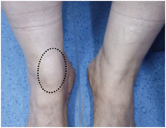
    Figure 1: Clinical image showing the swelling at the level of the right

midfoot (circle). Note the normal aspect in the contralateral side.
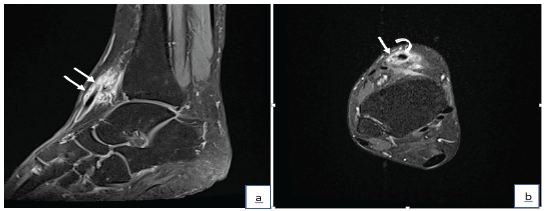
    Figure 2: MRI according to sagittal (a) and axial T1 FS (b) with gadolinium

injection showing a hypersignal surrounding the anterior

tibialis tendon at the level of the midfoot (straight arrows). Note

the normal appearance of ATT hypointense with normal thickness

(curved arrow).
Conflict of Interest
The authors declare that they have no competing interests.
References