
Case Report
Austin J Clin Case Rep. 2023; 10(1): 1271.
A Curious Case of Misapprehension – Dermoid Cyst Masquerading as a Ruptured Ectopic Pregnancy
Sahni A, Aboda A*, Abi-Sen S and McCully B
Department of Obstetrics & Gynaecology, Mildura Base Public Hospital, Australia
*Corresponding author: Ayman AbodaDepartment of Obstetrics & Gynaecology, Mildura Base Public Hospital, Australia
Received: December 01, 2022; Accepted: January 12, 2022; Published: January 19, 2023
Abstract
We discuss the case of a 26-year-old woman who presented with pelvic pain and recent onset vaginal bleeding in early pregnancy. Transvaginal USS identified significant free fluid in the pelvis associated with a right adnexal mass and an empty uterus. A ruptured ectopic pregnancy was suspected, and the patient was taken to theatre for an emergency laparoscopy. Paradoxically, the procedure found no signs of intra-abdominal blood loss. Instead, a large, unruptured 20cm ovarian cyst filled the right adnexa and Pouch Of Douglas (POD), extending posteriorly towards the pelvic brim. It was a right Ovarian, Dermoid cyst. Surprisingly, the dilemma is not unique. We identified similar reports in the literature where unsuspected ovarian pathology is discovered at the time of early pregnancy surveillance, often masquerading as an acute complication of early pregnancy. In this report, the patient went on to have a spontaneous miscarriage, and the cyst was later removed by mini- laparotomy. We present this case as an opportunity to highlight the epidemiology of adnexal pathology presenting for the first time in early pregnancy and to help broaden clinical suspicion when considering potential diagnoses at the time of presentation.
Keywords: Ectopic Pregnancy; Free Fluid; Dermoid Cyst.
Introduction
A teratoma is a tumour of germ cell origin. They most commonly arise in the ovary and are inimitable in their proclivity to contain tissues such as hair, teeth, fat and occasionally, bone [1]. This is because the cells of origin are totipotential which means disorders of growth may lead to differentiation into any number of tissue lineages [2]. Characteristically, these include skin and epidermal appendages, and for this reason, the tumors are often called Dermoids [1]. They are most often found in the ovary but may occur throughout the body. They are usually benign and small growing and are rarely associated with symptoms. Occasionally, however, they may reach a size of more than 15 cm and, in this setting, present with abdominal discomfort or bloating or, more rarely, with the acute complications of torsion or rupture [3-4].
In this case report, a large, unruptured Dermoid was identified in early pregnancy. Whilst this is not an unusual finding, the size and radiological impression at presentation were certainly novel and, as we shall see, curiously deceptive such that a diagnosis of ruptured ectopic pregnancy seemed irrefutable based on findings that noted, most significantly, a large volume of free intra-abdominal fluid. This was later found to be erroneous and was, in fact, the contents of the unruptured cyst. Our intention is to demonstrate the paradox of unexpected pathology and thus the importance of broad, clinical cognizance that is able to include the improbable, such that when it is found, we are able to respond appropriately, with sufficient dexterity of thought and action, to allow swift adaptation of clinical strategy to best suit the new situation. Our report outlines the subsequent care of this patient and the definitive management of this unsuspected presentation. It also alludes to lessons that can be learnt in the broader context of care for women in a primary health setting where the diagnosis of progressive disease can be aided by the provision of timely pelvic surveillance.
Case Presentation
A 26-year-old lady presented acutely to the emergency department of a rural general hospital with recent onset pelvic pain. On arrival, she was noted to have moderate discomfort associated with vaginal bleeding. The patient had previously noted two positive home pregnancy tests. The patient was from Malaysia, working in remote western Victoria as a seasonal fruit picker. She spoke little English and had no support persons with her. With the assistance of a phone interpreter, we discovered that this was her first pregnancy and that her last menstrual period was on 14/1/22. The pregnancy though unexpected, was not undesired.
Abdominal examination demonstrated moderate tenderness above the symphysis with voluntary guarding to palpation over the right iliac Fossa. Vaginal inspection by speculum showed a small amount of blood from the cervix. Digital examination confirmed general tenderness but did not detect any specific masses. The cervix was noted to be closed. Vital signs remained stable afterwards. A Transvaginal Ultrasound (TVUSS) demonstrated an empty Uterus adjacent to a large, complex hypoechoic area in the right adnexa with internal echoes and potential sac-like structures. Additionally, there was a large volume of free fluid estimated to be at least 1 litre. A diagnosis of hemoperitoneum and likely ruptured ectopic pregnancy was suspected. (Figures 1 & 2). On examination, she was not distressed. She had a moderate tachycardia of 105 bpm, and her blood pressure was normal at 116/70 mmHg, with no postural drop. She was well perfused with warm peripheries and normal oxygen saturation on room air. She was a febrile. Her hemoglobin was 122g/dl, platelet count 188, and she was COVID-19 PCR negative. She had had an earlier serum hCG reported 2220 units. The repeat estimation at the time of admission was 1880.
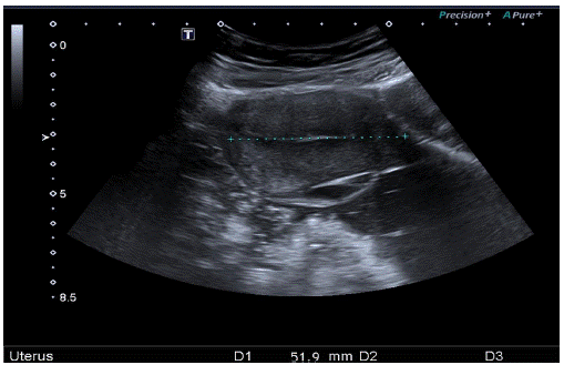
Figure 1: Empty Uterus.
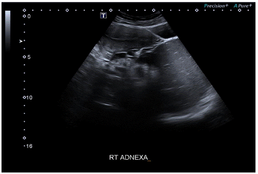
Figure 2: Pelvic free fluid.
The patient was treated as a likely ruptured ectopic pregnancy. A haemorrhagic corpus luteum was also considered. With consent, an emergency laparoscopy was arranged using a direct umbilical port entry. Paradoxically, there was no evidence of haemorrhage. The pelvis was dry, and the uterus and tubes were normal however, a large, right-sided ovarian cyst was identified, filling the Pouch of Douglas and extending across the midline up along the posterior abdominal wall (Figure 3). The cyst was smooth-walled, intact and showed no evidence of torsion. No further action was taken. A Dilation and Curettage (D&C) were performed, having noted the falling hCG titres. Currettings were sent for histopathology.
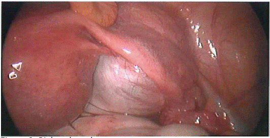
Figure 3: Right adnexal cyst.
Postoperatively, the initial images from the ultrasound were reviewed. It was noted that an examination of the upper abdomen, and in particular, Morrison's pouch, was not performed. Follow- up examination of the chest, abdomen and pelvis by CT scan showed fat and solid material with at least three calcified tooth-like structures in a large pelvic cystic mass. The appearances were consistent with an Ovarian Dermoid Cyst. There was a small amount of free fluid but no ascites or lymphadenopathy (Figure 4). A simple, 5x4 cm cyst was also noted on the left ovary.
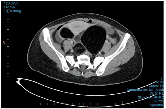
Figure 4: CT Pelvis.
Histopathology of endometrial curetting reported secretory change with Arias Stella's reaction. Serial b-HCG continued to downtrend, becoming negative 4 weeks later. Tumour markers showed Ca 125 at 87 U/ml (<35), Ca 19.9 at 131 U/ml (<35), AFP of 2.5, and a CEA of 2.6 (<0.5).
The patient discharged for outpatient review. Repeat tumour markers reported CEA 2.3, CA125 59, CA 19-9 29. The findings were discussed with Gynae-oncology. The patient elected to proceed to cystectomy with conservation of anatomy if possible. She was admitted 2 weeks later. A laparoscopic entry was converted to a transverse mini-laparotomy in the early procedure to improve surgical access. The ovarian cyst was identified in the right adnexa. It measured 18x10x8 cm and extended posteriorly to the midline, filling the POD and up towards the pelvic brim along the posterior abdominal wall. There was no distinguishable normal ovarian tissue. The right fallopian tube was intimately adherent and could not be separated. The Dermoid was removed without rupture (Figure 5). The uterus was normal. Two small 3x2 cm uncomplicated cysts were also identified in the left ovary. These were aspirated for cytology but otherwise left intact. Histology confirmed a mature cystic teratoma surrounded by a fallopian tube. Cytology from both cysts in the remaining left ovary was normal. The patient recovered fully. Repeat tumour markers 2 weeks later were normal.
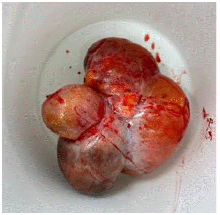
Figure 5: Surgical specimen – Dermoid cyst.
Discussion
A teratoma is a tumour of germ cell origin. They most commonly arise in the ovary in women aged between 20 and 40 years [1]. They are often called Dermoids because of their proclivity to contain epidermal appendages such as hair and teeth in a capsule surrounding fat and sebaceous tissue [5]. They are usually benign and rarely present with symptoms. Most remain small but occasionally may grow up to 15 cm or more prior to detection, which is then most often pre-empted by abdominal discomfort or bloating or, more rarely, with the acute complications of torsion or rupture [6,7]. They are most often discovered opportunistically at the time of investigation for other suspected pathology, such as appendicitis or inflammatory bowel disease, pelvic pain or infection, including endometriosis or PID (pelvic inflammatory disease) [8]. In this case presentation, the cyst was discovered following USS examination in the setting of pain and bleeding in early pregnancy. This is not an unusual scenario. What makes the case novel was the convincing duplicity of initial findings that led the clinical team to believe there was significant free fluid in the abdomen, a sine qua non of ruptured ectopic pregnancy. Not surprisingly, plans for immediate emergency surgery ensued. We learnt afterwards that this was in error.
Upon reflection, we noted that views of the upper abdomen, and in particular, of Morrison's pouch, were not performed. This was because the volume of pelvic free fluid was such that they had not been considered necessary. We imagine that had they been done, there would have been no evidence of free fluid, but it is unlikely that this would have changed our proposed management. It may, however, have made the surprise of the consequent findings somewhat less confounding when realized at the time of surgery. A lesson learnt from this case comes from the tumult of coming face to face with unexpected pathology or, more explicitly, of not finding the pathology that was expected. It highlights the importance of a broad, clinical perspective that allows room to consider the improbable, such that when it occurs, we are not left entirely dumbfounded. We can be forewarned, or, at the very least, mindful of the incongruous, such that we can reconcile the paradigms of preconceived planning and adjust to those that are more appropriate to the findings found. We can do what is necessary, we can adapt, and we can avoid the harm of abiding with what may no longer be required. We can keep patients safe. We protect consent, and we avoid the risk of doing greater harm from untimely or unnecessary action. In this case, the operative findings were acknowledged, the patient was kept safe, and a plan for definitive treatment was put in place with the patient’s consent at a time removed from the fracas of initial presentation.
Another lesson gleaned from this case is the indolence of ovarian pathology. In our report, as in many others, the cyst was discovered coincidentally as part of a wider search for other pathology. Whether or not it contributed to the symptoms of presentation is hard to say, particularly given its size and weight resting against the posterior pelvic wall. We acknowledge, however, that it is by serendipity that most ovarian cysts are brought to account. In the absence of this, they may persist for many years without detection. Fortunately, in the setting of benign disease, this rarely creates a consequence of mortal import, although we note that the discovery is rarely without cost. In our case, definitive management required surgical excision. Regrettably, this was not possible without the loss of the entire ovary on that side. Additionally, the right fallopian tube was so entwined with the mass that it could not be preserved undamaged. There was, thus, a loss of normal anatomy, ovarian reserve and tubal patency, which may have an untold effect on the patient in terms of future fertility and hormonal well- being. We might argue that this was unavoidable, but upon reflection, we might also ask if the diagnosis had been made sooner, and the tumour thus discovered at a smaller size, could the surgery have been performed without such detriment? Almost certainly so. This evokes the question of routine pelvic examination for women where the diagnosis of progressive disease can be aided by the provision of timely pelvic surveillance. If not for the benefits we have cited, one can imagine no greater profit than for the detection of pre-metastatic ovarian cancer. As women get older, the likelihood of malignant change in ovarian disease becomes ever more likely, and for as long as the disease remains hidden, as indeed for many it does, the potential for systemic spread is ever threatening. As part of our discussion, we stress the importance of primary health care, including pelvic surveillance, to avoid or at least mitigate the risk of abiding disease and, thus inevitably, the personal cost of missed opportunity to treat.
Conclusion
Finding a dermoid in early pregnancy is not rare; however, to find it masquerading as an acutely ruptured ectopic is novel. This report demonstrates the conflict that may arise when discovering unsuspected pathology and the importance of cognitive dexterity such that when it is encountered, we can respond appropriately to best meet the conditions and nuances required of the new situation. The case also highlights the importance of visualizing of Morrison’s pouch as an adjunct to decision-making when free fluid is discovered in the pelvis. Significantly, our report highlights the benefit of regular pelvic surveillance in primary health care to mitigate the effects of progressive disease whilst still asymptomatic. In our case, it was a Dermoid cyst, with effects limited to surrounding structures. This is rarely so with other ovarian diseases. We hope this report will help shift the balance and thus promote safety for women in community.
References
- Dermoid cyst S Shareef, L Ettefagh - Stat Pearls. 2021. ncbi.nlm.nih.gov
- Benign rapidly growing ovarian dermoid cysts: a case series. Qurieno Deguchy, Ghaneh Fananapazir, Michael orwin, Ramit Lamba, Eugenio Gerscovich, John McGahan, 2017.
- AA Tooley, P Tailor, AQ Tran, JA Garrity. Differentiating intradiploic orbital dermoid and epidermoid cysts utilizing clinical features and machine learning. Indian J Ophthalmol. 2022; 70: 2102-2106.
- Uyanikoglu H, Dusak A. A huge ovarian dermoid cyst: successful laparoscopic total excision. J Clin Diagn Res. 2017; 11: QD03-5.
- Hoang VT, CT Trinh, CH Nguyen, V Chansomphou, V Chansomphou, Tam Tran TT. Overview of epidermoid cyst. Eur J Radiol Open. 2019; 6: 291-301.
- S Mobeen, R Apostol. Ovarian Cyst. 2020.
- WH Lim, N Woods, VP Lamaro. Trends and outcomes of ruptured ovarian cysts - Postgraduate med. 2022; 98: e9.
- S Petousis, C Chatzakis, SC Westerway, Abramowicz JS, Dinas K, et al. World Federation for Ultrasound in Medicine Review Paper: Incidental Findings during Obstetrical Ultrasound. Ultrasound Med Biol. 2022; 48: 10-19.